Anatomy Exam 1 practice

0.0(0)
Studied by 0 people
call kaiCall Kai
Locked
learnLearn
examPractice Test
spaced repetitionSpaced Repetition
heart puzzleMatch
flashcardsFlashcards
GameKnowt Play
Card Sorting

1/51

encourage image

There's no tags or description

Looks like no tags are added yet.

Last updated 7:54 PM on 9/23/26
Name
Mastery
Learn
Test
Matching
Spaced
Call with Kai
Chat

No analytics yet

Send a link to your students to track their progress

52 Terms

1
New cards

Infraspinatus

In this posterior view of the right shoulder, what muscle has its proximal attachment to the areas painted in red and its distal attachment to the areas painted in blue, as indicated by the arrows?

Infraspinatus

Subscapularis

Supraspinatus

Teres major

<p>In this posterior view of the right shoulder, what muscle has its proximal attachment to the areas painted in red and its distal attachment to the areas painted in blue, as indicated by the arrows?</p><p>Infraspinatus </p><p>Subscapularis </p><p>Supraspinatus </p><p>Teres major</p>
2
New cards

Serratus anterior

In this lateral view of the right shoulder and chest, identify the muscle indicated by the arrows.

Coracobrachialis

Levator scapulae

Pectoralis major

Pectoralis minor

Serratus anterior

<p>In this lateral view of the right shoulder and chest, identify the muscle indicated by the arrows.</p><p>Coracobrachialis</p><p>Levator scapulae</p><p>Pectoralis major</p><p>Pectoralis minor</p><p>Serratus anterior</p>
3
New cards

Long head of biceps

In this anterior view of the right arm, identify the muscle indicated by the arrow.

Brachialis

Brachioradialis

Coracobrachialis

Long head of biceps

<p>In this anterior view of the right arm, identify the muscle indicated by the arrow.</p><p>Brachialis</p><p>Brachioradialis</p><p>Coracobrachialis</p><p>Long head of biceps</p>
4
New cards

Superior mesenteric artery

In this anterior view of the contents of the abdomen, the greater omentum and transverse colon have been reflected superiorly. Identify the vessel indicated by the arrow.

Inferior mesenteric artery

Intestinal artery

Portal vein

Superior mesenteric artery
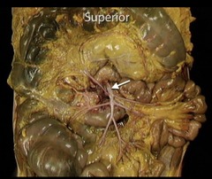
<p>In this anterior view of the contents of the abdomen, the greater omentum and transverse colon have been reflected superiorly. Identify the vessel indicated by the arrow.</p><p>Inferior mesenteric artery</p><p>Intestinal artery</p><p>Portal vein</p><p>Superior mesenteric artery</p>
5
New cards

Superficial palmar arch

In this view of the palm of the hand, identify the vessel indicated by the arrow.

Common palmar digital artery

Deep branch of ulnar artery

Deep palmar arch

Palmer digital artery

Superficial palmar arch

<p>In this view of the palm of the hand, identify the vessel indicated by the arrow.</p><p>Common palmar digital artery</p><p>Deep branch of ulnar artery</p><p>Deep palmar arch</p><p>Palmer digital artery</p><p>Superficial palmar arch</p>
6
New cards

Mesentery

In this anterior view of abdominal contents, identify the feature indicated by the arrow.

Gastrocolic ligament

Mesentery

Sigmoid mesocolon

Suspensory ligament of Trietz

<p>In this anterior view of abdominal contents, identify the feature indicated by the arrow.</p><p>Gastrocolic ligament</p><p>Mesentery</p><p>Sigmoid mesocolon</p><p>Suspensory ligament of Trietz</p>
7
New cards

Extensor carpi radialis longus

In this posterior view of a partially dissected right forearm, identify the muscle indicated by the arrows.

Brachioradialis

Extensor carpi radialis longus

Extensor carpi ulnaris

Flexor carpi radialis

<p>In this posterior view of a partially dissected right forearm, identify the muscle indicated by the arrows.</p><p>Brachioradialis</p><p>Extensor carpi radialis longus</p><p>Extensor carpi ulnaris</p><p>Flexor carpi radialis</p>
8
New cards

Parietal pericardium

In this anterior view of the heart in situ, identify the structure indicated by the arrow.

Epicardium

Parietal pleura

Parietal pericardium

Visceral pleura
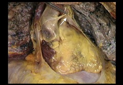
<p>In this anterior view of the heart in situ, identify the structure indicated by the arrow.</p><p>Epicardium</p><p>Parietal pleura</p><p>Parietal pericardium</p><p>Visceral pleura</p>
9
New cards

Arterial arcade

In this anterior view of abdominal contents, identify the vessel indicated by the arrow.

Arterial arcade

Inferior mesenteric artery

Intestinal artery

Superior mesenteric artery

<p>In this anterior view of abdominal contents, identify the vessel indicated by the arrow.</p><p>Arterial arcade</p><p>Inferior mesenteric artery</p><p>Intestinal artery</p><p>Superior mesenteric artery</p>
10
New cards

Extensor pollicis longus

In this lateral view of the right hand, identify the muscle whose tendon is indicated by the arrow.

Abductor pollicis longus

Extensor digitorum

Extensor pollicis brevis

Extensor pollicis longus

<p>In this lateral view of the right hand, identify the muscle whose tendon is indicated by the arrow.</p><p>Abductor pollicis longus</p><p>Extensor digitorum</p><p>Extensor pollicis brevis</p><p>Extensor pollicis longus</p>
11
New cards

Visceral pleura

What membrane is indicated by the arrow?

Diaphragmatic pleura

Hilum of lung

Parietal pleura

Pulmonary ligament

Visceral pleura

<p>What membrane is indicated by the arrow?</p><p>Diaphragmatic pleura</p><p>Hilum of lung</p><p>Parietal pleura</p><p>Pulmonary ligament</p><p>Visceral pleura</p>
12
New cards

Intercostal nerve

This is a transverse section of the thorax, which of the following structure is NOT located within the red circle?

Aorta

Azygos vein

Esophagus

Intercostal nerve
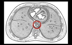
<p>This is a transverse section of the thorax, which of the following structure is NOT located within the red circle?</p><p>Aorta</p><p>Azygos vein</p><p>Esophagus</p><p>Intercostal nerve</p>
13
New cards

Arcuate line

Identify the structure(s). indicated by the arrows

Anterior layer of rectus sheath

Arcuate line

Linea alba

Semilunar line

Tendinous intersections

<p>Identify the structure(s). indicated by the arrows</p><p>Anterior layer of rectus sheath</p><p>Arcuate line</p><p>Linea alba</p><p>Semilunar line</p><p>Tendinous intersections</p>
14
New cards

Pronator quadratus

In this view of the right forearm, what muscle attaches to the areas painted in blue and red, as indicated by the arrows?

Biceps brachil

Brachioradialis muscle

Pronator quadratus

<p>In this view of the right forearm, what muscle attaches to the areas painted in blue and red, as indicated by the arrows?</p><p>Biceps brachil</p><p>Brachioradialis muscle</p><p>Pronator quadratus</p>
15
New cards

Cystic duct

In this close-up view of the dissected porta hepatis, identify the structure indicated by the arrow.

Common bile duct

Common hepatic duct

Cystic duct

Left hepatic duct

<p>In this close-up view of the dissected porta hepatis, identify the structure indicated by the arrow.</p><p>Common bile duct</p><p>Common hepatic duct</p><p>Cystic duct</p><p>Left hepatic duct</p>
16
New cards

Radial artery

In this anterior view of the right forearm and wrist, identify the vessel indicated by the arrow.

Basilic vein

Brachial artery

Cephalic vein

Radial artery

<p>In this anterior view of the right forearm and wrist, identify the vessel indicated by the arrow.</p><p>Basilic vein</p><p>Brachial artery</p><p>Cephalic vein</p><p>Radial artery</p>
17
New cards

Opening of coronary sinus

In this lateral view of the right side of the heart, identify the feature indicated by the arrow.

Fossa ovalis

Opening of coronary sinus

Opening of inferior vena cava

Opening of superior vena cava

<p>In this lateral view of the right side of the heart, identify the feature indicated by the arrow.</p><p>Fossa ovalis</p><p>Opening of coronary sinus</p><p>Opening of inferior vena cava</p><p>Opening of superior vena cava</p>
18
New cards

Flexor digitorum superficialis

In this anterior view of the right hand, identify the muscle whose tendon is indicated by the arrows.

Extensor digiti minimi

Extensor indicis

Flexor digitorum profundus

Flexor digitorum superficialis

<p>In this anterior view of the right hand, identify the muscle whose tendon is indicated by the arrows.</p><p>Extensor digiti minimi</p><p>Extensor indicis</p><p>Flexor digitorum profundus</p><p>Flexor digitorum superficialis</p>
19
New cards

Rhomboid major

In this posterior view of the right shoulder and back, identify the muscle indicated by the arrow.

Levator scapulae

Rhomboid major

Rhomboid minor

Serratus posterior superior

<p>In this posterior view of the right shoulder and back, identify the muscle indicated by the arrow.</p><p>Levator scapulae</p><p>Rhomboid major</p><p>Rhomboid minor</p><p>Serratus posterior superior</p>
20
New cards

Ulnar artery

In this anterior view of the elbow, identify the artery indicated by the arrow.

Brachial artery

Posterior interosseous artery

Posterior ulnar recurrent artery

Radial artery

Ulnar artery

<p>In this anterior view of the elbow, identify the artery indicated by the arrow.</p><p>Brachial artery</p><p>Posterior interosseous artery</p><p>Posterior ulnar recurrent artery</p><p>Radial artery</p><p>Ulnar artery</p>
21
New cards

Splenic

In this anterior view of the dissected upper abdomen, in which the stomach has been removed, identify the artery indicated by

Celiac trunk

Common hepatic

Left gastric

Splenic

<p>In this anterior view of the dissected upper abdomen, in which the stomach has been removed, identify the artery indicated by</p><p>Celiac trunk</p><p>Common hepatic</p><p>Left gastric</p><p>Splenic</p>
22
New cards

Teniae coli

In this view of the large intestine, identify the feature indicated by the arrow.

Epiploic (omental) appendices

Diverticulum

Mesoappendix

Plicae circulares

Teniae coli

<p>In this view of the large intestine, identify the feature indicated by the arrow.</p><p>Epiploic (omental) appendices</p><p>Diverticulum</p><p>Mesoappendix</p><p>Plicae circulares</p><p>Teniae coli</p>
23
New cards

Round ligament of the liver (ligamentum teres)

In this anterior view of the contents of the right upper abdomen, identify the structure indicated by the arrow.

Coronary ligament

Hepatoduodenal ligament

Left triangular ligament

Lesser omentum

Round ligament of the liver (ligamentum teres)

<p>In this anterior view of the contents of the right upper abdomen, identify the structure indicated by the arrow.</p><p>Coronary ligament</p><p>Hepatoduodenal ligament</p><p>Left triangular ligament</p><p>Lesser omentum</p><p>Round ligament of the liver (ligamentum teres)</p>
24
New cards

Radial collateral ligament

In this lateral view of the right elbow, identify the structure indicated by the arrow.

Anular ligament

Interosseous membrane

Olecranon

Radial collateral ligament

<p>In this lateral view of the right elbow, identify the structure indicated by the arrow.</p><p>Anular ligament</p><p>Interosseous membrane</p><p>Olecranon</p><p>Radial collateral ligament</p>
25
New cards

Papillary muscle

In this lateral view of the right side of the heart, identify the structure(s) indicated by the arrow.

Chordae tendineae

Papillary muscle

Pectinate muscle

Trabeculae carneae

<p>In this lateral view of the right side of the heart, identify the structure(s) indicated by the arrow.</p><p>Chordae tendineae</p><p>Papillary muscle</p><p>Pectinate muscle</p><p>Trabeculae carneae</p>
26
New cards

Recurrent motor branch of median nerve

In this palmar view of the right hand, identify the nerve indicated by the arrow.

Common digital branch of median nerve

Musculocutaneous nerve

Palmar digital branch of median nerve

Recurrent motor branch of median nerve

<p>In this palmar view of the right hand, identify the nerve indicated by the arrow.</p><p>Common digital branch of median nerve</p><p>Musculocutaneous nerve</p><p>Palmar digital branch of median nerve</p><p>Recurrent motor branch of median nerve</p>
27
New cards

Transverse pericardial sinus

Name the structure through which the index finger is passing?

Auricle

Coronary sinus

Oblique pericardial sinus

Transverse pericardial sinus

<p>Name the structure through which the index finger is passing?</p><p>Auricle</p><p>Coronary sinus</p><p>Oblique pericardial sinus</p><p>Transverse pericardial sinus</p>
28
New cards

Common bile duct

In this anterior view of the dissected upper abdomen, in which the stomach has been removed, identify the structure indicated by the arrow.

Common bile duct

Common hepatic duct

Cystic duct

Hepatic artery proper

<p>In this anterior view of the dissected upper abdomen, in which the stomach has been removed, identify the structure indicated by the arrow.</p><p>Common bile duct</p><p>Common hepatic duct</p><p>Cystic duct</p><p>Hepatic artery proper</p>
29
New cards

Lumbrical

In this anterior view of the right hand, identify the muscle indicated by the arrow.

Adductor pollicis

Flexor digitorum superficialis

Flexor pollicis brevis

Lumbrical

<p>In this anterior view of the right hand, identify the muscle indicated by the arrow.</p><p>Adductor pollicis</p><p>Flexor digitorum superficialis</p><p>Flexor pollicis brevis</p><p>Lumbrical</p>
30
New cards

Coronary sinus

In this posterior view of the heart, identify the vessel indicated by the arrows.

Anterior interventricular artery

Circumflex artery

Coronary sinus

Marginal artery

<p>In this posterior view of the heart, identify the vessel indicated by the arrows.</p><p>Anterior interventricular artery</p><p>Circumflex artery</p><p>Coronary sinus</p><p>Marginal artery</p>
31
New cards

Extensor digiti minimi

In this posterior view of the right forearm and hand, identify the muscle whose tendon is indicated by the arrows.

Abductor pollicis longus

Extensor digiti minimi

Extensor indicis

<p>In this posterior view of the right forearm and hand, identify the muscle whose tendon is indicated by the arrows.</p><p>Abductor pollicis longus</p><p>Extensor digiti minimi</p><p>Extensor indicis</p>
32
New cards

Tunica albuginea

In this sagittal section of the isolated testis, identify the structure indicated by the arrow.

Dartos muscle

Epididymis

Mediastinum of testis

Tunica albuginea

<p>In this sagittal section of the isolated testis, identify the structure indicated by the arrow.</p><p>Dartos muscle</p><p>Epididymis</p><p>Mediastinum of testis</p><p>Tunica albuginea</p>
33
New cards

Internal abdominal oblique

Identify the muscle indicated by the arrow.

External abdominal oblique

Internal abdominal oblique

Pyramidalis

Rectus abdominis

<p>Identify the muscle indicated by the arrow.</p><p>External abdominal oblique</p><p>Internal abdominal oblique</p><p>Pyramidalis</p><p>Rectus abdominis</p>
34
New cards

Brachialis

In this view of the right upper limb, what muscle has its proximal attachment to the area painted in red and its distal attachment to the area painted in blue, as indicated by the arrows?

Anconeus

Biceps brachii

Brachialis

<p>In this view of the right upper limb, what muscle has its proximal attachment to the area painted in red and its distal attachment to the area painted in blue, as indicated by the arrows?</p><p>Anconeus</p><p>Biceps brachii</p><p>Brachialis</p>
35
New cards

Right coronary artery

In this lateral view of the heart, identify the vessel indicated by the arrows. (The auricle has been removed for better viewing.)

Anterior interventricular artery

Circumflex artery

Left coronary artery

Right coronary artery

<p>In this lateral view of the heart, identify the vessel indicated by the arrows. (The auricle has been removed for better viewing.)</p><p>Anterior interventricular artery</p><p>Circumflex artery</p><p>Left coronary artery</p><p>Right coronary artery</p>
36
New cards

Superior mesenteric vein

In this close-up view of the structures on the posterior abdominal wall, identify the vessel indicated by the arrow.

Pancreas

Inferior mesenteric vein

Inferior phrenic vein

Portal vein

Superior mesenteric vein

<p>In this close-up view of the structures on the posterior abdominal wall, identify the vessel indicated by the arrow.</p><p>Pancreas</p><p>Inferior mesenteric vein</p><p>Inferior phrenic vein</p><p>Portal vein</p><p>Superior mesenteric vein</p>
37
New cards

Opponens pollicis

In this anterior view of the right hand, identify the muscle indicated by the arrow.

You can respond once

Abductor pollicis brevis

Adductor pollicis

First dorsal interosseous

Flexor pollicis brevis

Opponens pollicis

<p>In this anterior view of the right hand, identify the muscle indicated by the arrow.</p><p>You can respond once</p><p>Abductor pollicis brevis</p><p>Adductor pollicis</p><p>First dorsal interosseous</p><p>Flexor pollicis brevis</p><p>Opponens pollicis</p>
38
New cards

Left coronary artery

In this lateral view of the heart, identify the vessel indicated by the arrow.

Anterior interventricular artery

Circumflex artery

Left coronary artery

Posterior interventricular arterv

<p>In this lateral view of the heart, identify the vessel indicated by the arrow.</p><p>Anterior interventricular artery</p><p>Circumflex artery</p><p>Left coronary artery</p><p>Posterior interventricular arterv</p>
39
New cards

Epididymis

In this lateral view of the isolated testis, identify the structure indicated by the arrow.

Dartos muscle

Ductus deferens

Epididymis

Seminiferous tubules

<p>In this lateral view of the isolated testis, identify the structure indicated by the arrow.</p><p>Dartos muscle</p><p>Ductus deferens</p><p>Epididymis</p><p>Seminiferous tubules</p>
40
New cards

Middle colic

In this anterior view of the contents of the abdomen, the greater omentum and transverse colon have been reflected superiorly. Identify the artery indicated by the arrow.

Ileo-colic

Intestinal artery

Middle colic
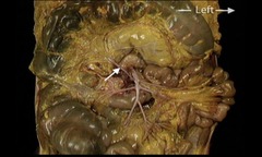
<p>In this anterior view of the contents of the abdomen, the greater omentum and transverse colon have been reflected superiorly. Identify the artery indicated by the arrow.</p><p>Ileo-colic</p><p>Intestinal artery</p><p>Middle colic</p>
41
New cards

left to right shunt

Congenital anomalies can be seen in the interatrial septum (patent fossa ovale) or interventricular septum. What is the common outcome of both?

left heart failure

left to right shunt

no common outcome

right to left shunt

<p>Congenital anomalies can be seen in the interatrial septum (patent fossa ovale) or interventricular septum. What is the common outcome of both?</p><p>left heart failure</p><p>left to right shunt</p><p>no common outcome</p><p>right to left shunt</p>
42
New cards

Tail of pancreas

In this anterior view of the dissected upper abdomen, in which the stomach and part of the liver has been removed, identify the structure indicated by the arrow.

Duodenum

Spleen

Tail of pancreas

<p>In this anterior view of the dissected upper abdomen, in which the stomach and part of the liver has been removed, identify the structure indicated by the arrow.</p><p>Duodenum</p><p>Spleen</p><p>Tail of pancreas</p>
43
New cards

Segmental bronchus

In this anterior view of an isolated pulmonary tree, identify the structure indicated by the arrow.

Carina

Primary bronchus

Secondary (lobar) bronchus

Segmental bronchus

<p>In this anterior view of an isolated pulmonary tree, identify the structure indicated by the arrow.</p><p>Carina</p><p>Primary bronchus</p><p>Secondary (lobar) bronchus</p><p>Segmental bronchus</p>
44
New cards

D

In this posterior view of the heart, choose the arrow that identifies the superior vena cava.

A

B

C

D

E

<p>In this posterior view of the heart, choose the arrow that identifies the superior vena cava.</p><p>A</p><p>B</p><p>C</p><p>D</p><p>E</p>
45
New cards

Scaphoid

In this skeleton of the right wrist, identify the bone indicated by the arrow.

Capitate

Hamate

Lunate

Scaphoid

<p>In this skeleton of the right wrist, identify the bone indicated by the arrow.</p><p>Capitate</p><p>Hamate</p><p>Lunate</p><p>Scaphoid</p>
46
New cards

Deep branch of radial nerve

In this lateral view of the right elbow and forearm, identify the nerve indicated by the arrow.

Deep branch of radial nerve

Median nerve

Musculocutaneous nerve

Superficial branch of radial nerve

<p>In this lateral view of the right elbow and forearm, identify the nerve indicated by the arrow.</p><p>Deep branch of radial nerve</p><p>Median nerve</p><p>Musculocutaneous nerve</p><p>Superficial branch of radial nerve</p>
47
New cards

Lateral cord of brachial plexus

In this anterior view of the right axilla, identify the structure indicated by the arrow.

Medial cord of brachial plexus

Musculocutaneous nerve

Lateral cord of brachial plexus

Posterior cord of brachial plexus

<p>In this anterior view of the right axilla, identify the structure indicated by the arrow.</p><p>Medial cord of brachial plexus</p><p>Musculocutaneous nerve</p><p>Lateral cord of brachial plexus</p><p>Posterior cord of brachial plexus</p>
48
New cards

Greater tubercle

In this view of the right humerus, identify the structure indicated by the arrow.

Bicipital groove

Deltoid tuberosity

Greater tubercle

Head

<p>In this view of the right humerus, identify the structure indicated by the arrow.</p><p>Bicipital groove</p><p>Deltoid tuberosity</p><p>Greater tubercle</p><p>Head</p>
49
New cards

Long thoracic nerve

In this anterior view of the right axilla, identify the structure indicated by the forceps and the arrow.

Axillary nerve

Long thoracic nerve

Subscapular nerve

Thoracodorsal nerve

<p>In this anterior view of the right axilla, identify the structure indicated by the forceps and the arrow.</p><p>Axillary nerve</p><p>Long thoracic nerve</p><p>Subscapular nerve</p><p>Thoracodorsal nerve</p>
50
New cards

Superficial branch of radial nerve

In this posterior view of the right hand, identify the nerve indicated by the arrow.

Deep branch of radial nerve

Median nerve

Musculocutaneous nerve

Superficial branch of radial nerve

<p>In this posterior view of the right hand, identify the nerve indicated by the arrow.</p><p>Deep branch of radial nerve</p><p>Median nerve</p><p>Musculocutaneous nerve</p><p>Superficial branch of radial nerve</p>
51
New cards

Median nerve

In this anterior view of the right elbow and forearm, identify the nerve indicated by the arrows.

Deep branch of radial nerve

Median nerve

Musculocutaneous nerve

Superficial branch of radial nerve
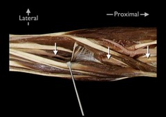
<p>In this anterior view of the right elbow and forearm, identify the nerve indicated by the arrows.</p><p>Deep branch of radial nerve</p><p>Median nerve</p><p>Musculocutaneous nerve</p><p>Superficial branch of radial nerve</p>
52
New cards

Acromion

Identify the bony feature indicated by the arrow

Acromion

Coracoid process

Glenoid fossa

Spine of scapula

Supraglenoid tubercle

<p>Identify the bony feature indicated by the arrow</p><p>Acromion</p><p>Coracoid process</p><p>Glenoid fossa</p><p>Spine of scapula</p><p>Supraglenoid tubercle</p>



