Necrosis components / Exudates
1/67
There's no tags or description
Looks like no tags are added yet.
Name | Mastery | Learn | Test | Matching | Spaced | Call with Kai |
|---|
No analytics yet
Send a link to your students to track their progress
68 Terms
necrosis cause: Ischemia, free radicals, toxins, burns, x-rays, Nutritional (WMD = Vit E + selenium deficiency)
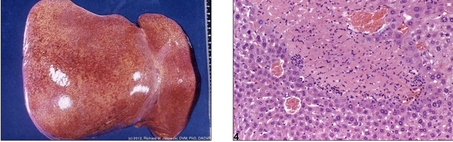
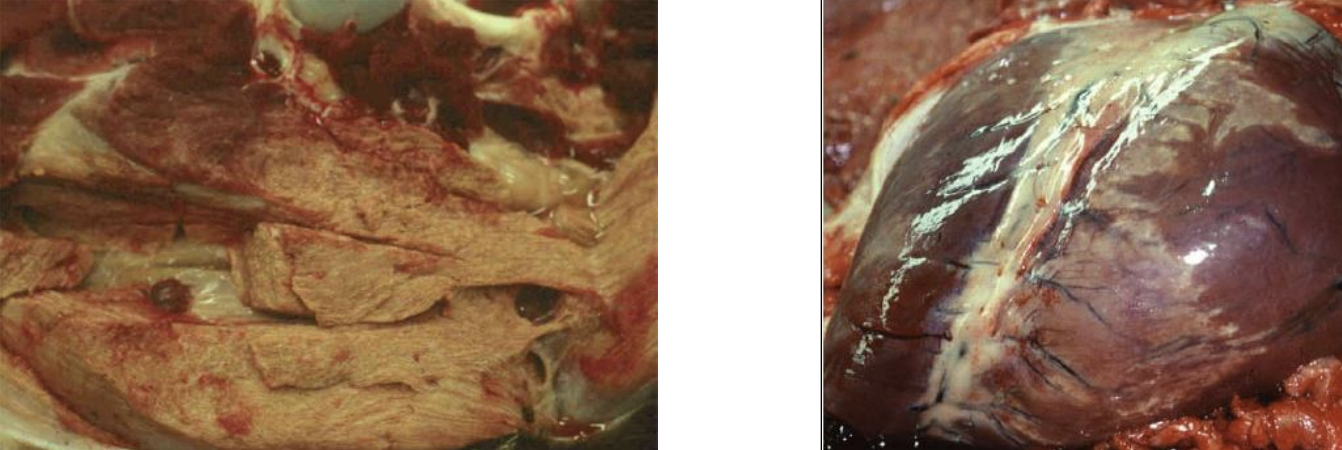
coagualative
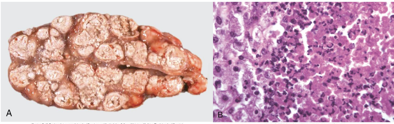
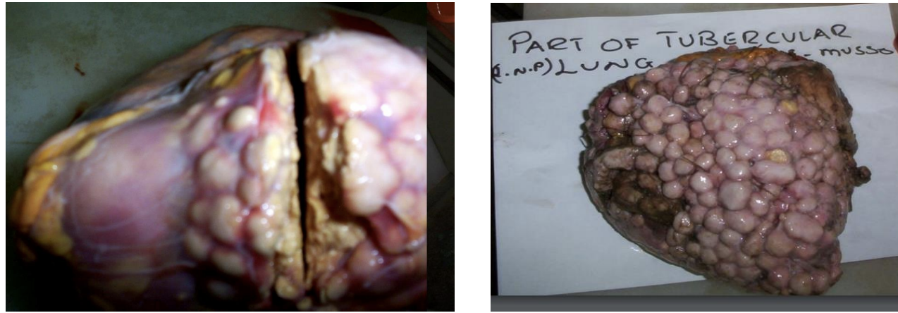
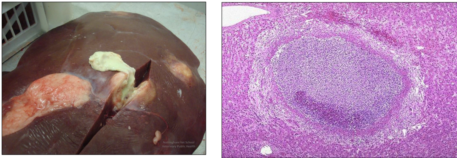
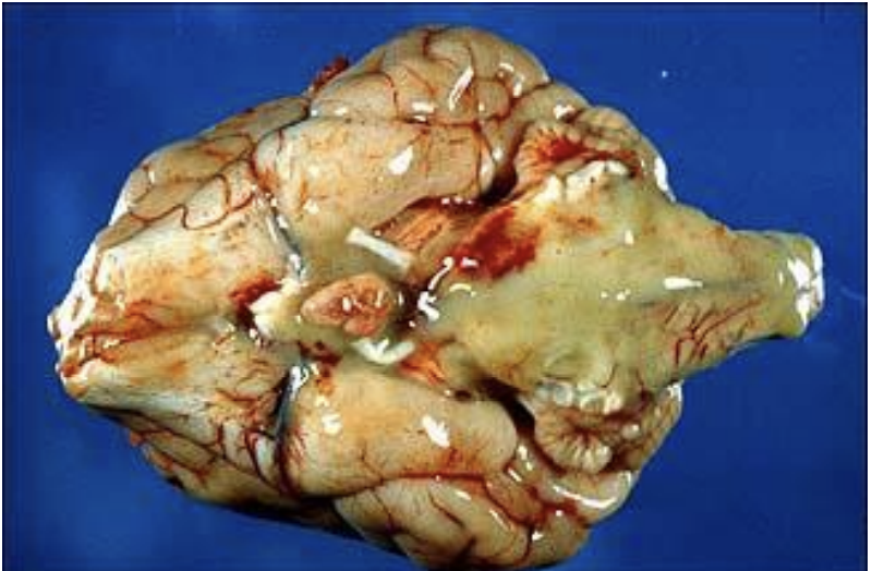
necrosis cause: Toxins of certain microorganisms (tuberculosis)
caseous
necrosis causee: ischemia, free radicals, toxins, burns, x-rays
liquefactive
necrosis cause: Infection, Coagulative + liquefactive combo, Distal extremities or dependent parts of organs
wet gangrene
necrosis cause: Loss of blood supply resulting in coagulative necrosis, Distal extremities or dependent parts of organs
dry gangrene
necrosis cause: Enzymatic, traumatic, idiopathic, nutritional (yellow fat dz, stealitis)
fat
necrosis gross appearance: Grey/white (unless mixed w/ blood), Depressed compared to surrounding tissue
coagulative
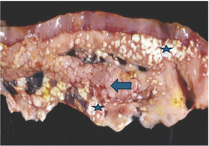
necrosis gross appearance: Cheese, milk curds, dry greasy, breaks easily
caseous
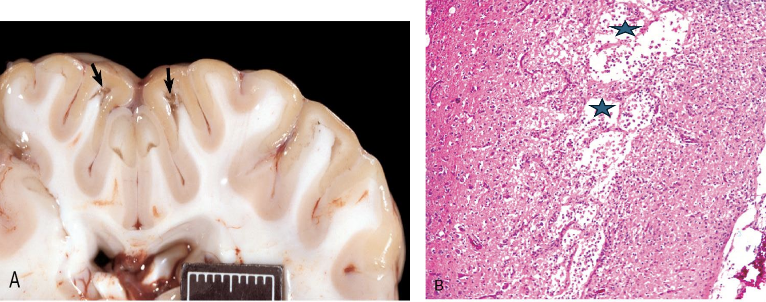
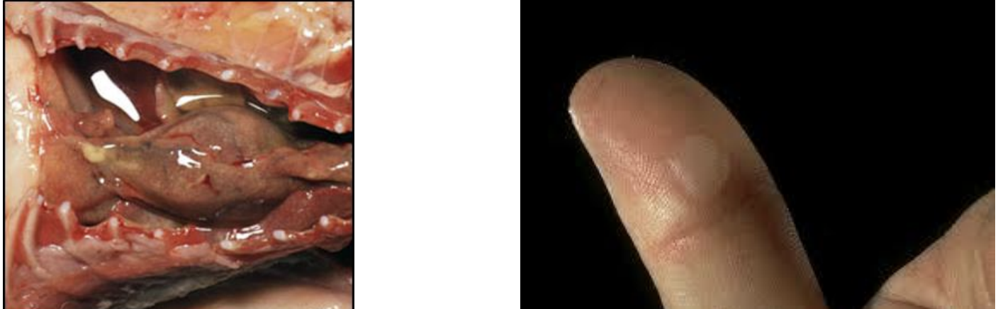
necrosis gross appearance: CNS! Abscesses/cavities containing yellowish fluid Necrotic tissue converted to liquid
liquefactive
necrosis gross appearance: Red+black WET tissue
wet gangrene
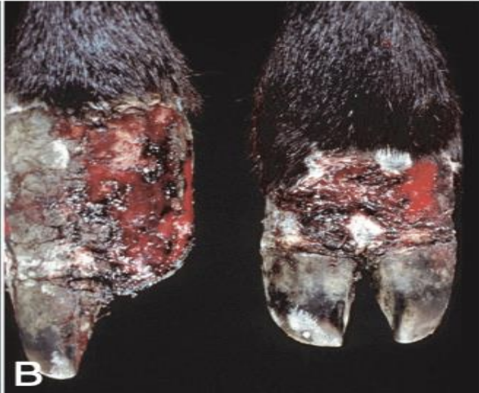
necrosis gross appearance: Dry, leathery, hard, MUMMIFICATION (no bacterial involvement)
dry gangrene
necrosis gross appearance: White, opaque, granular
fat
necrosis histological appearance: outline of necrotic cell = preserved
acidophilic/eosinophilic cytoplasm
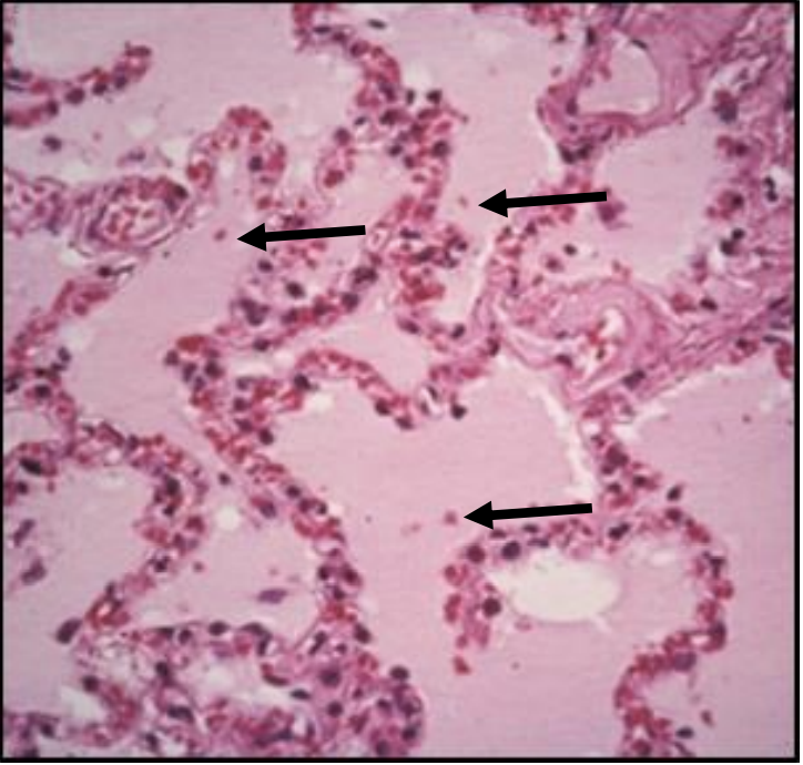
Nuclear changes
-Pyknosis - Shrunk, round dark
-Karyorrhexis - fragmented
-Karyolysis - Dissolution
coagulative
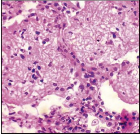
necrosis histological appearance:
Granular stains purple Loss of cell outline, normal tissue architecture |
caseous
necrosis histological appearance: Clear spaces with or without pink staining
Proteinaceous precipitate in necrotic area
Leukocytes present and produce hydrolytic enzymes
liquefactive
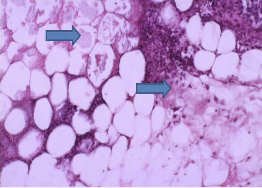
necrosis histological appearance: Shadowy outline w/ blue or purple (calcification) in areas of necrosis
Adipose tissue with saponification
fat
necrosis
caseous
necrosis
caseous
necrosis
liquefactive
necrosis
liquefactive
necrosis
fat
necrosis
fat
necrosis
dry gangrene
necrosis
wet gangrene
necrosis
coagulative necrosis
necrosis
coagulative necrosis

whats arrow 1
pyknosis
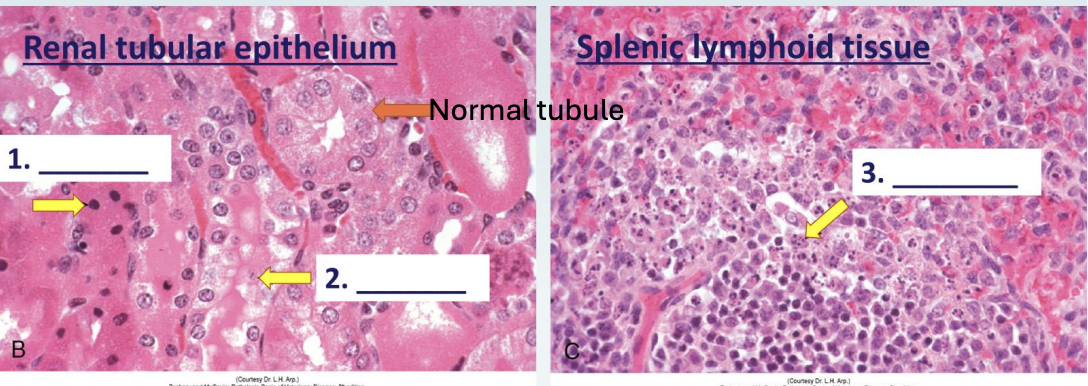
whats arrow 2
karyolysis

whats arrow 3
keryorrhetic
Cell shrinkage, fragmentation, NO INFLAMMATION
apoptosis
membrane rupture, INFLAMMATION, protein denaturation, dead contiguous cell
necrosis
extrinsic initiation phase apoptosis
death receptor initiated
intrinsic apoptosis initiation phase
mitochondrial
what activates apoptosis
capsase 3 cascade
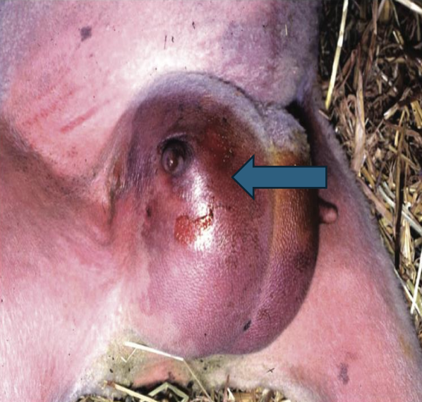
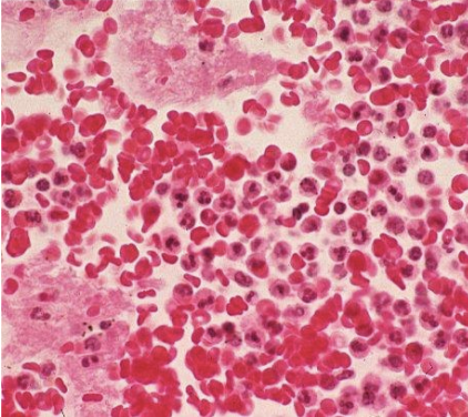
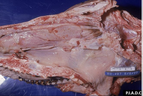
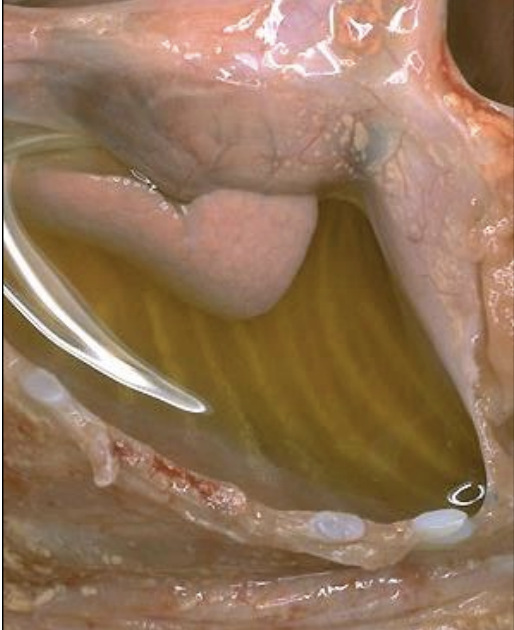
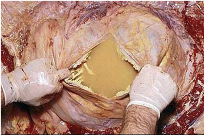
exudate: clear straw colored fluid in cavities, skin, lungs or mucosal surfaces
Microscopically: very few cells, homogenous, pink (many soluble proteins)
Often seen in mild/early injury that causes increased vascular permeability
serous
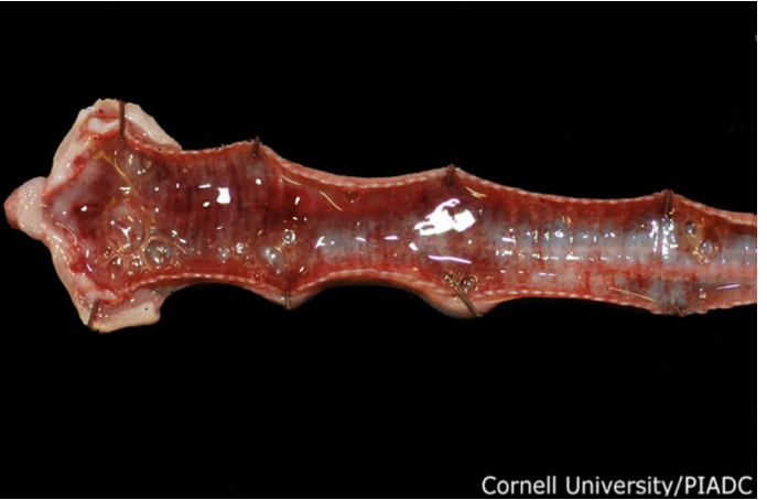
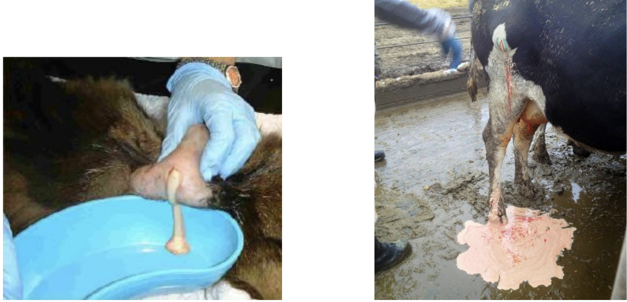
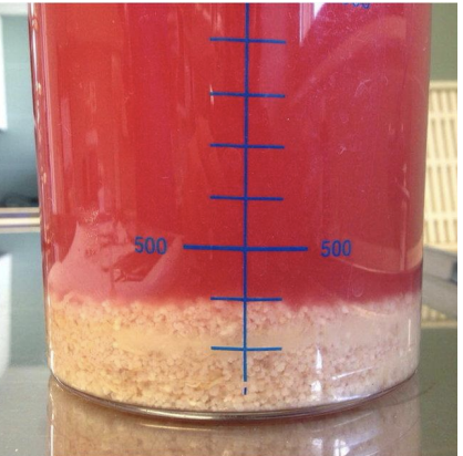
exudate: red tinged fluid because of widespread diapedesis/mild vascular damage
serohemorrhagic
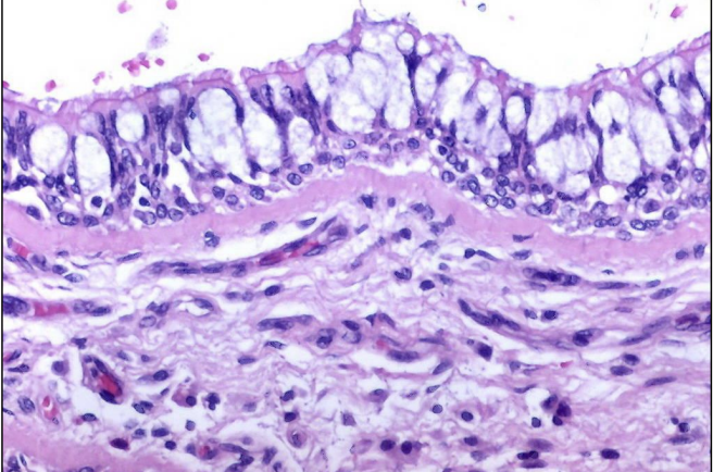
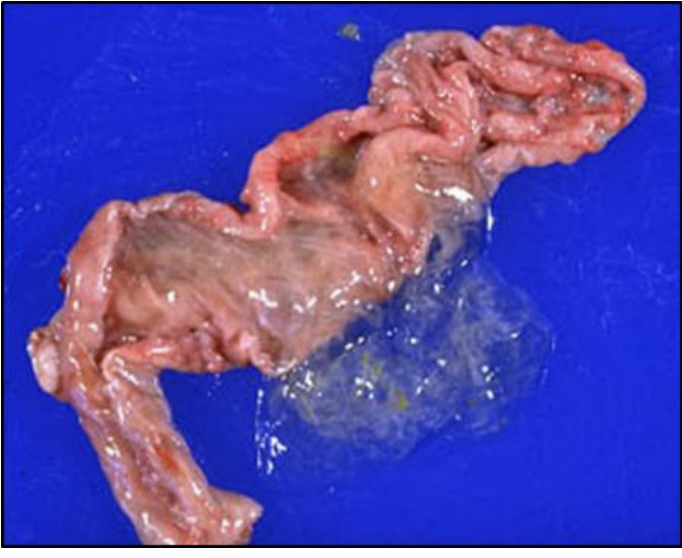
exudate: clear, snot-like consistency from mucous membranes in GI, Resp or urogenital tract
Goblet cell hypertrophy/hyperplasia
Microscopically: deep purple, trapped air, fat and leukocytes
catarrhal
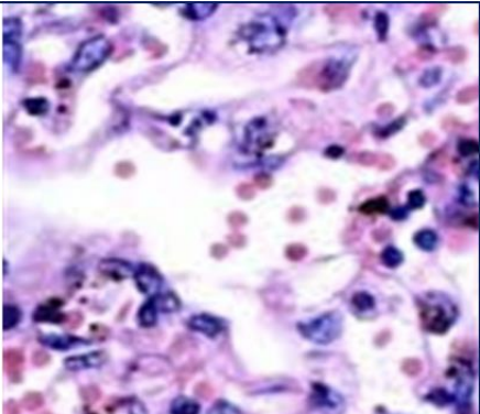
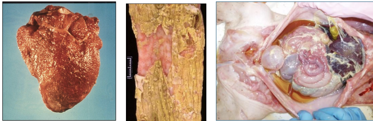
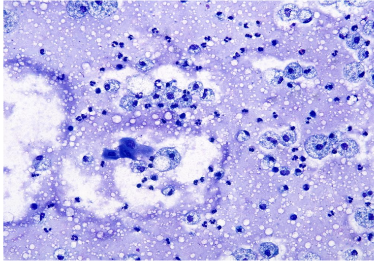
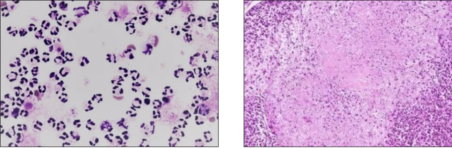
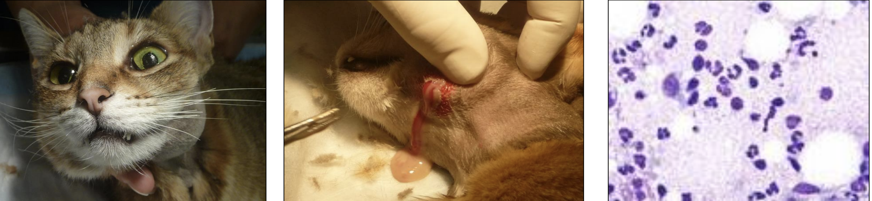
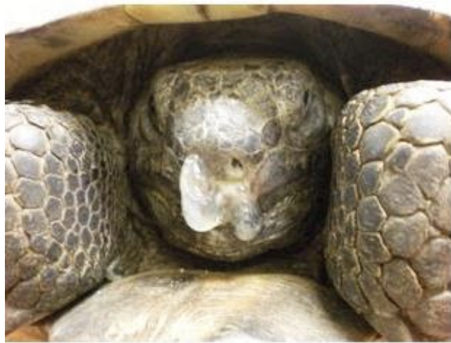
exudate: Thick, yellow, opaque → often bacterial component, (Pyo-) often prefix Associated with Abscesses
Microscopically: Neutrophils! With liquefactive necrosis due to granulation and digestion by non-specific enzymes
purulent / suppurative
con of purulent exudate
Massive destruction of host tissue
exudate: pale, flat, no fluid
lymphoplasmacytic
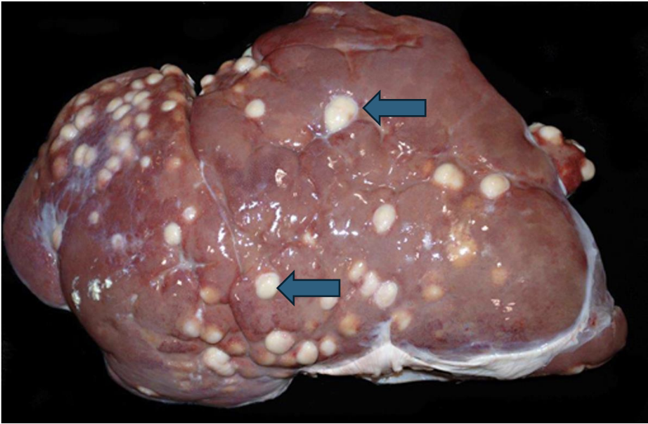
exudate: white, solid, raised tissue
distinct aggregates of macrophages that surround and wall off an inciting cause (pathogen, foreign material) → over time, becomes encapsulated with connective tissue (often has necrotic center)
granulomatous
2 types of chronic exudates
lymphoplasmacytic and granulomatous
hemorrhage by diapedesis
is the escape of blood cells through the walls of small vessels into surrounding tissues, often resulting in localized bleeding and tissue damage.
true hemorrhage
is the escape of blood from a vessel into body spaces or tissues, leading to significant bleeding. This can occur due to trauma, rupture of a blood vessel, or erosion of a vessel wall.
hemorrhage type
hemorrhage by diapedesis
hemorrhage type
true hemorrhage
exudate
serous
exudate
serous
exudate
fibrinous
exudate
fibrinous
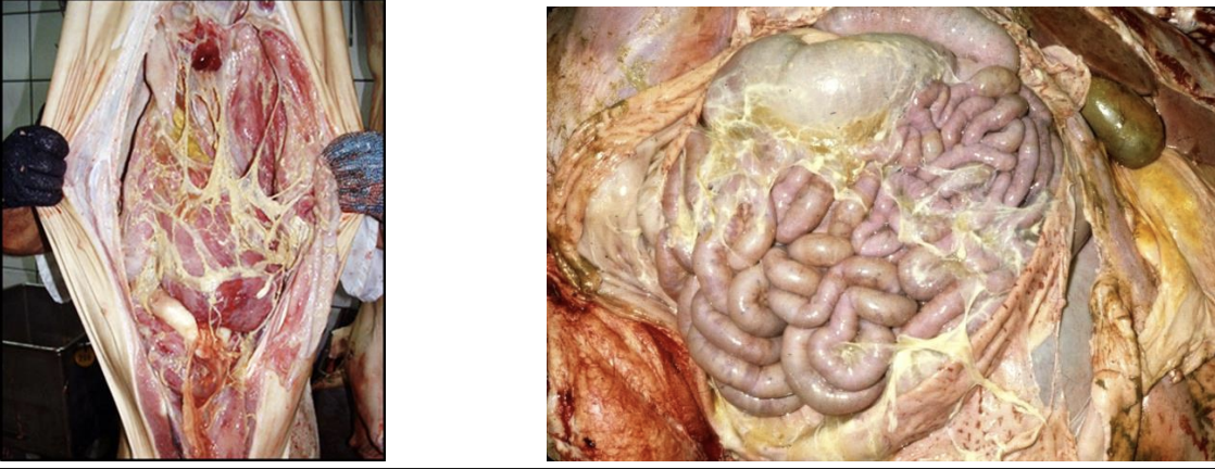
exudate
fibrinous
exudate that can be pulled off like a string; involved in clotting and is an acute exudate in inflammation
fibrinous
describes connective (scar) tissue, a chronic sequela of fibrin that is involved in healing -Cannot be pulled off
fibrous
exudate
catarrhal
exudate
catarrhal
exudate
catarrhal
exudate
catarrhal
exudate associated with Gastrointestinal tract Respiratory tract Urogenital tract
catarrhal
exudate
catarrhal
exudate
purulent/suppurative
exudate
purulent/suppurative
exudate
purulent/suppurative
exudate
purulent/suppurative
exudate
suppurative
exudate
serous
exudate
catarrhal
exudate
Fibrinopurulent
exudate
Serohemorrhagic