Vital Signs Lecture NURN 155 Exam 1
 Call Kai
 Learn
Learn Practice Test
 Spaced Repetition
 Match
Match Flashcards
 Knowt Play
1/57

There's no tags or description
Looks like no tags are added yet.
Name | Mastery | Learn | Test | Matching | Spaced | Call with Kai | Chat |
|---|
No analytics yet
Send a link to your students to track their progress
58 Terms
What vital signs are included in this PowerPoint? (The order)
Blood pressure (BP) Temperature (T) Pulse (P) Respirations (R) Pulse oximetry (Pulse Ox)
Why are vital signs assessed?
To: Establish baseline data Evaluate health status Detect changes Identify problems and guide plans/interventions
When should vital signs be obtained?
During: Admission and discharge, Home-health visits, Before, during, and after surgery/invasive procedures, Blood transfusions, Changes in condition, Medication administration, Procedures, Nonspecific symptoms of distress
What general nursing guidelines apply when obtaining vital signs?
Consider: Delegation, Interventions, Documentation, Analysis, Communication, Patient education.
What should the nurse consider when interpreting a vital sign?
Consider: Equipment, Baseline Patient history, Environment, Trends, and Other influencing factors. What needs to be documented/reported?
Exam mindset: Don't judge one number alone—compare it with the patient's baseline and trends.
What general information should be documented with vital signs?
Numeric value, Patient position, Site, and Relevant notes.
Accurate recording is mandatory: If it is not recorded, it was not done. II. Blood Pressure
What is blood pressure (BP)?
The force of blood against the walls of the arteries as blood is pumped from the heart.
Why are abnormal blood pressures important?
Hypertension: can weaken/damage the heart and increases risk for vascular disease, MI, CVA, renal disease, and dementia. Hypotension: can decrease oxygen and nutrient perfusion, which may lead to cellular death.
What do systolic and diastolic blood pressures represent?
Systolic: pressure in the arteries during ventricular contraction. Diastolic: lowest pressure between heartbeats during relaxation.
What are hypertension, hypotension, and orthostatic blood pressure?
Hypertension: consistent elevation of systolic or diastolic BP above 140/90 mmHg according to the terminology slide.
Hypotension: low blood pressure.
Orthostatic: drop in systolic BP of more than 20 mmHg when rising to sit or stand.
What physiological factors influence blood pressure?
Cardiac output, Peripheral vascular resistance, Blood volume, Blood viscosity, Arterial elasticity, Pulse pressure
What patient factors can influence blood pressure?
Age, Gender, Race, Social determinants, Diurnal rhythm, Weight, Exercise, Emotions, Stress III. AHA Blood Pressure Categories
What BP is classified as normal on the PowerPoint's AHA chart?
Systolic <120 AND diastolic <80 mmHg
What BP is classified as elevated?
Systolic 120–129 AND diastolic <80 mmHg
What is Stage 1 hypertension?
Systolic 130–139 OR diastolic 80–89 mmHg
What is Stage 2 hypertension?
Systolic ≥140 OR diastolic ≥90 mmHg
What does the AHA chart identify as severe hypertension or hypertensive emergency range?
Systolic >180 and/or diastolic >120 mmHg. The slide distinguishes severe hypertension from a hypertensive emergency based on whether concerning symptoms are present. Application: A BP of 136/76 = Stage 1 because the systolic value alone meets Stage 1 criteria. IV. Blood Pressure Equipment & Correct Technique
What equipment is used to manually assess blood pressure?
Aneroid sphygmomanometer, BP cuff with an inflatable bladder, Appropriate cuff size, Stethoscope.
How is the correct upper-arm BP cuff size determined?
Based on the midpoint circumference of the limb: Width = 40% of arm circumference Length = 80% of arm circumference
How should a BP cuff be positioned?
Snugly on the arm 1–2 inches above the inner elbow Bladder positioned over the artery Manometer at eye level Do not measure over clothing
What conditions help obtain an accurate blood pressure?
Quiet environment, bare arm. Correct cuff size. Patient sitting with feet flat. Back/feet supported, Legs uncrossed, arm supported at heart level. The patient remains quiet. Maintain standard precautions. Assess contraindications. On an initial assessment, take BP in both arms and use a manual cuff.
What common errors can falsely alter a BP reading?
Examples shown in the PowerPoint include: Cuff too small Cuff over clothing Back/feet unsupported Legs crossed Not resting before measurement Talking Labored breathing Full bladder Pain Arm above or below heart level Application: If a patient's feet are dangling and they are talking, correct those conditions before trusting the reading.
How is the manual two-step BP method performed?
Inflate while palpating radial/brachial pulse. Note where pulse disappears. Inflate 30 mmHg above that point. Slowly deflate and note when pulse returns. Fully deflate; wait 30 seconds. Reinflate 30 mmHg above the disappearance point. Deflate at 2–4 mmHg/sec. First sound = systolic. Last sound = diastolic. Continue about 10 mmHg below last sound, fully deflate, and document. A measurement may be repeated after about 30–60 seconds.
When and how is the one-step manual BP method used?
When and how is the one-step manual BP method used?
When is it used?
When the patient’s usual/baseline systolic BP is already known
May also be used in an emergency when a quicker BP measurement is needed
How is it performed?
Use the arm with the strongest brachial pulse.
Apply the correct-size cuff to the bare upper arm.
Place the stethoscope over the brachial artery.
Inflate the cuff to 30 mmHg above the known systolic BP.
Deflate slowly at 2–4 mmHg/second.
Listen for Korotkoff sounds:
First sound = systolic BP
Last sound = diastolic BP
Continue deflating about 10 mmHg below the final sound, then release the cuff completely.
Document the BP.
Example: Baseline BP is 120/80 → inflate the cuff to about 150 mmHg.
When should an electronic BP machine NOT be used according to the PowerPoint?
With Irregular heart rate, shivering, seizures, inability to cooperate, and Systolic BP <90 mmHg, use the proper cuff size.
How is systolic BP obtained by palpation or Doppler?
Inflate while palpating the radial/brachial artery until the pulse disappears. Inflate 30 mmHg above that point. Slowly deflate. The pressure at which the pulse returns = systolic BP by palpation. Doppler uses the same approach with a Doppler device.
How are orthostatic blood pressure and pulse measured?
Have the patient lie supine for at least 5 minutes. Measure and record supine BP and pulse. Ask the patient to stand/ sit up. Measure BP and pulse at 1 minute and 3 minutes. If unable to stand, use upright sitting. Observe for dizziness, lightheadedness, or weakness.
What are Korotkoff sounds and which phases identify BP?
These are the sounds heard while auscultating blood pressure.
Phase I: first sharp tapping = systolic
Phase II: soft swishing
Phase III: rhythmic tapping
IV: muffling/fading
Phase V: sounds disappear = diastolic VI. Pulse
What are tachycardia and bradycardia in an adult?
Tachycardia: usually >100 beats/min. (Fast/ rapid heart rate)
Bradycardia: usually <60 beats/min. (Slow heart rate)
Normal adult heart rate in the PowerPoint: 60–100 beats/min.
What are stroke volume and cardiac output?
Stroke volume (SV): blood pumped with each contraction. Cardiac output (CO): blood pumped in 1 minute SV × HR = CO.
How can a pulse be assessed and what characteristics are evaluated?
Assessment: Palpation Auscultation with stethoscope Doppler Assess: Rate Rhythm Volume/strength Equality
What is the 0–4+ pulse-strength scale?
0: absent 1+: weak/thready; easily obliterated 2+: normal; easily identified 3+: increased 4+: full/bounding; cannot be obliterated
What factors can affect pulse?
Gender Stress Exercise Food Fever Disease Blood loss Medications
What major pulse sites should you recognize?
Temporal, carotid, apical, brachial, radial, ulnar, femoral, popliteal, posterior tibial, and dorsalis pedis.
Which pulse sites have special clinical uses?
Carotid: closest to the heart; emergencies; never palpate both simultaneously.
Radial: wrist below the thumb; most common in adults.
Brachial: BP; also used in children/infants.
Femoral: emergencies/cardiac procedures.
Posterior tibial, popliteal, and dorsalis pedis: lower-extremity circulation.
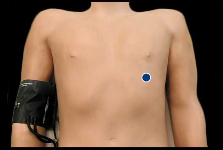
When should the apical pulse be used and where is it located?
Most accurate. Assesses rhythm. Use with an irregular rhythm
Location: 5th intercostal space, left midclavicular line. Document pulse site, rate, regular/irregular rhythm, strength, and bilateral symmetry.
What constitutes one respiration, and what is the normal adult respiratory rate?
One full inspiration + expiration = one respiration.
Normal adult rate: 12–20 breaths/minute.
What respiratory rates does the PowerPoint list by age?
Newborn: 35–40
Infant: 30–50
Toddler: 25–32
Child: 20–30
Adolescent: 16–20
Adult: 12–20 or 10-20.
(breaths/min)
What breathing patterns should you recognize visually from the PowerPoint?
Eupnea, Apnea, Tachypnea ,Bradypnea ,Agonal Cheyne-Stokes, Hyperpnea ,Shallow breathing, and Kussmaul's, Sighing. The PowerPoint shows these as waveform patterns rather than giving written definitions.

How should respirations be assessed?
Count them while appearing to palpate the radial pulse. Regular: count 30 seconds Irregular: count 60 seconds Observe rate, rhythm, depth, and accessory-muscle use Application: Don't announce that you're counting respirations; the slide specifically directs you to count while palpating the radial pulse.
What factors can affect respirations, and what is documented?
Factors: Age, Fever, Exercise, Pain, Stress, Smoking, Hemoglobin, Disease, Medications, and Position.
Document: Numeric Rate, Regular/irregular Depth Abnormalities VII. Pulse Oximetry
What does pulse oximetry measure, and what values does the PowerPoint identify?
It measures oxygen saturation of hemoglobin. 95–100% = normal, 70% or less = life-threatening.
Can detect hypoxemia before cyanosis
What should be documented with pulse oximetry?
SpO₂ % — oxygen saturation
Pulse rate/heart rate — usually displayed as beats per minute (bpm)
Sensor site — finger, toe, ear, etc.
Room air vs. supplemental oxygen
Patient assessment — always look at the patient, not just the monitor
What are hyperthermia, hypothermia, and diaphoresis?
Hyperthermia: elevated body temperature Hypothermia: lowered body temperature; <95°F Diaphoresis: sweaty/clammy Temperature sites: Axillary = underarm Rectal = rectum Tympanic = ear Temporal = forehead Oral=mouth
What temperature values should you know?
Normal: 36–38°C / 96.8–100.4°F
Core deep-body temperature: about 37.2°C / 99°F
Average oral/tympanic: 37°C / 98.6°F
Average rectal: 37.5°C / 99.5°F
Average axillary: 36.5°C / 97.7°F
What produces heat and what promotes heat loss?
Heat production / conserving heat
Normal metabolism → cells constantly make heat as they use energy.
Vasoconstriction → blood vessels near the skin narrow, so less warm blood reaches the skin and less heat escapes. This mainly conserves heat.
Voluntary movement → moving your muscles creates heat.
Shivering → rapid involuntary muscle contractions create extra heat.
Heat loss
Vasodilation / peripheral vasodilation → skin blood vessels widen, bringing warm blood closer to the skin so heat can escape.
Diaphoresis → sweating; sweat evaporates and cools the body.
Inhibition of heat production → the body reduces processes that generate extra heat.
Easy memory:
Cold → constrict + shiver = keep/make heat
Hot → dilate + sweat = lose heat
What factors affect body temperature?
Age, Diurnal variation, Exercise, Hormones, Stress, Environment, and Illness Temperature equivalents highlighted in the deck: •Normal •96.8 – 100.4 (36-38 °C)
What should you know about oral and rectal temperature measurement?
Oral
Comfortable and easily accessible
Most accurate surface temperature reading
Reflects rapid changes in core temperature
Can be affected by food, drinks, and smoking
Avoid with:
Oral surgery
Facial trauma
History of epilepsy
Shaking/chills
Infants
Rectal
Most closely reflects core temperature
Lubricate thermometer tip before insertion
Insertion depth:
Adult: 1 inch
Infant/child: ½ inch
Avoid with:
Rectal problems
Newborns
Children with diarrhea
What should you know about axillary, tympanic, and temporal temperatures?
Axillary — armpit
Safe, inexpensive, and noninvasive
Useful for newborns and uncooperative patients
Least accurate of the common temperature sites
May lag behind changes in core temperature
Usually takes longer to obtain
Scenario: A newborn needs a temperature → axillary is a safe option.
Tympanic — ear
Fast, accessible, and reliable when positioned correctly
Not affected by eating or drinking
Cerumen (earwax) or otitis media may affect the reading
Avoid the affected ear after ear surgery
Use a disposable probe/sensor cover
Scenario: An adult just drank hot coffee → tympanic can be used because oral intake does not affect it.
Temporal — forehead/temporal artery
Noninvasive, quick, and accessible
Closely reflects core temperature
Not affected by oral intake
Sweating (diaphoresis) and direct airflow from fans/AC may alter accuracy
Scenario: A patient is eating, so an oral temperature is inconvenient → temporal is a good alternative.
Easy memory:
Axillary = safest but least accurate
Tympanic = ear, fast
Temporal = forehead, fast; watch for sweat/airflow
What should the nurse do with abnormal vital signs?
Validate the finding
Recheck the vital sign to make sure it is accurate.
Especially important if the measurement was delegated to someone else.
Recognize critical changes
Compare the result with the patient’s baseline and assess for concerning symptoms.
Report abnormal findings promptly
Notify the appropriate healthcare professional when needed.
Intervene to protect the patient
Take appropriate nursing actions based on the abnormal finding and the patient’s condition.
Document accurately
Record the abnormal vital sign, reassessment, actions taken, and patient response.
Easy memory:
Validate → Recognize → Report → Intervene → Document
What adult normal values should you know from the final slide?
Adult normal values from the final slide:
Temperature: 36–38°C / 96.8–100.4°F
Heart rate: 60–100 beats/min
Respirations: 12–20 breaths/min
Blood pressure: <120/80 mmHg
Pulse oximetry: 95–100%
What are the blood pressure categories and their systolic/diastolic ranges?
What are the blood pressure categories and their systolic/diastolic ranges?
Normal:
Systolic <120 AND diastolic <80
Elevated:
Systolic 120–129 AND diastolic <80
Stage 1 Hypertension:
Systolic 130–139 OR diastolic 80–89
Stage 2 Hypertension:
Systolic ≥140 OR diastolic ≥90
Severe Hypertension:
Systolic >180 and/or diastolic >120
No symptoms: contact the healthcare professional promptly.
Hypertensive Emergency:
Systolic >180 and/or diastolic >120
WITH symptoms: requires emergency care/911.
Symptoms may include chest pain, shortness of breath, back pain, numbness, weakness, vision changes, or difficulty speaking.
Exam tip: If the systolic and diastolic values fall into different categories, classify the BP using the higher category.
What should the nurse teach or assess regarding blood pressure health promotion?
Normal BP values
Teach the patient what a normal blood pressure range looks like and when readings are considered elevated.
Risk factors for hypertension
Discuss factors that can increase blood pressure, such as family history, high-sodium diet, inactivity, smoking, stress, and certain health conditions.
Hypertension may have no symptoms
Explain that high blood pressure can be present without the patient feeling sick, which is why routine BP checks are important.
Benefits of treatment
Explain that controlling hypertension helps reduce the risk of complications such as stroke, heart disease, kidney damage, and vision problems.
Lifestyle changes
Encourage healthy habits such as regular activity, balanced nutrition, limiting sodium, maintaining a healthy weight, and following prescribed treatment.
Home BP monitoring
Teach the patient/caregiver how to correctly use an electronic home BP monitor and record readings.
Safety with low or orthostatic BP
Teach the patient to change positions slowly, especially when moving from lying to sitting or standing.
Encourage them to sit or lie back down if they become dizzy or lightheaded to help prevent falls.
Exam key: Patient teaching focuses on knowing normal values, preventing complications, monitoring BP correctly, and staying safe with abnormal BP.
What does the 0–4+ pulse strength scale mean?
0: Absent 1+: Weak/thready; easily obliterated 2+: Normal; easily felt and not easily obliterated 3+: Increased/strong; requires moderate pressure to obliterate 4+: Full/bounding; cannot be obliterated Memory: 2+ = normal pulse.
What four characteristics should be assessed when assessing a pulse?
Rate — how fast Rhythm — regular or irregular Volume/Strength — how strong the pulse feels Equality — whether pulses are equal on both sides
What peripheral pulse assessment sites are shown in the lecture?
Radial — wrist Brachial — inner arm/antecubital area Popliteal — behind the knee Dorsalis pedis — top of foot Posterior tibial — behind/near the medial ankle
How should the nurse assess respirations?
What should the nurse assess when counting respirations?
Rate: number of breaths per minute
Rhythm: regular or irregular breathing pattern
Depth: shallow, normal, or deep
Effort: observe for use of accessory muscles or signs of increased work of breathing
How long should respirations be counted?
Regular respirations: count for 30 seconds × 2
Irregular respirations: count for a full 60 seconds
Why may the nurse count respirations while appearing to continue checking the radial pulse?
Because patients may consciously change their breathing pattern if they know it is being observed. Continuing to hold the wrist makes it appear that the nurse is still checking the pulse, allowing the nurse to assess the patient's natural respiratory rate, rhythm, and depth.
Exam key: Observe respirations without drawing the patient’s attention to their breathing.
What important Fahrenheit/Celsius temperature equivalents should I know?
98.6°F = 37°C → normal body temperature
104°F = 40°C → very high fever