PSY 324 Purves et. al. Quizzes
1/113
There's no tags or description
Looks like no tags are added yet.
Name | Mastery | Learn | Test | Matching | Spaced |
|---|
No study sessions yet.
114 Terms
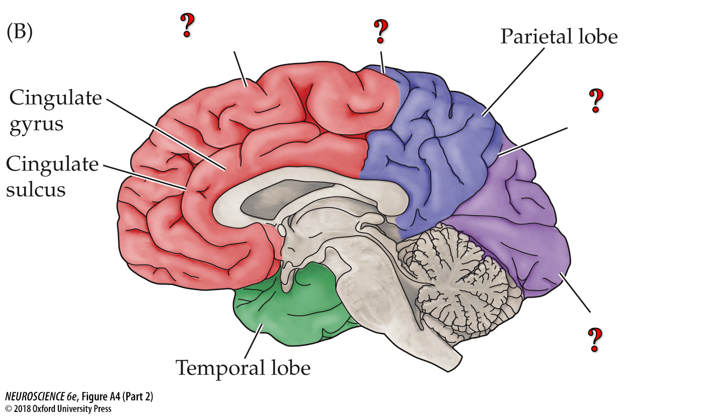
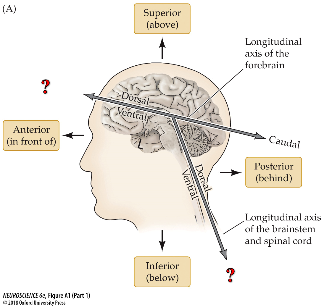
Fill in labels (labeled as “?”) from top to bottom
Frontal Lobe
Central Sulcus
Parieto-Occipital Sulcus
Occipital Lobe
The Frontal lobe, Central sulcus, Parieto-Occipital sulcus, and occipital lobe are all part of what structure?
Cerebral Cortex
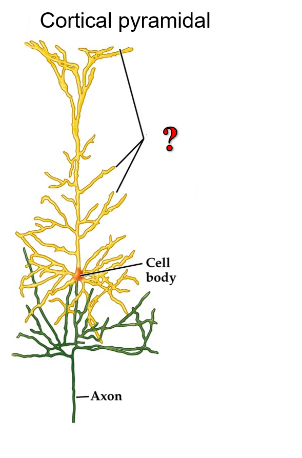
Fill in label (labeled as “?”)
Dendrites
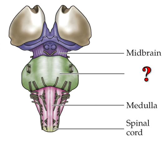
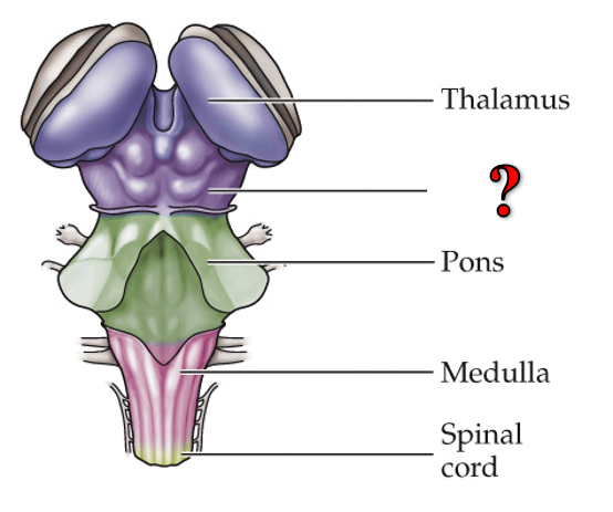
Fill the missing label
Pons
The Pons, Midbrain, Medulla, and Spinal cord are all part of what structure?
The brainstem
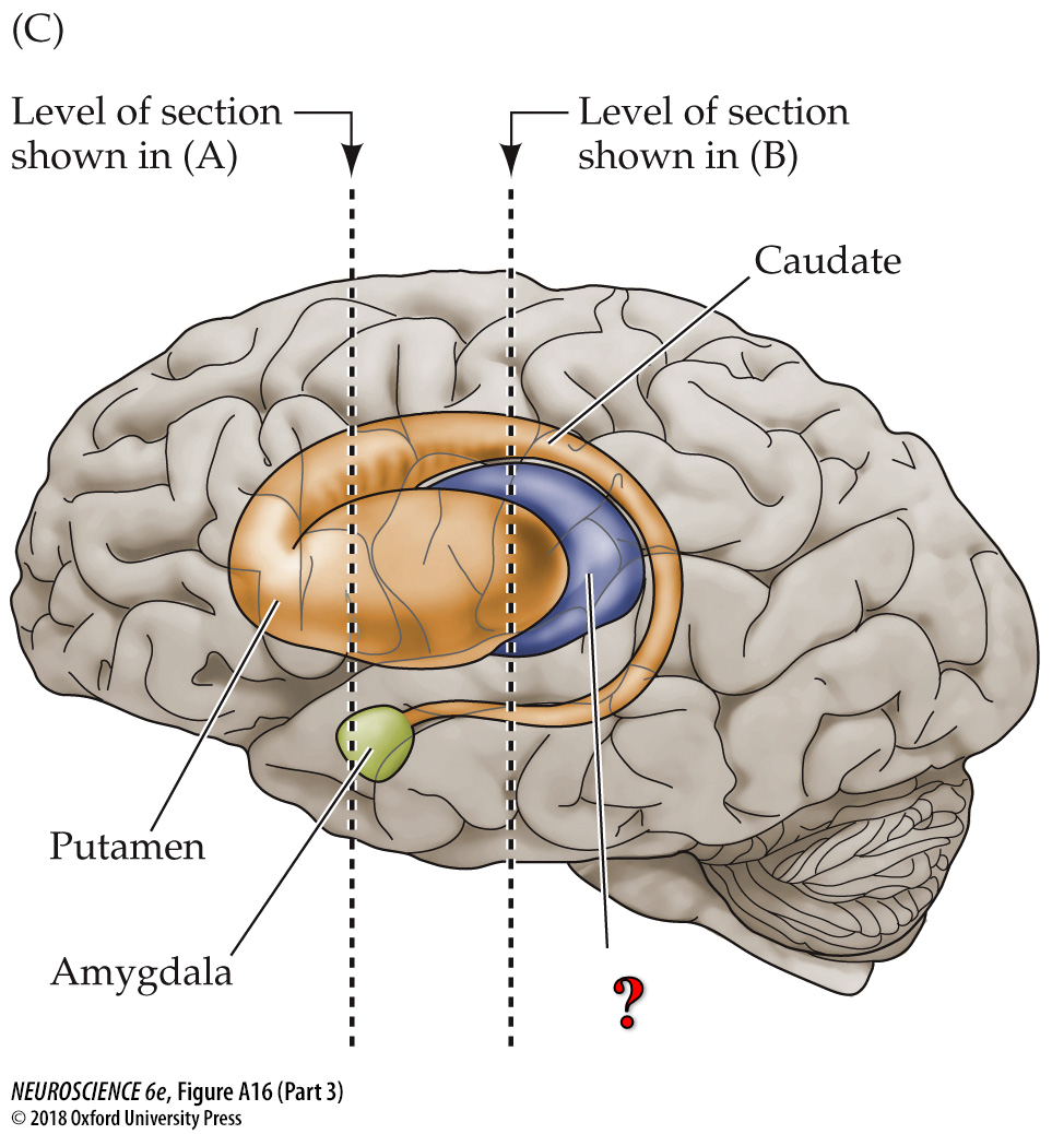
The Caudate, Putamen, and Globus Pallidus are all part of the ______.
The basal ganglia
Fill the missing label
The Thalamus
Fill in the blanks marked “?” from top to bottom.
Rostral
Caudal
Fill in the missing label
Midbrain
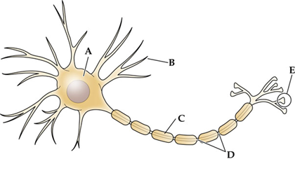
which structure is the main target for incoming signals recieved from the axons of other cells?
B- Dendrites
What are the components of the peripheral nervous system?
cranial and spinal nerves
The structural brain imaging technique that relies on the resonance frequences of atoms in a small magnetic field is
MRI

What structure insulates the axon allowing the action potential to travel more rapidly down its length?
C- myelin sheath

What structure contains the most concentrated number of synaptic vesicles and is the structure from which neurotransmitters are released?
E- terminal buttons
What type of cell produces myelin in the nerves of the peripheral nervous system?
Schwann cells
Which statement best describes most neurons?
They receive information via dendrites
What is the brain imaging technique that makes use of a narrow X-ray beam?
CT scan
What function is characteristic primarily of neurons only, and not glia?
Transmits action potentials
Which statement best describes the function of a neuron with multiple highly branched dendrites and one axon?
it integrates information from many neurons.
What areas do retinal axons project to?
hypothalamus, thalamus, pretectum
Visual area located most anteriorly is the
MT (V5)
The strictly monocular portion of the visual field is represented exclusively by what region of the retina?
nasal
In what layer of V1 does the koniocellular pathway terminate?
2/3
Ocular dominance columns occur in what layer of V1?
4
A person with cerebral achromatopsia has trouble
recognizing colors of objects
Cerebral achromatopsia is due to damage of the
extrastriate cortex
the visual area that is located most caudally in the brain is
V1
Neurons in striate cortex are not tuned to which property of a stimulus?
speed
Retinal ganglion cell axons cross at the
Optic chiasm
What percent of ganglion cells cross at the optic chiasm?
60%
How are upper motor neurons in the superior colliculus organized?
As a topographical map of eye movement vectors
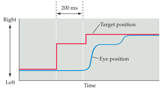
A subject in an eye movement experiment is instructed to focus on a specific target. After the start of the scene, the target is moved. The target is moved again (solid red line in figure) 200 ms later. What would a graph showing eye position in relation to target position look like?
The eye position should change about 200 ms after the target changes each time
What is the function of tiny saccades and drift during visual perception?
To change retinal stimulus during fixation, preventing retinal adaptation
How do vergence movements differ from other eye movements?
In vergence movements, the eyes move in difference directions
Where the lower motor neuron cell bodies that innervate the lateral rectus muscle are located
Abducens nucleus
The frontal eye fields influence horizontal eye movement by innervating the
Superior colliculus and the paramedian pontine reticular formation (PPRF)
what eye movement dysfunction is associated with Huntington’s disease?
inability to initiate voluntary saccades
What type of eye movement is affected by damage to the dorsal visual stream in the parietal lobe?
smooth pursuit movements
What muscle is responsible for abduction of the eye?
Lateral rectus
What eye movement dysfunction is associated with Schizophrenia?
impairment in smooth pursuit movements
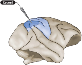
Which behavioral task would lead to increased firing rate in the neurons in the location outlined in the image?
Attending a visual target
what structure loses input if the output from neocortical layer 6 is blocked?
Thalamus
A husband reports that his wife has begun acting strangely. For example, she refuses to eat the food on the left side of her plate at meals, claiming she has finished all her food. She also has neglected to put her left arm in her shirt the last few days and has begun putting her makeup on only the right side of her face. Hearing these symptoms, where would you expect to find damage in the woman’s nervous system?
Right posterior parietal cortex
What lobe of the cerebral cortex is important for selecting and planning appropriate behavioral responses?
frontal lobe
What neocortical layer would be affected if thalamic output was disrupted?
Layer 4
How does each hemisphere of the brain control attention?
Left- controls right visual field
Right- controls left and right visual fields
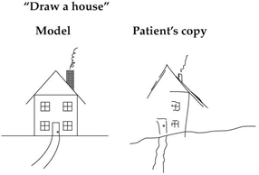
A patient is asked to draw a house by copying the model picture shown above. His version is shown on the right. What disorder do you suspect the patient has?
Contralateral neglect syndrome
What structure would be affected if neocortical output from Layer 5 was disrupted?
Striatum
What characteristic led 20th century scientists to dividing the brain into 50 regions?
Histological (microscopic) features
How many layers are in the neocortex?
6
What sensory areas are associated with the parietal lobe?
somatosensory and visual
What lobe is responsible for recognizing a face?
temporal
What lobe would be responsible for participating in a delayed response task?
Frontal
What is the parietal lobe important for?
attending to stimuli
What is the temporal lobe important for?
object and condition recognition
Major function of the temporal lobe
recognition and identification of stimuli
What is the main symptom of prosopagnosia
the inability to recognize faces
Damage to what region leads to language related agnosia?
lateral surface of the left temporal lobe
A patient is shown an image of a face during an MRI. What region would you expect to see an increase in neural activity?
Right fusiform gyrus
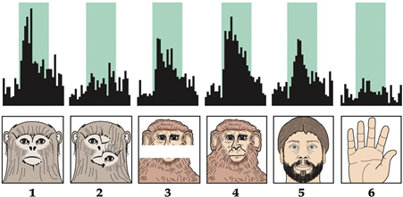
The graphs represent a neuron firing in a monkey brain recorded while presenting the corresponding images. What region was the neuron located in?
Inferior temporal cortex
what symptom would you expect a patient with a right temporal lobe lesion to exhibit?
Deficit in facial recognition
what association cortex is involved in recognizing objects?
Temporal
what association cortex is involved in deciding what to do with the object?
frontal
A teacher at a local school feels frustrated whenever the seasons change because her students begin wearing different shoes to school. This troubles her because she usually identifies her students by their footwear, and without that clue, she must wait for each student to talk before she knows who they are. Considering her symptoms, where would you expect to find damage in her nervous system?
right fusiform gyrus
What structure is important for the production of almost all vocalizations?
Larynx
what do offspring of deaf parents exhibit in sign language that is analogous to a verbal action of children in hearing families?
babbling
what do lesions in Wernicke’s and Broca’s areas produce in deaf people?
deficits analagous to those caused by lesions in hearing people
What would a lesion in the right hemisphere do to a deaf person?
affect emotional tone of singing
True or false: People who are deaf show lateralization of function similar to that of hearing people
true
What did studies of deaf patients with left/right hemisphere lesions demonstrate?
the language centers of the brain are specialized for pairing symbols with meaning, rather than being specialized for heard/spoken language
Where is the visual word form area located?
Left occipito-temporal sulcus
which element of language is mainly controlled by the right hemisphere?
prosody

The figure shows an fMRI scan of a subject completing a task. What are they doing?
Reading something out loud
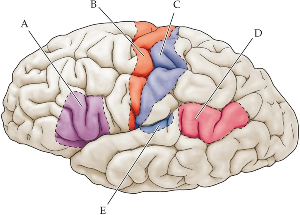
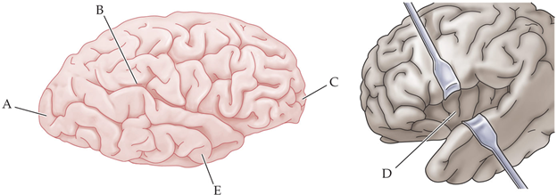
Label areas A, D, and E
A- Broca’s Area
D- Wernicke’s Area
E- Primary Auditory Cortex (A1)
Who developed the method of clinically assessing language lateralization in humans by anesthetizing one hemisphere?
John Wada
After a stroke, an individual has difficulty understanding speech, but can form and speak words, albeit nonsensically. Which disorder do these symptoms implicate?
Wernicke’s Aphasia
What are phonemes?
the precepts elicited by different speech sounds
The (Zhang et al., 2013) paper's results show slower times after ball heading on __ task
the Anti-point task
The (Zhang et al., 2013) paper measures sensorimotor and cognitive behavior using:
A tablet
Heading a ball in soccer is typically considered a ____ head impact.
subconcussive
True or False: The (Zhang et al., 2013) paper provides definitive evidence that the cognitive slowing that occurs after soccer playing with ball heading is only a transient effect and not a longer-lasting change or brain injury.
False
True or False: In the Zhang et al. (2013) paper, all participants were male
False
In the Zhang et al. (2013) paper, data was collected from ____ different participants
24
The (Zhang et al., 2013) paper examines behavior in:
Humans
A subject is instructed to focus on an image of a person. While the subject examines the image, an auditory stimulus is presented from the same location. What does this test measure?
Supra-modal attention
What region is likely to cause hemi-spatial neglect syndrome when damaged?
B- Right parietal lobe
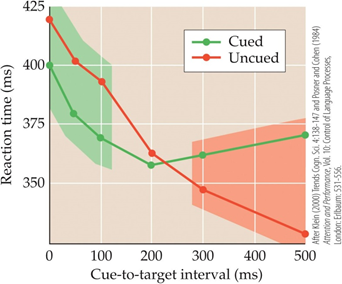
While subjects are focusing on a blank screen, a flash of light unexpectedly appears in one of two locations. Following the flash, a target image on the screen. Half the time the target image is in the same location as the flash, and half the time it is on the other side of the screen. The data collected are presented in the graph. What phenomenon occurs when the target is presented more than 300ms after the cue?
Inhibition of return
(takes longer for to respond to stimulus in location that was already attended to)
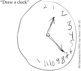
A subject is instructed to draw a clock from memory. Her version is shown in the figure. What would the patient’s diagnosis most likely be?
Hemispatial Neglect Syndrome
Which evidence suggests that unattended information is processed in the brain and that filtering occurs late in the sensory processing pathway?
A person attends to his name when it is mentioned in an unattended conversation
A patient experienced a severe head trauma and is now unable to point to an object space under visual guidance and has difficulty moving his eyes toward a target. What would the most likely diagnosis for this patient be?
Balint’s Syndrome
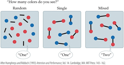
A patient is presented with the images in the figure one at a time and asked to report how many colors she can see. When the two colors are presented as seperate objects, as in the Random or Single display, she reports that she can only see one color. When the two colors are presented as part of the same object, however, she reports seeing both colors. What would the most likely diagnosis of this patient be?
Balint’s Syndrome
When subjects are presented with a picture of a person, they tend to focus their attention on the face and eyes of the individual. However when they are asked to draw conclusions about the individual in the image, like wealth, they shift their gaze to look at the clothes or surrounding environment. What does this test measure?
Overt Attention
When subjects are focusing on a blank screen, a flash of light unexpectedly appears in one of two locations. Following the flash, a target image appears on the screen. Half of the time the target image is in the same location as the flash, and half the time it is presented on the other side of the screen. What does this test measure?
Exogenous attention
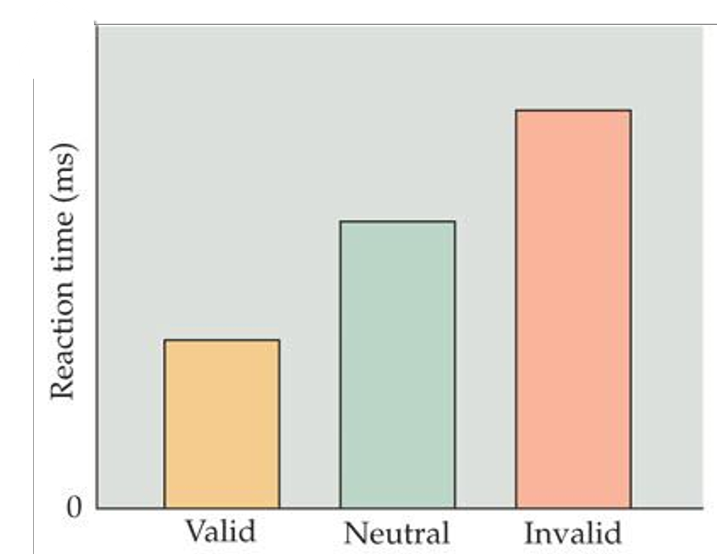
Subjects are asked to focus on a center point until a shape appears, and then name the shape. Before the shape appears, an arrow appears either pointing in the direction the shape will appear (valid), pointing in both directions (neutral), or pointing in the opposite direction from which the shape will appear (invalid). How would the subjects’ reaction time for naming the shape differ among these conditions?
invalid= highest reaction time
valid= lowest reaction time
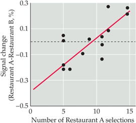
Subjects indicated whether they prefer French fries from Restaurant A or B. They are then given French fries from the restaurant they prefer while being evaluated using fMRI. The figure shows the activation of a particular brain region in response to tasting fries from Restaurant A compared to Restaurant B (y axis) plotted against the number of times a subject chose A over B (x axis). Which brain region showed differences in activation?
Ventromedial prefrontal cortex
Which change or deficiency was not reported in early case studies of individuals with frontal lobe damage?
Difficulty with intellectual functioning
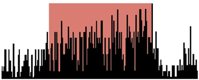
While a monkey participates in a delayed response task, data from a prefrontal cortical neuron is collected. Each bar in the graph represents an action potential in the neuron, and the red box is the time the screen is closed.
short-term memory and planning
Why did the medical community stop performing leukotomies around 1950?
Effective psychotropic drugs were developed
What symptom would you expect bilateral lesions to the dorsolateral prefrontal cortex to produce in a monkey?
Delayed or abolished success during the delayed response task
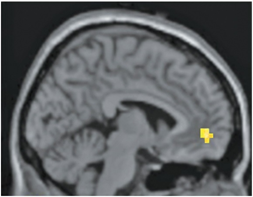
The activated region in the fMRI scan above is responsible for which function?
The planning and execution of appropriate behavior