Radiologic Evaluation of the Spine
1/118
There's no tags or description
Looks like no tags are added yet.
Name | Mastery | Learn | Test | Matching | Spaced | Call with Kai |
|---|
No analytics yet
Send a link to your students to track their progress
119 Terms
Why are C-spine images ordered?
post trauma (canadian c-spine rule)
pain (neck, UE, headache)
pre-operativelu
suspected malignancy
suspected anomaly or abnormality
suspected instability
What is the purpose of the Canadian C-Spine Rules?
to determine if cervical spine imaging is needed after trauma
What high-risk factors in the Canadian C-Spine Rule requires cervical spine imaging?
age > 65 y/o
dangerous mechanism of injury
paresthesias in extremities
if any are present = radiography indicated
What are the low-risk factors in the Canadian C-Spine Rule that allow assessment of cervical ROM?
simple read-end MVC
delayed onset of neck px
sitting position in ED
ambulatory at any time after injury
absence of midline C-spine tenderness
if none are present = radiography indicated
If a patient has at least one low-risk factor in the Canadian C-Spine Rule, what should you assess next?
assess active cervical rotation (45 degrees to the left and right)
if can rotate bilaterally 45 degrees = no imaging indicated
if can’t rotate bilaterally 45 degrees = cervical spine imaging indicated
Which radiographic views best visualize the intervertebral foramina in the cervical and thoracic spine?
cervical: right and left oblique views
thoracic: lateral view
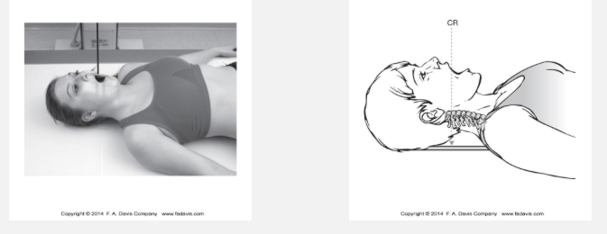
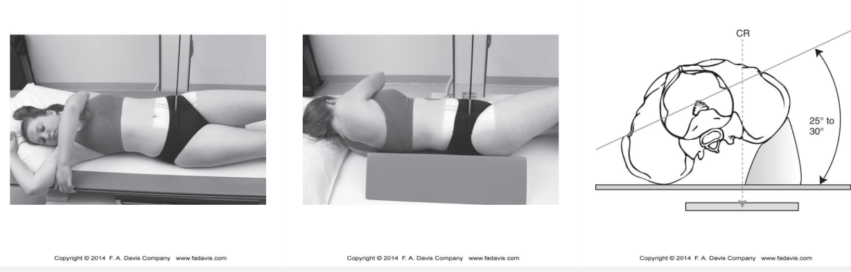
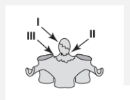
What structures are visualized on the AP open-mouth (odontoid) cervical spine radiograph?
dens and body of C2 (axis)
angle of mandible
anterior arch of atlas
posterior arch of atlas
lateral AA facet joints
atlas TP
C2 spinous process
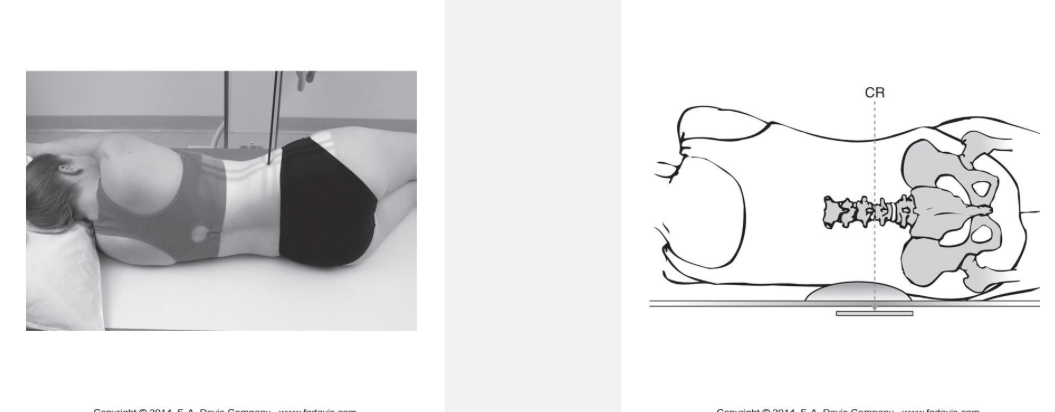
Where is the central ray (CR) directed for the AP open-mouth (odontoid) view?
through the center of the open mouth
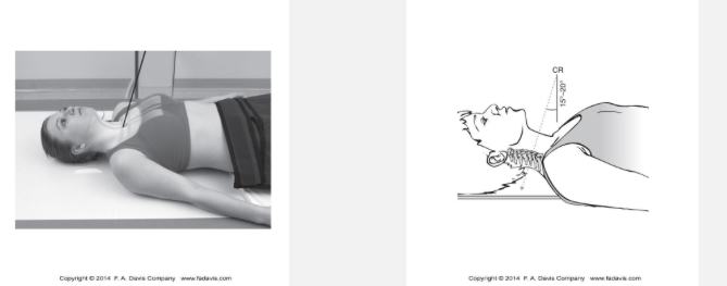
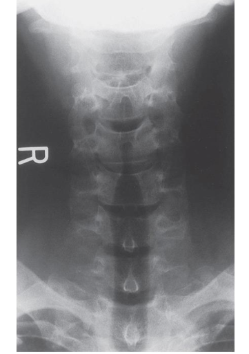
What structures are visualized on an AP lower cervical spine radiograph?
lower 5 cervical vertebrae
upper thoracic vertebrae & associated ribs
medial clavicle
trachea
mandible obscured the upper cervical vertebrae (C1-C2)
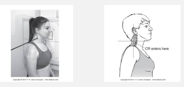
What structures are visualized on a lateral cervical spine radiograph?
all 7 cervical vertebrae (C1-C7)
intervertebral disc spaces
articular pillars (stacked facets) & facet joints
spinous processes
prevertebral soft tissues
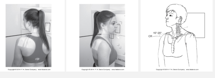
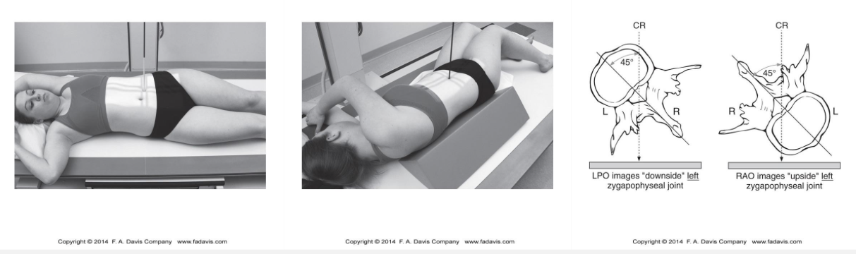
What is visualized on an oblique cervical spine radiograph?
intervertebral foramina
uncovertebral joints (b/t vertebral bodies)
facet joints
pedicles
obtained at 45 degrees from lateral view
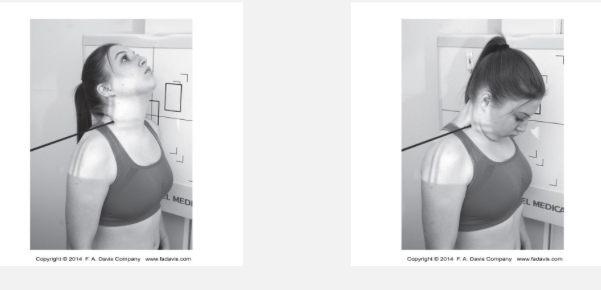
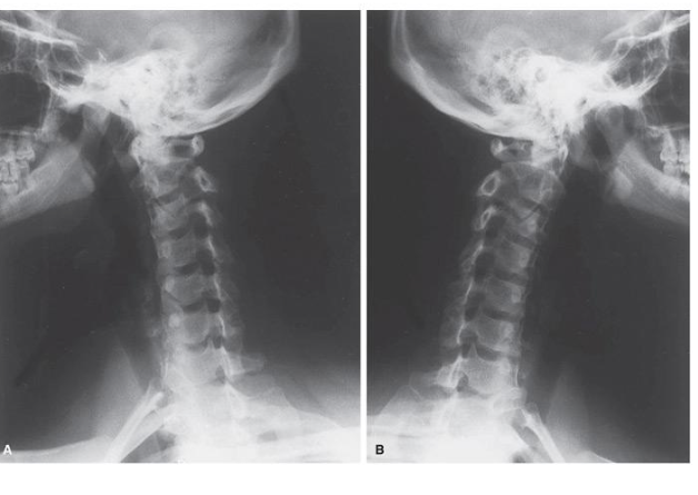
What is a lateral cervical spine view with stress?
non-routine radiograph used to assess joint alignment while soft tissue structures are stressed; may be used to evaluate ligamentous instability or abnormal motion
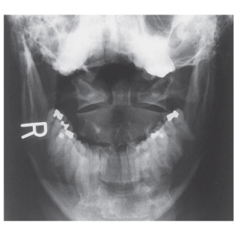
What view is this?
AP open mouth (odontoid)
What view is this?
oblique (intervertebral foramina)
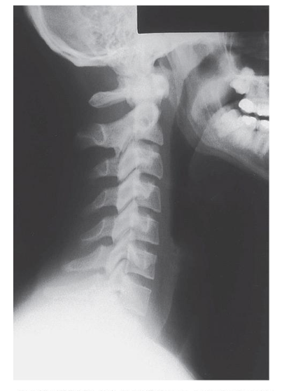
What view is this?
lateral (facet joints, no IF)
What view is this?
AP lower cervical (SP leading to sternum, clavicle, 1st rib)
What are common indications for a CT image of the spine?
acute trauma in adults
degenerative conditions and OA
post-operative evaluation of bone graft or fusion
infectious processes of the spine
image guidance for spinal interevntions (i.e., biopsy or injections)
neoplastic conditions & complications
inflammatroy lesions and crystal deposition disease (i.e., gout or arthritis)
congenital or developmental spine abnormalities (i.e., scoliosis, spondylolysis)
spinal cord syrinxes & other masses
MRI contraindicated
What are common indications for a MRI image of the spine?
degenerative disk disease
extradural soft tissue and bony neoplasm
intradural extramedullary masses
intradural masses or leptomeningeal disease
intramedullary tumors
treatment fields for radiation therapy
intrinsic spinal cord pathology (demyelination & inflammatory)
spinal vascular malformations or subarachnoid hemorrhage
syringohydromyelia
post-operative intraspinal fluid/soft-tissue changes
meningeal abnormalities
spinal infections
pre-operative assessment
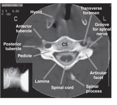
What spinal structures are best visualized in the axial/transverse CT plane?
spinal canal & cord
nerve roots
pedicles
facet joints
vertebral body cross-sections
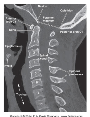
What spinal structures are best visualized in the sagittal CT plane?
vertebral alignment
vertebral bodies
spinous processes
intervertebral disc spaces
spinal curvatures
spondylolisthesis
compression fractures
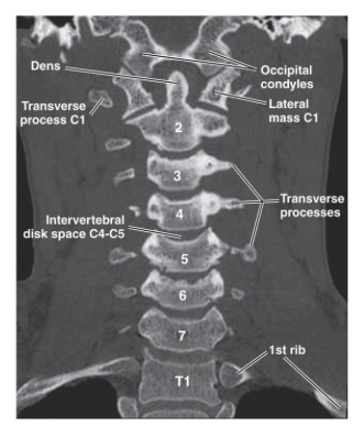
What spinal stuctures are best visualized in the coronal plane?
scoliosis
lateral alignment
vertebral body symmetry
transverse processes
What are the ABCDS when interpreting spine imaging?
A: alignment and anatomy
B: bone density
C: canal space
D: disc integrity
S: soft tissue
What MRI planes and sequences are commonly used for spine imaging?
planes: sagittal and axial
T1-weighted: anatomy and structural detail
T2-weighted: detecting fluid abnormalities (edema, inflammation, disc abnormalities, CSF)
What is the signal intensity of the CSF on T1 and T2 MRI?
T1 = low (dark)
T2: high (bright)
What is the signal intensity of the intervertebral discs on T1 and T2 MRI?
T1: intermediate (gray)
T2: high (bright)
What is the signal intensity of cortical bone and ligaments on T1 and T2 MRI?
T1: low (dark)
T2: low (dark)
What is the signal intensity of the spinal cord on T1 and T2 MRI?
T1: intermediate (gray)
T2: intermediate (gray)
What is the signal intensity of muscle on T1 and T2 MRI?
T1: intermediate (gray)
T2: intermediate (gray)
What is the purpose of the Swimmer’s lateral view?
to better visualize the lower cervical spine and upper thoracic spine by elevating one arm overhead to remove shoulder superimposition
When is a thoracic spine oblique view used?
to visualize the facet joints
Waht is a thoracolumbar view used for?
to view the thoracolumbar junction (a commong injury site), using a coned/narrowed exposure field
Why is an RAO oblique view used for sternum imaging?
to project the sternum over the homogenous density of the heart, improving contract and reducing superimposition from thoracic spine
Which view is commonly used to visualize the sternoclavicular (SC) joints?
RAO oblique view
Why is an oblique view needed for sternum radiographs?
to avoid superimposition of the thoracic spine over the sternum
How are rib radiographs obtained?
anterior, posterior, and axillary
right or left side
upper ribs (1-9)
lower ribs (8-12)
What structures are visualized on an AP thoracic spine radiograph?
vertebral bodies
intervertebral disc spaces
pedicles
spinous processes
transverse processes
articular processes
costovertebral joints
posterior ribs
What structures are visualized on a lateral thoracic spine radiograph?
vertebral bodies
intervertebral disc spaces
intervertebral foramina
upper thoracic vertebrae may be partially obscured by the shoulders
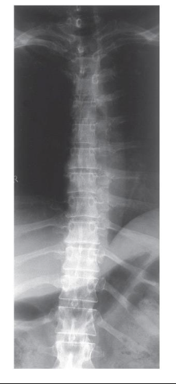
What view is this?
anteroposterior (AP)
vertebral bodies, transverse processes, ribs
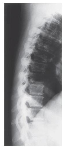
What view is this?
lateral
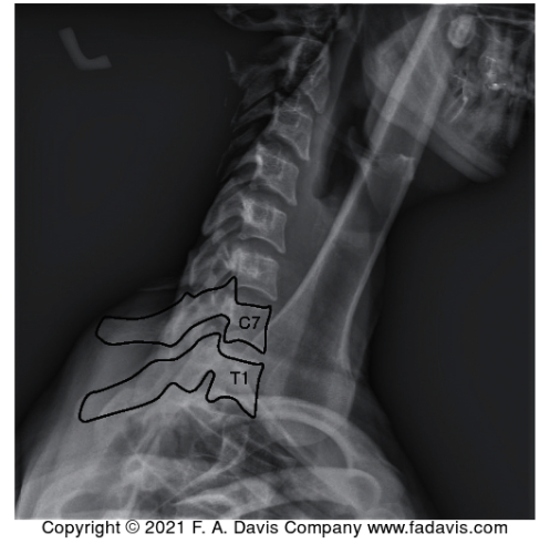
What view is this?
swimmer’s
What structures can be visualized in an AP lumbar spine radiograph?
all 5 lumbar vertebrae (L1-L5)
sometimes sacrum and coccyx
vertebral bodies
pedicles
spinous processes
transverse processes
IV disc spaces
What structures can be visualized in a lateral lumbar spine radiograph?
vertebral bodies
intervertebral disc spaces
pedicles
spinous processes
intervertebral foramina
lumbosacral articulation
What structures can be visualized in an oblique lumbar spine radiograph?
facet joints
superior and inferior articular processes
pars interarticularis
pedicles
Why is a lateral L5-S1 spot view obtained?
to better visualize the L5-S1 junction by reducing exposure to surrounding structures and overcoming superimposition from the ilia
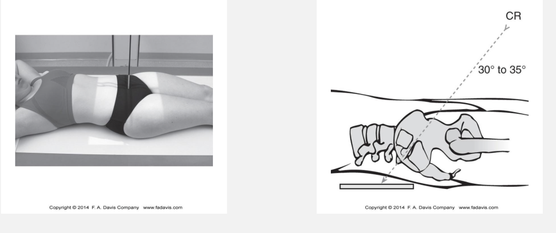
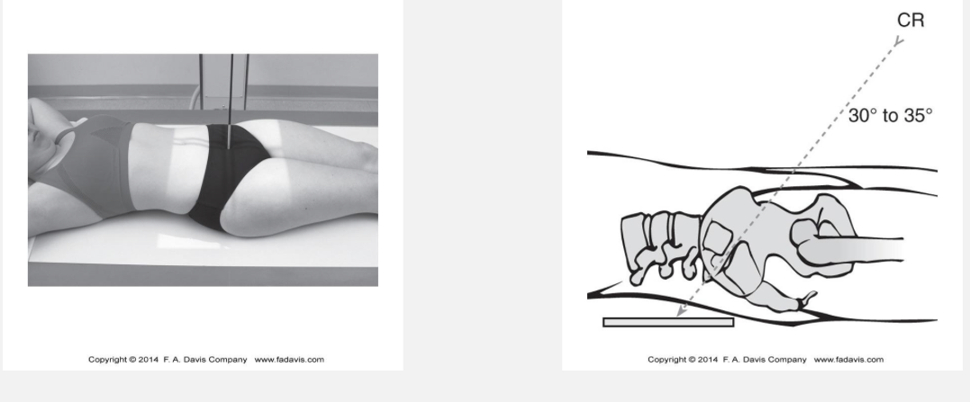
What is visualized on an AP axial/sacroiliac radiograph?
bilateral SI joints
What is visualized on an oblique sacroiliac radiograph?
an individual sacroiliac joint
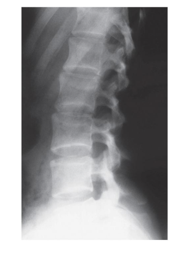
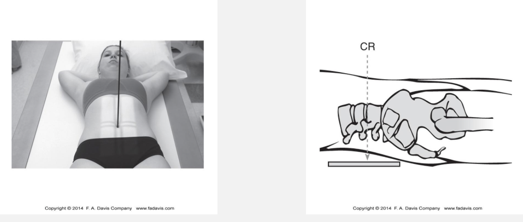
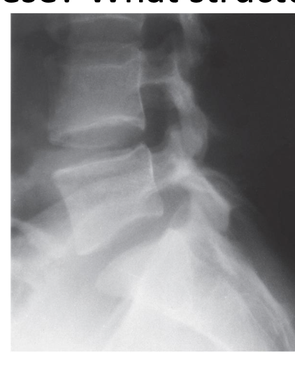
What view of the lumbar spine is this?
lateral
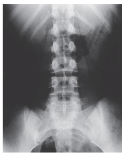
What view is this?
anteroposterior (AP)
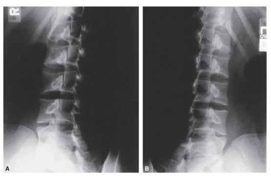
What view is this?
oblique
What view of the lumbar spine is this?
lateral L5-S1
What is a stable fracture?
no ligament disruption; no bone or joint displacement
What is an unstable fracture?
displacement of potential for displacement; fracture-dislocation or bilateral facet dislocation
What immobilization devices are commonly used for stable vs unstable cervical fractures?
cervical collar (C-collar): typically used for stable fx
halo immobilizer: used for unstable fx
What is a Clay-Shoveler’s fracture?
avulsion fx of a spinous process (common at C6, C7, T1); stable fx
What is the mechanism of a Clay-Shoveler’s fracture?
hyperflexion of the neck or forceful contraction of the trapezius and rhomboid muscles (often during heaving labor of UE)
What radiographic view is most useful for identifying a Clay-Shoveler’s fracture?
lateral cercial spine view
What is an odontoid fracture?
fracture of the dens of C2, accounting for ~20% of cervical fractures
What are common mechanisms of a dens fracture?
motor vehicle accidents (MVA) or falls that cause extreme force on the dens or alar ligaments
What are the three types of dens fractures?
type I, type II, type III

What is a type I dens fracture?
avulsion fracture of dens tip due to alar ligament stress

What is a type II dens fracture?
fracture at the junction of the dens and C2 body (difficult to heal)
What is a type III dens fracture?
fracture extending below the junction into the C2 vertebral body
What imaging is used to identify a dens fracture?
radiograph (AP open-mouth/odontoid) or a CT
What is the attachment and function of the alar ligament?
attaches the dens to the occipital bone; provides restraint against excessive rotation of the head and upper cervical spine
What is a teardrop fracture?
a triangular fragment of the vertebral body separates; unstable
What is a teardrop fracture often associated with?
ligament rupture
disc injury
spinal cord injury (SCI)
What is the mechanism of a teardrop fracture?
hyperflexion and compression force
What is a wedge fracture?
an anterior compression fracture in which the vertebral body becomes wedge-shaped; usually stable
What is the mechanism of a wedge fracture?
hyperflexion and compression forces
What is a Jefferson fracture?
a burst fracture of the naterior and posterior arches of C1 (atlas)
What is the mechanism of a Jefferson fracture?
axial compression (diving accidents)
What imaging is used to identify a Jefferson fracture?
AP open-mouth (odontoid) view and CT
What is a burst fracture?
an axial compression fracture in which the vertebral body is crushed and fragments may be displaced outward (comminuted)
What is the mechanism of a burst fracture?
axial compression, causing the intervertebral disc to transmit force into the vertebral body resulting in “bursting”
What is a Hangman’s fracture?
a fracture of the bilateral pars interarticularis/pedicles
What is the mechanism of a Hangman’s fracture?
hyperextension of the cervical spine
What is the Denis Three-Column Model and what is it used for?
a model to assess thoracic and lumbar spine fracture stability; anterior, middle, and posterior column
What structures are included in the anterior column of the Denis Three-Column Model?
anterior longitudinal ligament
anteriro 2/3 of vertebral bodies
anterior annulus fibrosus
anterior intervertebral disc
What structures are included in the middle column of the Denis Three-Column Model?
posterior longitudinal ligament
posterior 1/3 of vertebral bodies
posterior annulus fibrosus
posterior intervertebral discs
What structures are included in the posterior column of the Denis Three-Column Model?
everything posterior to posterior longitudinal ligament
posterior ligamentous structures
pedicles
laminae
facets
transverse processes
spinous processes
How does the Denis Three-Column Model determine fracture stability?
stable: injury to one column
potentially unstable: injury to two columns (depends)
unstable: injury to all three columns
What is a flexion-compression fracture of the thoracic/lumbar spine?
an anterior wedge compression fracture caused by compression of the anterior column with varying involvement of the middle and posterior columns
What is the mechanism of a flexion-compression fracture?
flexion combined with axial compression
What are the three patterns of flexion-compression injury?
anterior column failure only
anterior column failure & posterior column ligamentous failure
failure of all three columns
What is the first pattern for a flexion-compression injury?
only anterior column failure
middle and posterior columns remain intact
<50% loss of vertebral body height
stable fracture
What is the second pattern for a flexion-compression injury?
both anterior column & posterior ligamentous failure
>50% loss of vertebral body height
potentially unstable fracture
What is the third pattern for a flexion-compression injury?
failure of all 3 columns
anterior wedging
posterior vertebral body disruption
unstable fracture
risk of SC, nerve root, or vascular injury from displaced fracture fragments
What is an axial compression injury of the thoracic/lumbar spine?
an injury caused by axial compression that produces a burst fracture
What are the five subtypes of axial compression fractures?
fx of both endplates
fx of superior endplate
fx of inferior endplate
burst rotation fx
burst lateral flexion fx
What is the most common subtype of axial compression fractures?
fracture of the superior endplate
What is a Chance (seatbelt) fracture?
a flexion-distraction injury where the anterior vertebral body is compressed/crushed while the posterior vertebral body is pulled apart
What is the mechanism of a Chance fracture?
caused by hyperflexion over a fixed restraint (horizontal split of entire vertebrae)
What is a flexion-distraction injury?
mechanism that produces a Chance (seatbelt) fracture; characterized by failure of the posterior column
What are the two types of flexion-distraction injuries?
classic chance fracture
flexion-distraction subtype
What is a rotation fracture-dislocation?
involves failure of the posterior and middle columns with varying degrees of anterior column injury; unstable (multiple columns disrupted)
What is the mechanism of a rotation fracture-dislocation?
lateral flexion and rotation, with or without a posterior-to-anterior force
What structures are commonly disrupted in a rotation fracture-dislocation?
posteriro ligaments
articular facets
middle column structures
often portions of the anterior column
What is the characteristic radiographic finding of a rotation fracture-dislocation?
a “slice” appearance, caused when the upper vertebral body rotates and carreis part of the lower vertebral body with it
Is a rotation fracture-dislocation stable or unstable?
unstable
What type of joint is formed by an intervertebral disc?
a symphysis joint (fibrocartilage)