Axial Skeleton Bones and Bony Markings
 Call Kai
 Learn
Learn Practice Test
 Spaced Repetition
 Match
Match Flashcards
 Knowt Play
1/163

There's no tags or description
Looks like no tags are added yet.
Name | Mastery | Learn | Test | Matching | Spaced | Call with Kai | Chat |
|---|
No analytics yet
Send a link to your students to track their progress
164 Terms
Axial skeleton
Division of the skeleton including the skull, vertebral column, and thoracic cage

Long bones
Bones longer than they are wide, with a shaft and two ends (e.g., femur)

Short bones
Roughly cube-shaped bones (e.g., carpals, tarsals)

Flat bone
Thin, flattened bones often serving protective functions (e.g., skull bones)

Irregular bone
Bones with complex shapes that don't fit other categories (e.g., vertebrae)

Sutural/Wormian bones
Small extra bones found within the sutures of the skull

Condyle
A rounded articulating surface at the end of a bone

Facet
A small, smooth flat articulating surface

Head
A rounded articulating projection at the end of a bone, often on a narrow neck

Trochlea
A pulley-shaped articulating surface

Alveolus
A deep pit or socket, especially for a tooth

Fossa
A shallow depression in a bone

Sulcus
A groove or furrow in a bone, often for a vessel, nerve, or tendon

Canal
A tunnel-like passage through a bone
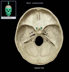
Fissure
A narrow slit-like opening in a bone

Foramen
A rounded opening through a bone for vessels or nerves
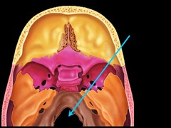
Meatus
A canal-like opening or passageway
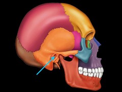
Sinus
An air-filled cavity within a bone

Crest
A narrow ridge of bone

Epicondyle
A projection above or on a condyle

Line
A low ridge of bone, less prominent than a crest
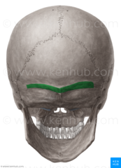
Process
Any bony projection

Ramus
An arm-like branch off the body of a bone
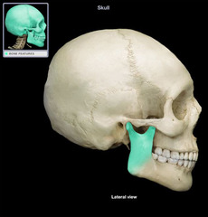
Spine
A sharp, slender bony projection

Tubercle
A small rounded bony projection

Frontal bone
Single cranial bone forming the forehead
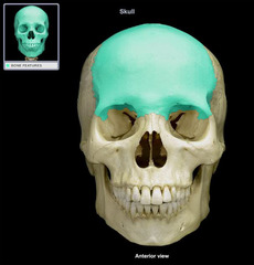
Orbital surface of frontal bone
Portion of the frontal bone forming the roof of the eye orbit

Glabella
Smooth area on the frontal bone between the eyebrows

Supraorbital margin
The upper rim of the eye orbit on the frontal bone

Supraorbital notch (or foramen)
Opening in the supraorbital margin for nerves and vessels

Parietal bone
Paired cranial bones forming the roof and upper sides of the skull

Temporal bone
Paired cranial bones forming the lower sides of the skull

Carotid canal
Opening in the temporal bone through which the internal carotid artery passes

Internal and external auditory (acoustic) meatus
Canals in the temporal bone leading to the middle/inner ear (internal) and ear canal (external)

Jugular fossa
Depression in the temporal bone that houses the jugular vein

Mandibular fossa
Depression on the temporal bone where the mandible articulates

Mastoid part of temporal bone
Region of the temporal bone behind the ear containing air cells

Mastoid process
Bony projection on the temporal bone behind the ear
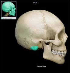
Styloid process
Slender pointed projection from the temporal bone
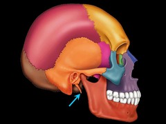
Squamous part of temporal bone
Thin, flat, scale-like portion of the temporal bone

Petrous part of temporal bone
Dense, pyramid-shaped portion of the temporal bone housing the inner ear

Tympanic part of temporal bone
Portion of the temporal bone surrounding the ear canal
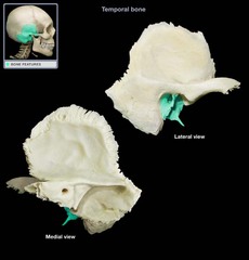
Zygomatic process of temporal bone
Projection of the temporal bone that joins the zygomatic bone

Occipital bone
Single cranial bone forming the posterior and base of the skull

Basilar part of occipital bone
Anterior portion of the occipital bone that joins the sphenoid

Cerebellar fossa
Depression on the internal occipital bone

External occipital protuberance
Bump on the posterior occipital bone at the midline

Foramen magnum
Large opening in the occipital bone for passage of the spinal cord

Grooves for superior sagittal sinus
Internal groove on the occipital bone

Groove for transverse sinus
Internal groove on the occipital bone
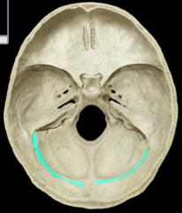
Internal occipital crest
Ridge on the internal occipital bone along the midline

Internal occipital protuberance
Internal bump on the occipital bone where sinus grooves converge

Occipital condyle
Rounded projection on the occipital bone that articulates with the atlas (C1)
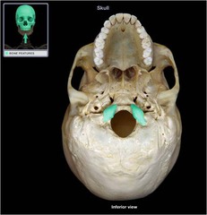
Sphenoid bone
Single butterfly-shaped bone forming part of the central cranial floor

Anterior and posterior clinoid process
Projections on the sphenoid bone surrounding the pituitary gland

Body of sphenoid
Central portion of the sphenoid bone

Dorsum sellae
Posterior wall of the sella turcica on the sphenoid bone

Foramen ovale
Opening in the sphenoid bone for the mandibular nerve

Foramen rotundum
Opening in the sphenoid bone for the maxillary nerve
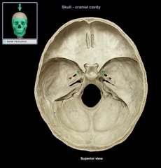
Foramen spinosum
Opening in the sphenoid bone for the middle meningeal artery
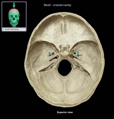
Greater wing
Lateral projection of the sphenoid bone forming part of the orbit and cranial floor

Hypophysial fossa
Depression in the sella turcica that holds the pituitary gland

Lateral pterygoid plate
bony plate extending from the sphenoid, muscle attachment

Medial pterygoid plate
Medial bony plate extending from the sphenoid, muscle attachment

Optic canal
Opening in the sphenoid bone for the optic nerve

Sella turcica
Saddle-shaped depression on the sphenoid bone holding the pituitary gland

Superior orbital fissure
Opening in the sphenoid bone for nerves and vessels entering the orbit

Ethmoid bone
Single irregular bone forming part of the nasal cavity and orbit walls

Cribriform plate and cribriform foramina
Perforated portion of the ethmoid bone allowing olfactory nerve fibers to pass

Crista galli
Upward projection of the ethmoid bone for dura mater attachment

Ethmoidal sinus (cells)
Air-filled spaces within the ethmoid bone
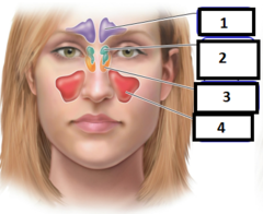
Middle nasal concha
Curved bony shelf of the ethmoid bone projecting into the nasal cavity

Orbital plate
Portion of the ethmoid bone forming part of the medial orbit wall

Perpendicular plate
Portion of the ethmoid bone forming the upper nasal septum
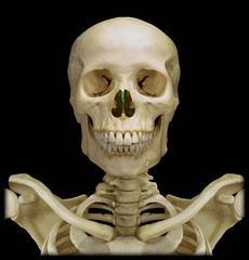
Coronal suture
Joins the frontal bone to the parietal bones

Sagittal suture
Joins the two parietal bones at the midline

Squamous suture
Joins the parietal bone to the temporal bone

Sphenosquamous suture
Joins the sphenoid bone to the squamous part of the temporal bone
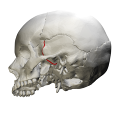
Occipitomastoid suture
Joins the occipital bone to the mastoid part of the temporal bone
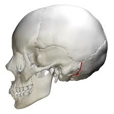
Lambdoidal suture
Joins the parietal bones to the occipital bone

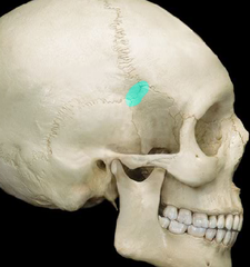
Pterion
H-shaped junction of the frontal, parietal, temporal, and sphenoid bones
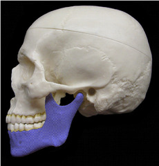
Mandible
Single bone forming the lower jaw, the only movable skull bone
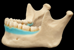
Alveolar process (mandible)
Ridge of the mandible containing the tooth sockets
Angle (mandible)
Point where the body and ramus of the mandible meet
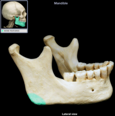
Body (mandible)
Horizontal portion of the mandible
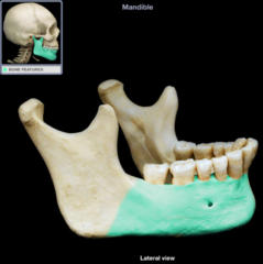
Condylar process
Projection of the mandible that articulates with the temporal bone

Coronoid process (mandible)
Projection of the mandible for temporalis muscle attachment

Head (mandible)
Rounded end of the condylar process that forms the jaw joint

Mandibular foramen
Opening on the inner mandible for nerves and vessels

Mandibular notch
Depression on the mandible between the condylar and coronoid processes

Mental foramen
Opening on the mandible below the premolars for nerves and vessels

Maxilla
Paired bones forming the upper jaw
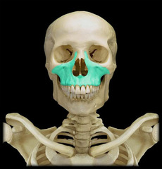
Alveolar process (maxilla)
Ridge of the maxilla containing the tooth sockets
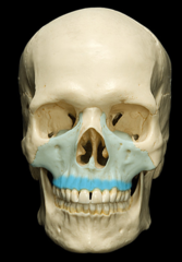
Anterior nasal spine
Pointed projection of the maxilla at the base of the nasal cavity

Maxillary sinus
Air-filled cavity within the maxilla

Orbital surface of maxilla
Portion of the maxilla forming the floor of the eye orbit

Palatine process
Portion of the maxilla forming part of the hard palate

Infraorbital foramen
Opening below the orbit on the maxilla for nerves and vessels

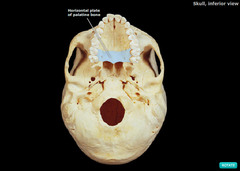
Palatine bone
Paired bones forming the posterior hard palate and part of the nasal cavity
Zygomatic bone
Paired bones forming the cheekbones
