4. Structure and Function of Immune System - Secondary Lymphoid Organs
1/30
Name | Mastery | Learn | Test | Matching | Spaced | Call with Kai |
|---|
No analytics yet
Send a link to your students to track their progress
31 Terms
List the secondary lymphoid organs.
Spleen
Lymph nodes
Mucosa associated lymphoid tissues
Which secondary lymphoid organs are the most highly organized ones?
Lymph nodes and spleen
Less-organized lymphoid tissues are collectively called
mucosal associated lymphoid tissue (MALT)
MALT includes
Peyer’s patches (in small intestine)
Tonsils
Appendix
Numerous lymphoid follicles within lamina propria of intestines
Mucous membranes of upper airways (bronchi associated lymphoid tissues (BALT))
Lymphoid tissues in genital tract
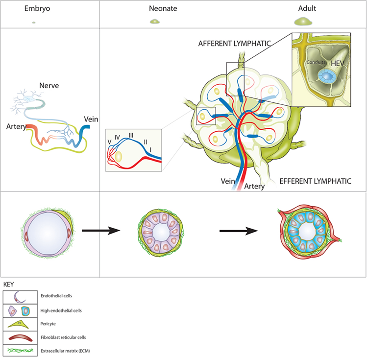
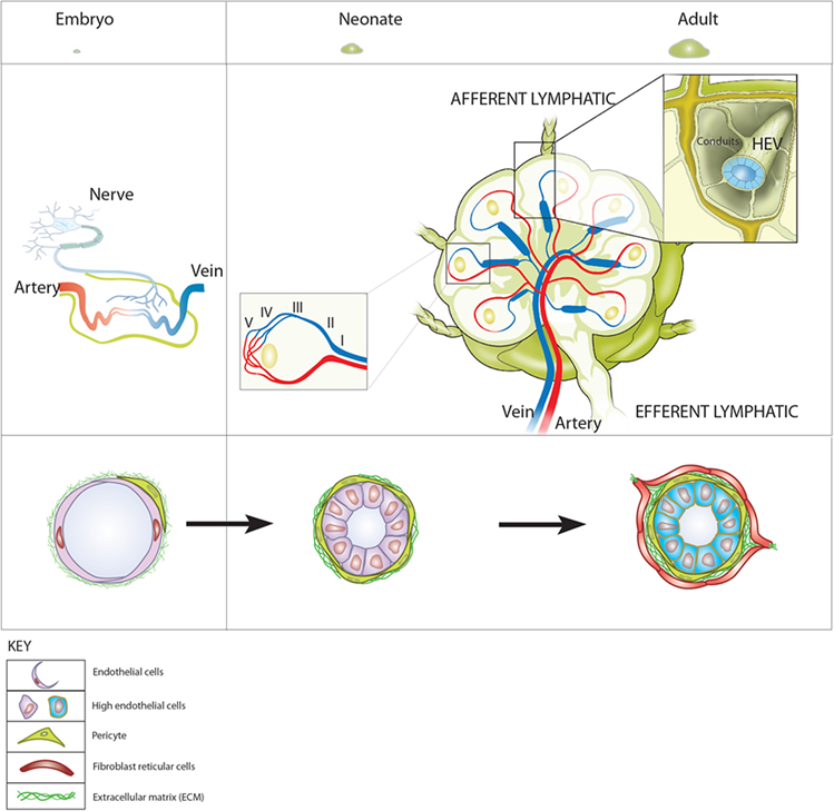
Secondary lymphoid organs are richly supplied with _____ vessels and _______ vessels, that facilitate movement of ____________, __________, and _________ cells into and out of these organs. Specialized regions of the vasculature called _____ ____________ ________ permit the movement of cells between the _____ and the ________ or _______ through which they are passing.
blood vessels
lymphatic vessels
lymphocytes
monocytes
dendritic
high endothelial venules
blood
tissues
organs
What processes does the leukocyte-rich nature of secondary lymphoid tissues facilitate?
Cellular interaction
Exchange regulatory signals
Undergo further development
Proliferation before re-entering circulation
What is the largest secondary lymphoid organ?
Spleen
What is the role of the spleen?
It clears particulate matter/blood borne antigens/pathogens through the stimulation of T and B cells
The spleen is the major site of immune responses to blood-born antigens
Where is the spleen situated?
Below the diaphragm, on the left side of the abdomen
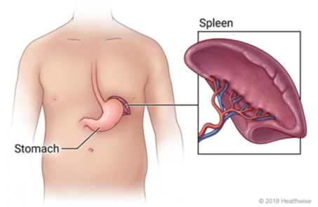
How much does the adult spleen measure and weigh?
Measures 5 inches in length and weighs 150g
What are the 2 compartments the spleen is divided into?
Central white pulp and outer red pulp
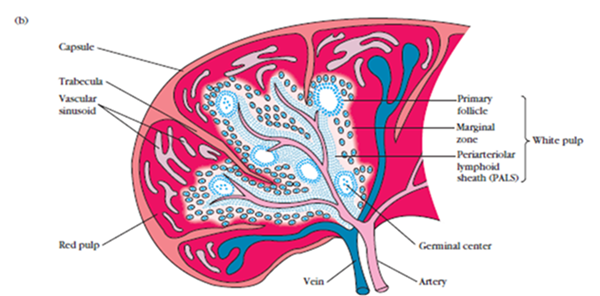
Describe the capsule of the spleen.
Spleen is surrounded by capsule that extends a number of projections (trabeculae) into interior to form the compartmentalized structure
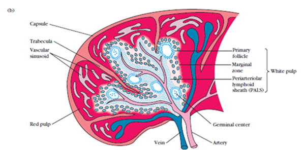
Describe the white pulp of the spleen.
Central densely populated area which contains T and B cells
Has 2 parts:
Periarteriolar lymphoid sheath (PALS)
Marginal zone
Describe the periarteriolar lymphoid sheath (PALS) of the white pulp of the spleen.
Rich in T cells
Surrounds the branches of the splenic artery
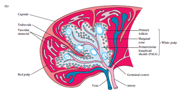
Describe the marginal zone of the white pulp of the spleen.
Located peripheral to PALS
Populated by B cells, lymphoid follicles (primary and secondary), and macrophages
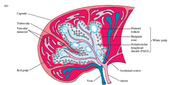
Describe the red pulp of the spleen.
Area that surrounds the sinusoids
Filled with RBC’s
Older and defective RBCs are destroyed here
Describe what happens where there are defects in the spleen and what they are caused by.
As the spleen is the site of destruction for most microbes, functional or structural abnormalities of spleen or splenectomy, esp in children, often leads to increased incidence of bacterial sepsis
Caused primarily by capsulated bacteria e.g.
Streptococcus pneumoniae
Neisseria meningitidis
Haemophilus influenzae
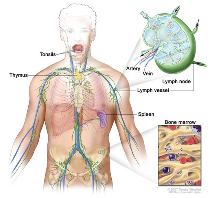
Describe the shape, arrangement and location of lymph nodes.
Small, bean-shaped organs
Occur in clusters or chains
Distributed along the length of lymphatic vessels
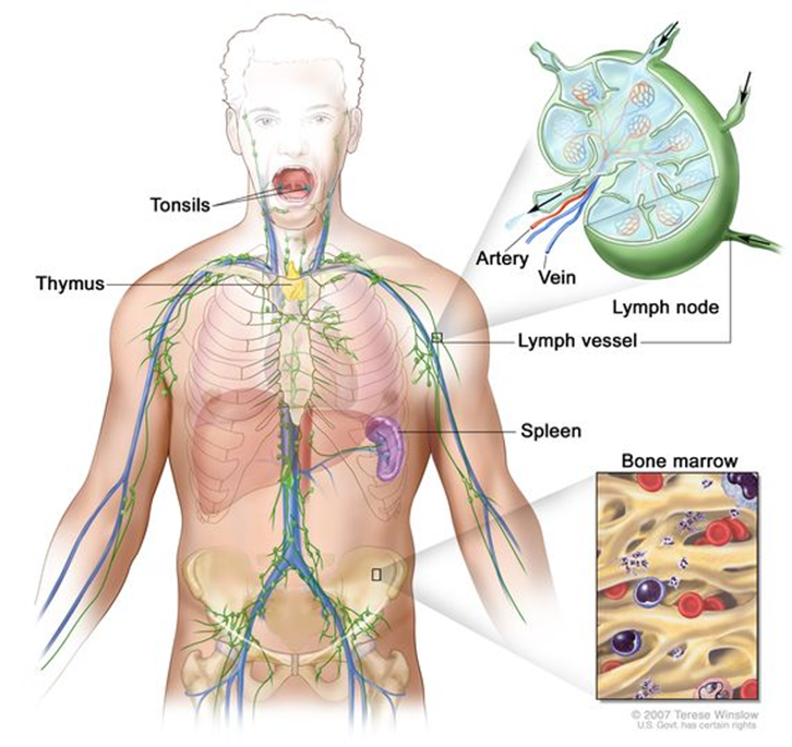
Describe the role of lymph nodes.
Sites where immune responses are mounted to antigens in lymph
Act as physiological barriers; they filter the microbial antigens carried to lymph node by activating the T and B cells
Are the first organized lymphoid structure to encounter antigens that enter through tissue spaces
If any particular antigen is brought in the lymph, it will be trapped by phagocytic and dendritic cells
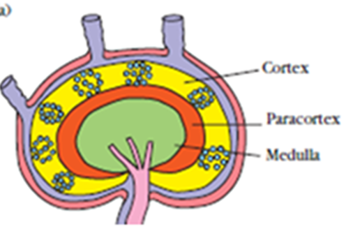
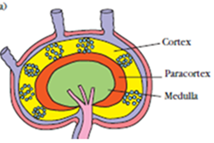
Lymph nodes are divided into 3 parts. List them.
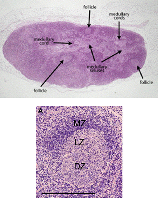
Cortex (B cell area)
Medulla (B cell area)
Paracortex (T cell area)
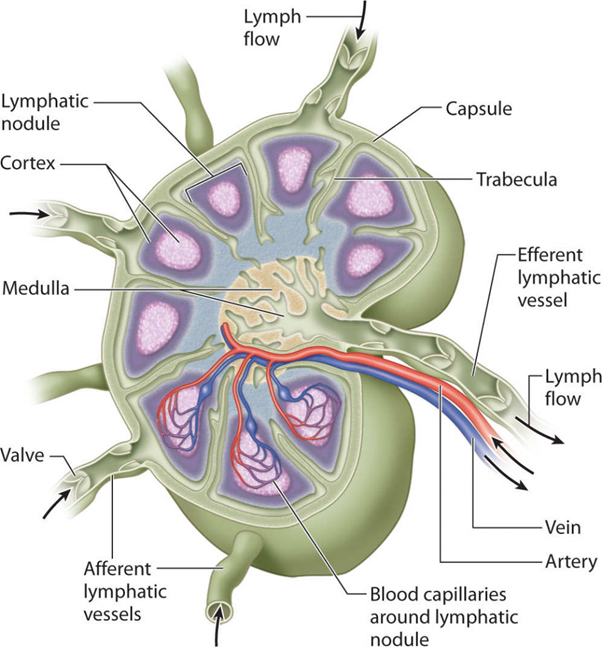
Describe the cortex of lymph nodes.
Surrounded by capsule and intervened by trabeculae
Contains lymphoid follicles that are composed of mainly B cells and few special types of dendritic cells (called follicular dendritic cells)
What are the two main types of lymphoid follicles?
Primary lymphoid follicles
Secondary lymphoid follicles
Describe primary lymphoid follicles.
Found before the antigenic stimulus
Smaller in size
Mainly contain resting B cells
Describe secondary lymphoid follicles and what occurs in them.
Following contact with an antigen, resting B cells in primary follicles start dividing and become activated
The activated B cells differentiate rapidly into plasma cells (produce antibodies) and memory B cells (which remember the antigenic structure and become activated on subsequent antigenic exposure)
Follicles become larger in size and are called secondary lymphoid follicles
Secondary follicles have two areas. Name and describe them.
Central area is called germinal center; contains dividing B cells of various stages
Germinal center has 2 zones: light and dark zones; site where activation of B cells takes place
Peripheral zone is called mantle area; contains activated B cells
Describe the paracortical area of the lymph node.
Present in between cortex and medulla
Is the T-cell area of lymph node
Rich in naive T-cells
Also contains macrophages and interdigitating dendritic cells, which trap the antigens and present to T-cells
Describe the medulla of the lymph node.
Innermost area of lymph node
Rich in B cells; mainly plasma cells
After infection, lymph leaving a node is enriched with antibodies newly secreted by medullary plasma cells and also higher conc. of lymphocytes
Describe mucosal associated lymphoid tissue (MALT).
Mucous membranes lining intestine, respiratory and urogenital tract (total surface area of about 400 m2) are major entry sites for most pathogens
Defense mechanisms needed in mucosal sites to prevent microbial entry
Group of lymphoid tissues lining mucosal sites are collectively known as MALT
Structurally, MALT may be arranged in 2 types. Describe them.
Loose clusters of lymphoid cells - usually found in lamina propria of intestinal villi
Lymphoid tissues arranged as organized structures - such as tonsils, appendix, and Peyer’s patches
Describe MALT in intestinal mucosa and describe its layers.
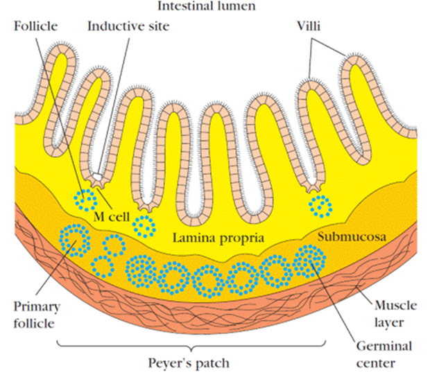
Lymphoid tissues lining intestinal mucosa are best studied in MALT
Present in different layers in intestinal wall: submucosa, lamina propria, epithelial layer
Submucosa: contains Peyer’s Patches (PP is a nodule of 20-40 lymphoid follicles)
Lamina propria: contains loose clusters of lymphocytes (B-cells, plasma cells, T-helper cells) and macrophages
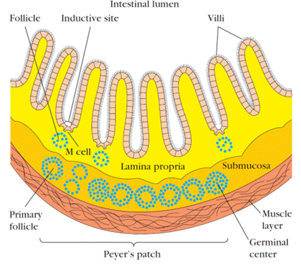
Epithelial layer: contains few specialized lymphocytes called intraepithelial lymphocytes (IELs) and modified epithelial cells called M-cells
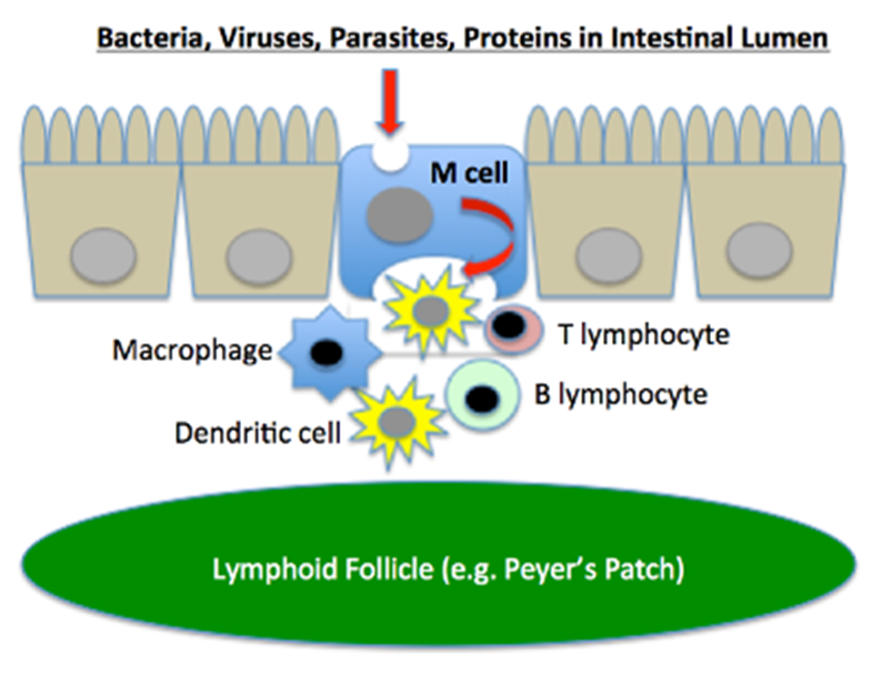
Describe M-cells and their role.
Specialized flattened epithelial cells
Do not have microvilli
Have deep invaginations or pockets in basolateral side; contain B-cells, T-cells, and macrophages
M-cells act as portal of entry of a number of microbes such as Salmonella, Shigella, Vibrio, and Polio virus
Invading microbes are taken up by M-cells, then transported in a vesicle, and delivered to the basolateral pockets
