15. CONTROL AND COORDINATION
1/32
There's no tags or description
Looks like no tags are added yet.
Name | Mastery | Learn | Test | Matching | Spaced |
|---|
No study sessions yet.
33 Terms
What are the features of the endocrine system, and how do hormones like ADH, glucagon, and insulin function?
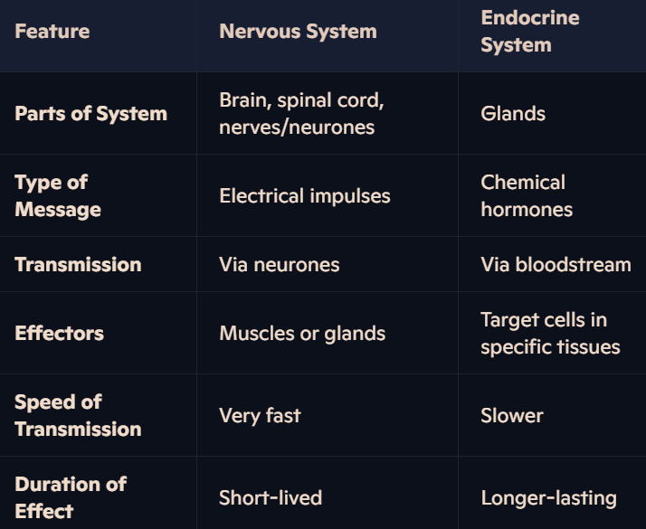
Definition: The endocrine system consists of glands producing and releasing hormones (chemical messengers) into the bloodstream to regulate specific target organs.
General Features:
Hormones control functions that do not require instant responses.
Hormones alter the activity of specific target organs with complementary receptors.
Receptors: Found on the cell surface membrane (peptide hormones like ADH, glucagon, insulin) or inside cells (steroid hormones).
Blood Supply: Endocrine glands have a rich blood supply to quickly transport hormones to target cells.
Specific Hormones:
ADH:
A peptide hormone.
Regulates water reabsorption in kidneys by increasing the permeability of collecting ducts via aquaporins.
Glucagon:
Secreted by α cells of the pancreas.
Stimulates glycogen breakdown (glycogenolysis) in liver cells to increase blood glucose concentration.
Insulin:
Secreted by β cells of the pancreas.
Stimulates glucose uptake in muscle, liver, and fat cells.
Promotes glycogenesis (conversion of glucose to glycogen).
Mechanism:
Peptide hormones (e.g., ADH, glucagon, insulin) are water-soluble and bind to cell-surface receptors.
Steroid hormones (e.g., testosterone, oestrogen) are lipid-soluble and bind to intracellular receptors.
How do hormones interact with target cells?
Peptide Hormones:
Bind to receptors on the cell surface membrane.
Activate second messengers to transfer signals within the cytoplasm.
Steroid Hormones:
Cross the phospholipid bilayer due to lipid solubility.
Bind to receptors in the cytoplasm or nucleus, directly influencing gene expression.
What are the differences between the nervous and endocrine systems?
Why are endocrine glands highly vascularized?
Endocrine glands require a rich blood supply to rapidly deliver hormones into the bloodstream for transport to target organs.
Hormones travel in blood plasma to reach cells with complementary receptors.
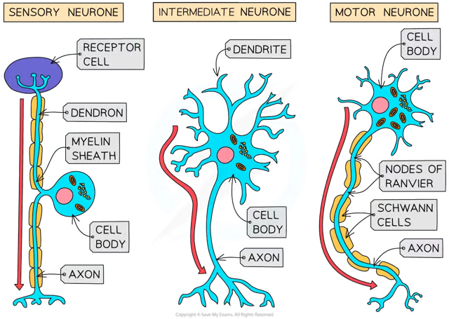
What are the features and functions of sensory and motor neurones?
Sensory Neurones:
Structure:
Cell body branches off the middle of the neurone.
Located near the source of stimuli or in a ganglion of the spinal nerve.
Long axon insulated by a myelin sheath with nodes of Ranvier.
Dendrites connect to receptor cells, forming networks for impulse transmission.
Function:
Carry impulses from receptors to the CNS (brain or spinal cord).
Motor Neurones:
Structure:
Large cell body located in the brain or spinal cord.
Contains highly branched dendrites for communication with other neurones.
Long axon insulated by a myelin sheath to transmit impulses efficiently.
Function:
Carry impulses from the CNS to effectors (muscles or glands).
Intermediate Neurones: Found entirely within the CNS, connecting sensory and motor neurones.
What is the role of sensory receptor cells in detecting stimuli?
Definition: Sensory receptor cells are transducers that convert energy from stimuli (e.g., light, sound, pressure) into electrical impulses within sensory neurones.
Location: Found in sense organs like the eyes, skin, and taste buds.
Types of Receptors:
Specialised Receptors: Light receptors in the eyes, chemoreceptors in taste buds.
Sensory Neurone Ends: Some touch receptors act directly as the ends of sensory neurones.
Response Mechanism:
Weak Stimulus: Insufficient depolarisation; impulse not transmitted.
Strong Stimulus: Sufficient depolarisation activates sensory neurones to send impulses to the CNS.
Establishes a generator potential when stimulated.
How do sensory, intermediate, and motor neurones work together in a reflex arc?
Pathway of a Reflex Arc:
Stimulus Detection: Sensory receptors detect stimuli (e.g., pain from a pin).
Impulse Transmission:
Sensory neurones send impulses to the spinal cord (coordinator).
Intermediate neurones pass impulses to motor neurones.
Effector Response: Motor neurones carry impulses to muscles or glands (effectors).
Outcome: Reflex action occurs (e.g., leg withdrawal from sharp object).
Feature: Reflex arcs bypass conscious brain regions, ensuring quicker responses.
How does the structure of myelinated neurones facilitate impulse transmission?
Axon: Long fibre insulated by a myelin sheath made by Schwann cells.
Nodes of Ranvier: Uninsulated gaps along the axon where impulses jump, speeding transmission.
Dendrites: Provide communication networks by connecting to other neurones.
Function: Efficient transmission of electrical impulses across long distances in the body.
What are Pacinian corpuscles, and what is their role in detecting stimuli?
Definition: Pacinian corpuscles are pressure receptors found deep in the skin, especially in fingers, soles of feet, joints, tendons, and ligaments.
Response to Pressure:
Detect changes in pressure, leading to the establishment of a generator potential.
Activate sensory neurones to transmit impulses based on pressure intensity.
Variation in Sensitivity:
Fingertips have a large number of Pacinian corpuscles, making them highly sensitive.
Backs of fingers have fewer receptors and lower sensitivi
How is an action potential triggered in a sensory neurone using a chemoreceptor in a human taste bud as an example?
Detection of Stimulus: Chemoreceptor detects chemical substances (taste molecules) and converts stimulus energy into an electrical signal.
Sequence of Events:
Depolarisation: Chemoreceptor cells are stimulated, leading to depolarisation and the establishment of a generator potential.
If the stimulus strength is sufficient, the sensory neurone is activated, transmitting impulses to the CNS.
Action Potential Generation:
Sodium ions enter through open sodium channels in response to the stimulus.
Depolarisation triggers voltage-gated sodium channels to open, allowing more sodium ions to enter (positive feedback).
When the threshold value (-50mV) is reached, many sodium channels open, creating an action potential.
Action potential travels along the sensory neurone towards the CNS.
How is the resting potential maintained in neurones?
Resting Potential: Electrical potential difference of -70mV between inside and outside of axon membrane.
Factors Maintaining Resting Potential:
Sodium-Potassium Pumps: Actively transport 3 sodium ions out and 2 potassium ions into the axon using ATP, maintaining ion gradients.
Impermeability to Sodium Ions: Sodium ions cannot diffuse back into the axon when at rest.
Negatively Charged Molecules: Large anions inside the axon attract potassium ions and reduce their diffusion out.
Voltage-Gated Channels: Closed channels prevent passive movement of ions.
What are the steps in the generation and transmission of an action potential?
Stimulus Detection: Sodium channels open, sodium ions enter, causing depolarisation.
Depolarisation: If the potential difference reaches the threshold (-50mV), more sodium channels open. The inside of the axon reaches +30mV, generating an action potential.
Propagation:
Sodium ions diffuse along the axon, triggering depolarisation at adjacent sections.
Action potential moves in one direction due to refractory periods in previous sections.
How is the resting potential restored during the refractory period?
Repolarisation:
Sodium channels close, stopping sodium influx.
Potassium channels open, allowing potassium ions to diffuse out, restoring negative potential (-70mV).
Hyperpolarisation: Brief phase where potential becomes more negative than -70mV due to excess potassium efflux.
Restoration:
Potassium channels close, and sodium-potassium pumps re-establish the original resting potential.
Ensures action potentials remain discrete and travel in one direction.
What is saltatory conduction, and how does it enable rapid impulse transmission in myelinated neurones?
Mechanism:
Myelin sheath insulates the axon, preventing ion diffusion and depolarisation along covered areas.
Depolarisation and action potentials only occur at the nodes of Ranvier.
Local currents cause action potentials to jump between nodes (saltatory conduction).
Advantages:
Speed: Increases impulse transmission speed (up to 50 times faster than unmyelinated neurones).
Efficiency: Reduces energy use by concentrating depolarisation at nodes.
What factors influence the speed of impulse transmission in neurones?
Presence or Absence of Myelin:
In myelinated neurones, the axon is insulated by a myelin sheath, which prevents ion movement across most of the axon membrane.
Depolarisation (and thus action potentials) only occur at the nodes of Ranvier, where the axon is not insulated.
This results in saltatory conduction, where the action potential appears to "jump" from one node to the next.
Effect: The impulse travels much faster compared to unmyelinated neurones because fewer depolarisation events are required. Transmission speed in myelinated neurones can be up to 50 times faster than in unmyelinated ones.
In unmyelinated neurones, depolarisation must occur continuously along the entire length of the axon, significantly slowing conduction.
Axon Diameter:
Neurones with thicker axons have a greater surface area, allowing for faster ion diffusion.
Increased ion diffusion leads to quicker depolarisation and action potential propagation.
Effect: Larger axons conduct impulses more rapidly than thinner ones due to reduced resistance to ion flow and enhanced depolarisation efficiency.
Temperature:
Higher temperatures increase the kinetic energy of ions, speeding up diffusion and enzyme activity (e.g., for ion pumps).
This enhances the speed of depolarisation and repolarisation processes.
Effect: Impulse transmission is faster at optimal physiological temperatures (within limits).
What is the refractory period, and what happens during this phase in a neurone?
Definition: The refractory period is the time during which a section of the axon membrane is unresponsive after generating an action potential.
Steps of Refractory Period:
Closure of Sodium Channels: Stops sodium ions from entering the axon.
Opening of Potassium Channels: Potassium ions diffuse out, causing repolarisation and returning the potential difference to -70mV.
Hyperpolarisation: Brief phase where the membrane becomes more negative than -70mV.
Resting State Restoration: Potassium channels close, sodium-potassium pumps re-establish the resting potential.
Key Features:
During this phase, the axon section cannot generate another action potential.
Why is the refractory period important for action potentials?
Ensures Discrete Action Potentials: Prevents action potentials from merging into one another, maintaining their integrity as distinct events.
Direction of Impulse:
Ensures action potentials move forward along the axon.
The region behind the action potential is in recovery (refractory), preventing backward transmission.
Efficient Transmission: Guarantees successful propagation of impulses in one direction only, crucial for proper nerve impulse coordination.
How does the refractory period determine the frequency of impulses?
Minimum Time Between Action Potentials: The refractory period sets the minimum time required for one action potential to end before another begins.
Maximum Frequency: Limits how frequently impulses can occur at a specific location along the axon:
Range: Between 500 to 1000 impulses per second.
Impact on Communication: The length of the refractory period regulates how rapidly signals can be sent, ensuring precise control of response rates in nervous system coordination.
What are the types of refractory periods, and how do they contribute to impulse transmission?
Absolute Refractory Period:
Occurs immediately after the action potential.
Sodium channels are completely closed, and no new action potential can be generated, ensuring discrete signals.
Relative Refractory Period:
Occurs during repolarisation and hyperpolarisation.
A stronger-than-usual stimulus is required to generate a new action potential.
Allows the nervous system to adjust the frequency of impulses based on stimulus intensity.
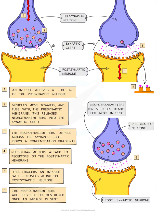
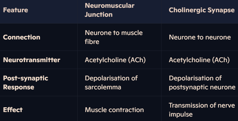
What is the structure of a cholinergic synapse?
Definition: A synapse is the junction between two neurones, where electrical impulses are transmitted chemically across a small gap known as the synaptic cleft.
Key Components:
Presynaptic Neurone:
Contains synaptic vesicles filled with the neurotransmitter acetylcholine (ACh).
Voltage-gated calcium ion channels are embedded in its membrane.
Synaptic Cleft:
A tiny gap (~20-40 nm wide) separating the presynaptic and postsynaptic neurones.
Neurotransmitters diffuse across this space.
Postsynaptic Neurone:
Contains receptor proteins specific to ACh on its membrane.
Sodium ion channels are part of these receptor proteins.
Supporting Structures: Surrounding tissue fluid and enzymes like acetylcholinesterase to regulate synaptic activity.
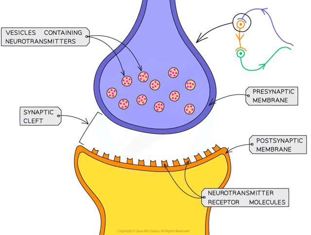
What is the basic mechanism of synaptic transmission at a cholinergic synapse?
Electrical impulses cannot jump across synapses, so transmission occurs chemically.
Arrival of Impulse: An electrical impulse reaches the axon terminal of the presynaptic neurone.
Neurotransmitter Release: ACh is released from synaptic vesicles into the synaptic cleft.
Diffusion Across Cleft: ACh molecules diffuse across the cleft.
Receptor Binding: ACh temporarily binds to receptor proteins on the postsynaptic membrane.
Impulse Restart: This binding causes sodium ion channels to open, triggering depolarisation and generating a new impulse in the postsynaptic neurone.
What is the detailed role of calcium ions (Ca²⁺) in synaptic transmission?
Action Potential Arrival: Depolarisation of the presynaptic membrane opens voltage-gated calcium channels.
Calcium Influx: Calcium ions diffuse down their electrochemical gradient from the surrounding tissue fluid (high Ca²⁺ concentration) into the cytoplasm of the presynaptic neurone (low Ca²⁺ concentration).
Vesicle Fusion: Calcium ions stimulate synaptic vesicles containing ACh to fuse with the presynaptic membrane.
Neurotransmitter Release: ACh is released into the synaptic cleft via exocytosis.
Importance of Calcium Ions: Essential for triggering vesicle fusion and ensuring ACh release, without which synaptic transmission would not occur.
How is the neurotransmitter acetylcholine (ACh) recycled after synaptic transmission?
ACh Breakdown: The enzyme acetylcholinesterase hydrolyses ACh into acetate and choline in the synaptic cleft.
Preventing Overstimulation: This breakdown ensures sodium ion channels on the postsynaptic membrane close, preventing continuous depolarisation.
Recycling:
Choline is reabsorbed by the presynaptic neurone.
Choline reacts with acetyl coenzyme A to reform ACh.
Re-synthesised ACh is packaged into vesicles for reuse.
Why is the cholinergic synapse highly efficient for signal transmission?
Speed: The entire sequence of synaptic transmission takes only 5-10 ms.
Specificity: ACh binds only to complementary receptors on the postsynaptic membrane, ensuring accurate signal relay.
Regulation:
Calcium ions control vesicle fusion and neurotransmitter release.
Acetylcholinesterase prevents overstimulation by rapidly breaking down ACh.
Recycling: Efficient reuse of choline conserves resources for continued neurotransmission.
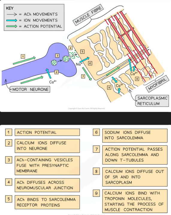
What are the roles of neuromuscular junctions, the T-tubule system, and sarcoplasmic reticulum in stimulating contraction in striated muscle?
Neuromuscular Junction:
An action potential arrives at the presynaptic membrane of a motor neurone.
Calcium ions diffuse into the neurone, stimulating vesicles containing acetylcholine (ACh) to fuse with the membrane.
ACh is released into the neuromuscular junction and binds to receptors on the sarcolemma of the muscle fibre.
This opens sodium ion channels, depolarising the sarcolemma and generating an action potential.
T-Tubules:
Transverse tubules (T-tubules) are deep folds in the sarcolemma that carry the action potential towards the centre of the muscle fibre.
Sarcoplasmic Reticulum (SR):
Action potentials stimulate voltage-gated calcium channels in the SR membrane to open.
Calcium ions diffuse into the sarcoplasm, surrounding myofibrils, and initiate contraction by binding to troponin on actin filaments.
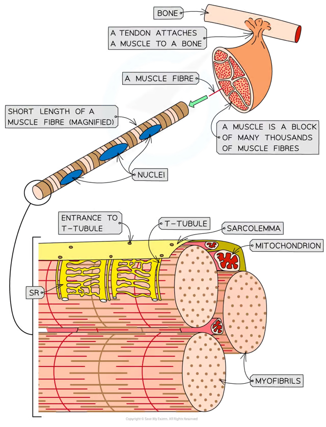
What is the ultrastructure of striated muscle, and how is it organised?
Muscle Fibre Components:
Sarcolemma: Cell surface membrane, with T-tubules that fold inwards.
Sarcoplasm: Cytoplasm containing mitochondria and myofibrils.
Sarcoplasmic Reticulum (SR): Specialized endoplasmic reticulum storing calcium ions.
Myofibrils:
Bundles of protein filaments (actin and myosin) responsible for contraction.
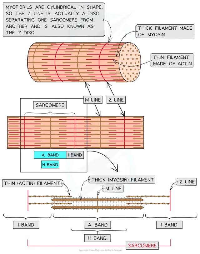
Sarcomere Structure
Sarcomere Structure:
Z Line: Attachment for actin filaments, marking the boundaries of a sarcomere.
M Line: Attachment for myosin filaments at the center of the sarcomere.
I Band: Contains only thin actin filaments.
H Band: Contains only thick myosin filaments.
A Band: Overlap of actin and myosin filaments.
Function: Sarcomeres shorten during contraction, creating movement.
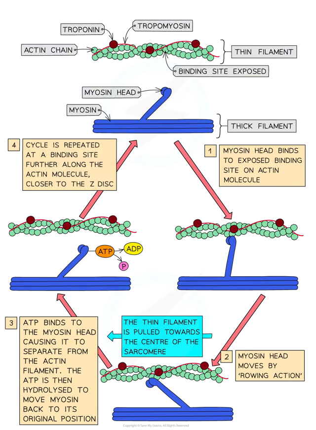
What is the sliding filament model of muscular contraction, and what roles do troponin, tropomyosin, calcium ions, and ATP play?
Triggering Contraction:
An action potential arrives at the neuromuscular junction.
Calcium ions are released from the sarcoplasmic reticulum into the sarcoplasm.
Troponin and Tropomyosin:
Calcium ions bind to troponin, causing it to change shape.
This moves tropomyosin, exposing myosin-binding sites on actin filaments.
Formation of Cross-Bridges:
Globular heads of myosin bind to actin at the exposed sites, forming cross-bridges.
Myosin heads pull actin filaments towards the sarcomere's center.
Role of ATP:
ATP binds to myosin heads, causing them to detach from actin.
ATP hydrolysis provides energy for the myosin heads to reset and attach to new sites on actin, continuing the contraction.
Sarcomere Shortening:
The Z lines are pulled closer together as actin slides over myosin, shortening the sarcomere and contracting the muscle.
How do structural features of actin and myosin facilitate contraction in the sliding filament model?
Actin Filaments:
Two chains of globular actin molecules twisted together.
Tropomyosin wraps around actin chains, with troponin attached at intervals.
Myosin Filaments:
Fibrous proteins anchored at the M line.
Globular heads extend outward, binding to actin during contraction.
Function: Myosin heads repeatedly attach, pull, and detach from actin in cycles, powered by ATP hydrolysis.
What similarities and differences exist between neuromuscular junctions and cholinergic synapses?
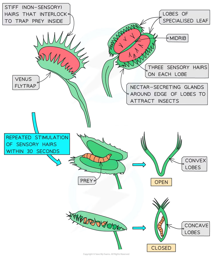
How does the Venus flytrap respond to stimulation of sensory hairs, and how does the trap close?
Response Mechanism:
Sensory Hairs: Three stiff hairs detect touch on each lobe.
Receptor Potential: Calcium ion channels open in cells at the base of the sensory hair, allowing calcium ions to enter and generate a receptor potential.
Action Potential: If two hairs are touched simultaneously or one is touched twice within 30 seconds, an action potential propagates across the cells of the trap.
Trap Closure: Cells at the base of the trap change shape, causing the lobes to fold along the midrib and capture the prey.
Ongoing Stimulation: Moving prey further stimulates sensory hairs, causing calcium ions to enter gland cells, stimulating exocytosis of digestive enzymes.
Digestion: The trap remains closed for up to a week while prey is digested and nutrients absorbed.
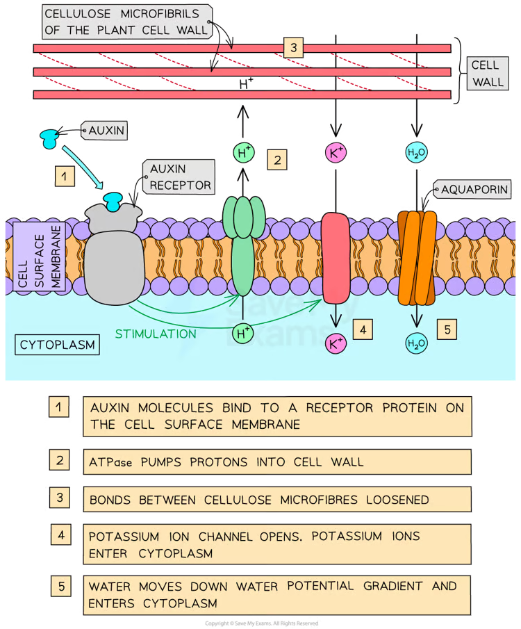
What is the role of auxin in elongation growth in plants?
Auxin Definition: IAA (indole 3-acetic acid) is a plant growth regulator synthesized in meristem tips of roots and shoots.
Mechanism of Elongation Growth:
Proton Pump Activation: Auxin binds to receptors on cell surface membranes and stimulates ATPase proton pumps to acidify cell walls.
Cell Wall Loosening: Lowered pH activates expansin proteins, which weaken bonds between cellulose microfibrils.
Water Absorption: Potassium ions enter cytoplasm, reducing its water potential and causing water uptake via osmosis.
Cell Wall Stretching: Increased internal pressure stretches the cell wall, elongating the cell.
Function: Increases shoot and root length by promoting elongation.
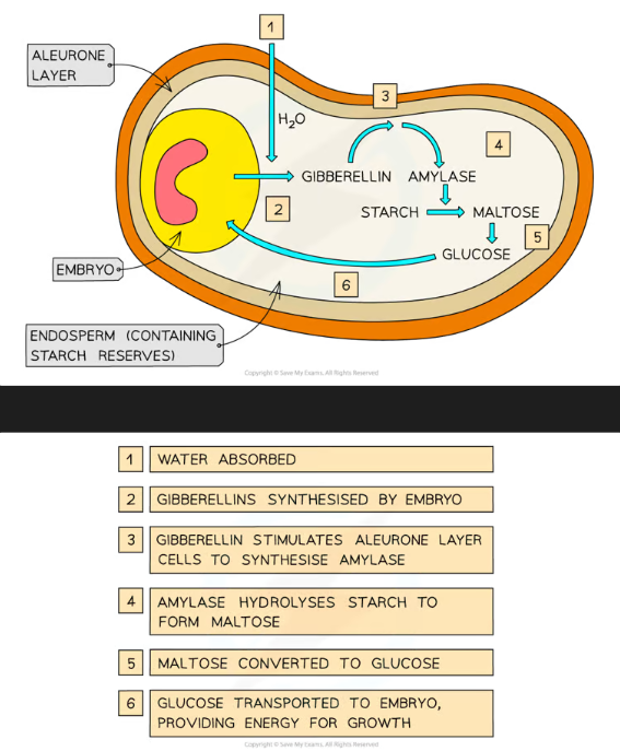
How does gibberellin regulate germination in barley seeds?
Dormancy: Barley seeds remain dormant until conditions such as water availability signal germination.
Process of Germination:
Water Absorption: Stimulates gibberellin production by the embryo.
Gibberellin Action:
Diffuses into the aleurone layer and increases transcription of mRNA for amylase synthesis.
Amylase hydrolyses starch in the endosperm, producing maltose and converting it to glucose.
Glucose Use: Glucose is respired by the embryo, providing energy for growth.
Outcome: Gibberellin enables seedling development by supplying energy.