Critical care exam 2
 Call Kai
 Learn
Learn Practice Test
 Spaced Repetition
 Match
Match Flashcards
 Knowt Play
1/172

There's no tags or description
Looks like no tags are added yet.
Name | Mastery | Learn | Test | Matching | Spaced | Call with Kai | Chat |
|---|
No analytics yet
Send a link to your students to track their progress
173 Terms
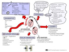
ischemic stroke
a type of stroke that occurs when the flow of blood to the brain is blocked due to plaque or emboli

examples of ischemic stroke
Large Artery Atherosclerosis, Cardioembolic, Lacunar stroke, Crytogenic stroke.

TIA
signs and symptoms of a stroke are present due to decreased cerebral blood flow, but symptoms resolve within 24 hours. found with CT scan

TIA time frame
5-10 mins symptoms less than 24 hours
-decrease in cerebral perfusion
What causes a TIA?
carotid artery stenosis
s/s of a TIA
monocular blindness, temp focal neurologic deficit, no permanent deficits

TIA treatment
carotid endarterectomy (cleaning out the arteries)

intracerebral stroke
-most common type of hemorrhagic stroke
-Rupture of small vessels inside the brain, most deadly type of stroke (bleeding of the brain tissue)
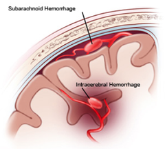
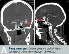
ruptured cerebral aneurysm
dilation of a section of an artery that causes the vessel wall to weaken (circle of willis). seizures can occur within the first 12 hours of rupture. risk of vasospasm.
hemorrhagic stroke
uncontrolled bleeding into intracerebral or subarachnoid space or both

arteriovenous malformation (AVM)
miscommunication between the artery and the vein.
-blood is being shunted to the veins w/o capillary use
-can present as tumor. Put the vesicles at risk for rupturing

NIHSS
National Institutes of Health Stroke Scale

What is the NIHSS used for?
To determine severity of stroke:
LOC, orientation, CN, speech, motor, visual

physical assessment of a stroke
-neuro checks and knowing where the stroke is in the brain will help with the assessment
-weakness or numbness of one side of body, slurred speech, visual disturbance, dizziness, incoordination, ataxia, vertigo, sudden onset of severe headache /assess airway, breathing, circulation

stroke diagnostics
CT without contrast, EKG, NIH, CBC, MRI, PT/INR, BMP, cardiac enzymes.
ischemic stroke treatment
tPA can be given as treatment within 4.5 hrs of symptom onset. permissive HTN with tPA is SBP

when is the nurse able to give TPA?
less than 4.5 hours from the onset, pt must be older than 18, CT scan shows ischemia, measurable deficits, the pt cannot have hemorrhage or bleeding, recent head trauma or surgery
when is the nurse NOT able to give TPA?
more than 4.5 hrs from onset, rapidly moving, current hemorrhage or bleeding, recent head trauma, recent cns surgery, severe HTN
Hemorrhagic stroke treatment
control HTN, HOB at 30-45 degrees, (MAP

What are the possible complications of stroke?
hydrocephalus (increased ICP), Seizures can onset within 7 days, infection, intracerebral hemorrhage.

focal seizures
one side of the brain, will experience an aura and loses awareness
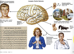
focal motor seizures
twitching, shaking, stiffening, or rubbing hands.

Focal non-motor seizure
-Other symptoms occur first
-Changes in sensation, emotions, thinking or experiences

alcohol withdrawl seizures
brief, generalized, 30-50% become delerium tremors within 24 hr of last intake.. Can also be seen with barbituates or benzos
Focal aware seizure
no loss of consciousness BUT communication may not be possible

focal unaware seizure
awareness impaired
Generalized seizures
sudden! both sides of brain
-impaired awareness
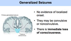
generalized motor seizure
stiffening (tonic) , jerking (clonic)

Generalized non-motor seizure
Absence seizures, lapses in awareness (staring into space), repeated movements like lip-smacking
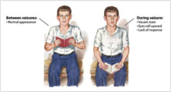
What is the assessment of a patient with seizures?
neuro (types of movements, aura, pupil changes, response to speech and touch, awareness of surroundings, LOC), respiratory, cardiovascular.
What should the nurse do when a patient is experiencing a seizure?
maintain airway and oxygenation, pad bedrails, turn pt to side.

What is the treatment for seizures?
lorazepam (sedation), levetiracetam, phenytoin
status epilepticus
A seizure that last more than 30 min or repeated seizure without recovery for more than 30 min
-typically generalized tonic/clonic

Causes of status epileptics
Tonic/clonic, Irregular epileptic drug use, ETOH withdrawal, drug withdrawal, head trauma, infection
What are some causes of a spinal cord injury (SCI)?
MVC, falls, violence, sports, diving, traumas

complete spinal cord injury
total loss of sensory and motor function

partial or incomplete spinal cord injury types
central, anterior, brown-sequard,
central spinal cord injury
-incomplete loss of motor function
C for "cellar"- the injury sustained by old ladies who fall down the cellar stairs. The motor supply of the arms is damaged, but not the legs, so the old lady can stand up and get out of the cellar, but can't use her hands.

Anterior spinal cord injury
damage to the front of the spinal cord.
-loss of motor function, pain, and temp
-can still feel light touch

Brown-Sequard spinal cord injury
Damage to one side of the spinal cord, causing weakness or paralysis on the same side of the injury.
-Loss of pain and temp sensation on the opposite side.

What is the relationship of location of injury to functional loss?
the more functionality there is lost the higher the injury is on the spinal cord.
what medications are given for spinal cord injuries?
corticosteroids, muscle relaxers, vasoconstrictors

corticosteroids for SCI
used for anti inflammatory and edema reducing effects, may interfere with healing.
muscle relaxers for SCI
Diazapam=used for clients with upper motor neuron injuries, helps control muscle spasticity.
vasoconstrictors for SCI
used to maintain perfusion to spinal cord + CPP (goal is to maintain map >85 for the first week post spinal cord injury)
-Norepinephrine + dopamine
What are the different system complications associated with spinal cord injury?
respiratory, neuro, hemodynamic, bowel, bladder, skin, psychosocial.

spinal shock
complete loss of motor, sensory, and reflex. hypotension, usually occurs with injury above T6, can last weeks to months

neurogenic shock
Disruption of ANS below level of injury
-Loss of sympathetic input: vasodialation, hypotension, bradycardia, hypothermia

treatment for neurogenic shock
Pacemaker if symptomatic bradycardia
-Resolves with sympathetic tone
-vasoconstrictors

autonomic dysreflexia
a life-threatening emergency in spinal cord injury patients (ABOVE T6) that causes a hypertensive emergency; it occurs AFTER spinal shock has resolved
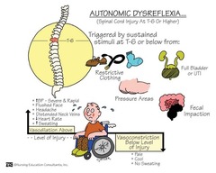
treatment for autonomic dysreflexia
-TREAT WHATS CAUSING THE ISSUE
-Control their temp and relieve tight clothing, kinks in catheter, urine blockage, elevate HOB.
-HTN: SBP above 150 treat with nitrates and nifedipine

what is a halo?
Static traction, headpiece with 4 pins into skull to give more mobility. Monitor neuro status or changes in movement

how should the nurse care for a patient with a halo?
do not hold or pull, assess skin integrity.Pin care: sterile pin site care as prescribed, keep key close in case of cardiac arrest, notify PCP if halo loosens or redness/swelling/drainage. Use foam to relieve pressure, keep vest lining dry
What is the management of tongs?
Weights attaches as countertractionMonitor neuro status, must hang freely, ropes remain in pulley, maintain body alignment, do not remove traction

What are the pharmacological and mechanical treatments for lethal arrhythmias?
Pharmacological: Epinephrine, amiodarone, vasopressin.
Mechanical: CPR, maintain airway, defibrillation
What are the dysrhythmia's effect on hemodynamics?
lowering CO because of the decreased filling time from increased heart rate
What are the mechanisms of neurogenic shock?
widespread vasodilation/ caused by spinal cord injuries, anesthesia complications / correct with fluids and pressors, cardiac parameters all low
What are the causes of elevated ICP?
increased brain volume (cerebral edema),
increase in CSF
or an increase in blood volume.
How is Cerebral Perfusion Pressure (CPP) calculated?
MAP-ICP=CPP

What is the Glasgow Coma Scale?
A scale used to assess a patient's neuro function by assessing their eye opening, verbal response, motor response.

What GCS score is consistent with a coma?
3-8

As the nurse what should you do if your pt has a GCS of less than 5?
call the Indiana donor network (IDN) for possible organ donation.
Decorticate posturing
arms flexed at the elbows inward and bent in toward the CORe of the body and the legs are internally rotated.
-this means the brainstem is not affected

Decerebrate posturing
$$$you can't celebrate with decerebrate
$WORST kind
arms, legs, and neck with the jaw clenched. The ares are extended straight to the sides of the hips. the head and neck is being arched backwards

how should the nurse posture a patient with increased ICP?
keep the HOB elevated to 30 degrees if appropriate. Avoid trendelenburg position

nursing care for a patient with increased ICP
-prevent flexion of the head, neck, hips.
-maintain neutral position of the head
-prevent shivering
-decrease stimuli
normal ICP
0-15 mmHg
What is considered an elevated ICP?
greater than 20 mmhg for 5 minutes or more
craniotomy
cutting into the skull
removes bone fragments, hematomas, foreign objects

mannitol
osmotic diuretic that is used to decrease ICP. causes diuresis
corticosteroids
-sone or -barbital
decreases brain activity and cerebral metabolism + decreases pressure
-must taper of
-reduce the leakiness in the blood-brain barrier.
What are anticonvulsants used for in ICP?
phenytoin, fosphenytoin, levetiracetam
given prophylacticly to prevent seizures
Why are antipyretics and muscle relaxers given for ICP?
given to decrease temperature, decrease metabolism, decrease shivering.
examples of meds: acetaminophen, diazepam
What paralytic agents are given for ICP and what is the assessment for them?
cisatracurium.
train of four assessment is done and 2 out of 4 twitches are goal.
What are the sedation agents given for ICP and what assessment is done for them?
propofol, lorazepam. BIS monitoring is done for these medications and goal is 40-60
What are the analgesics given for ICP and what assessments are done for them?
fentanyl and codeine.
-pain assessments are done for these medications using the FLACC scale or non-verbal pain scale. Respiratory assessments are also important.

intraventricular Catheter/external ventricular drain
allows drainage

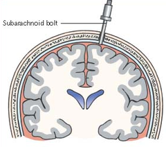
subarachnoid bolt
placed just through skull to monitor ICP but does not allow for CSF drainage
epidural sensor
no drainage

parenchymal fiberoptic catheter
•Placed directly into brain tissue just below subarachnoid space
•Very accurate
•Does not require fluid-filled transducer
•No CSF can be withdrawn

What is a subarachnoid bleed?
A brain bleed in the subarachnoid space where the CSF circulates

Uncal herniation
MOST COMMON.
pushes on the midbrain

s/s of cerebellar Tonsillar herniation
alterations in resp. and cardiac functions- rapidly progressing to cardiac arrest, Headache and neck stiffness, reduced muscle tone

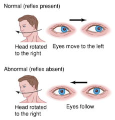
What are dolls eyes and what does it tell us?
eyes that move in the opposite direction when the head is turned
-this means the brainstem is intact
What are the most concerning signs and symptoms of a closed head injury?
changes in LOC, posturing changes, cushing's triad (bradycardia, widened pulse pressure, irregular respirations)
basilar skull fracture
linear fracture at the base of the skull
-check for halo sign

S/S of basilar skull fracture
raccoon eyes (bruising around eyes), battles sign (bruising behind the ears), CSF leaking from the ears (otorrhea) or norse (rhinorrhea)

acute subdural hematoma
Within 48 hours and almost always seen with cortical or brainstem injury.
Risk of death is high from injury to brain tissue and expanding hematoma.
Surgical intervention probable.

chronic subdural hematoma
2 weeks to several months after. Seen in the elderly, chronic alcohol abusers, and on anticoagulants/antiplatelets
From low velocity impact.

What should be monitored while giving mannitol?
hourly neuro assessments, ICP, CPP, serum osmolarity Q6H, electrolytes (K+, Na, chlor), ABGs, I/Os, VS, renal function
What is the priority intervention for a severe TBI?
Reduce ICP, maintain airway, maintain CPP, prevent secondary brain injury, normothermia, surgical interventions (craniotomy or canioectomy, burr holes), HOB 30 degrees
What interventions should you avoid with TBIs?
Trendelenburg, suctioning, pressure on abdomen, valsalva, straining, coughing, sneezing, monitor V/S, avoid narcotics, maintain nutrition.
how should the nurse monitor a patient that has received TPA?
maintain BP less than 185/110, they are at risk for hemorrhage at the area of infarction for the first 36 hours, nuro check q15 min for the first 2 hours, hold anticoagulants + anti platelets for 24 hours.
normal CPP
60-100
what is the goal for CPP in a patient with a brain injury?
above 70
what kind of patients is the parenchymal fiberoptic catheter used in?
Useful for patients with compressed or dislocated ventricles

What causes a subarachnoid hemorrhage?
ruptured aneurysm caused by a traumatic injury. this increases the risk of vasospasm.
BARBITURATE COMA (BI)
-phenobarbital
this is used when other modes of treatment is not working
Brain herniation
Increasing intracranial pressure related to the presence of lg pocket of blood (hematoma)
-CAUSES THE BRAIN TO MOVE

signs that the brainstem is affected
-negative for dolls eyes (eyes don't move when the head is turned)
-pupils are fixed and dilated
-babinski reflex: we will see toes that fan out when stimulated (we want the toes to curl in)
-vomiting
-widening pulse pressure
-seizures