Calf Management
 Call Kai
 Learn
Learn Practice Test
 Spaced Repetition
 Match
Match Flashcards
 Knowt Play
1/46

There's no tags or description
Looks like no tags are added yet.
Name | Mastery | Learn | Test | Matching | Spaced | Call with Kai | Chat |
|---|
No analytics yet
Send a link to your students to track their progress
47 Terms
Clinical Exam of a Calf
Systematic approach
Describe 10 differences from adult cattle
Systematic Approach: Similar to cows
Distance
Back
Left
Head and neck
Right
Differences to Adult Cattle: No udder, rumen auscultation, rectal OR vaginal exam in calves
History (milk intake easier than pasture intake)
Behaviour (more playful)
Normal: Adequate reaction to acoustic and optical stimuli, very BAR
→ Adequate reaction → Delayed reaction → Reaction to ONLY painful stimuli (eg. venipuncture) → No reaction to painful stimuli
Symmetry (underdeveloped rumen → different to adult abdomen shape)
Faeces VERY important to examine
Rub perineum to stimulate defaecation (may not be observed if very liquid and shoots out)
Palpate navel
Palpate joints
Pinging still important
Reflexes: Suckling and palpebral
Suckling = Strong → Weak → Absent/chewing movements
Management: Small teat openings and more frequent feeds to satisfy innate desire to suckle
Test with fingers (should be painful) + observe tail wag
Palpebral = Eyelids closed immediately and fully → Delayed closure → Delayed closure and NOT fully → NOT closed at all
Strength and posture
Normal: Calf standing by itself → after encouragement → Steady after lifting → Unsteady but can correct position if forced → Unsteady and unable to correct position → Sternal recumbency → Lateral recumbency
Encourage standing by rubbing hair over the back in the opposite direction
Ears (more prone to ear infections
TPR of calves
RR = 24 - 36/min
HR = 80 - 100 bpm
T = 38.5 - 39.5˚C
List 9 DDx for calf disease
Diarrhoea
Navel ill
Calf pneumonia
Rumen drinking/acidosis
Joint ill
Meningitis/salt poisoning/PEM (see “Systemic Diseases”)
Abomasal disease (ulcers, bloat and displacement)
Calf diphtheria
Other
Atresia ani/colonic aplasia
Contracted flexor tendons
Bovine neonatal pancytopenia
Overview of Umbilical Diseases
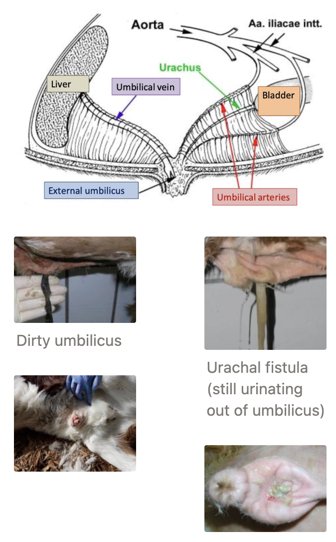
Normal umbilicus structure
Describe normal umbilicus involution
5 Types of umbilical diseases
Recommendations for umbilical care
Structure:
Urachus = To bladder
ONE umbilical vein = To liver
TWO umbilical arteries = To aorta (snap back after birth)
Normal Involution:
Umbilical cord ruptures at birth ~4 fingers away from skin
Rupture at skin = Risk of infection
Dries out within 4d (determine age or signs of infection)
Sloughs off at ~14d
Scab present until 3 - 4w
Types of Umbilical Diseases:
Uncomplicated umbilical hernia
Complicated umbilical hernia
Umbilical remnant infection (infected vein, arteries or urachus)
Umbilical abscess/chronic omphalitis
Urachal cyst/rupture
Umbilical Care: ASAP after birth before AND after transport
Iodine/chlorhexidine spray/dip to completely cover navel
Handle navel gently and as little as possible
Repeat ≥SID until navel dries out
Navel Ill
5 Risk factors
Diagnosis (2)
6 Clinical signs
5 Sequelae
2 Treatments of umbilical infections
2 Treatments of umbilical abscess
Risks:
Poor/no navel treatment
Insufficient colostrum intake
Low calf pick-up frequency (2x minimum and 4x ideal)
Poor general hygiene (calf pen, calving paddocks and trailer)
Overstocking
Diagnosis: History and general clinical exam (fever and inappetence) →
Further examination of umbilicus
Distance Examination
Length? Dry? Smell? Fistula? Discharge?
External Umbilicus
Size
Texture
Pain
Hernial ring
Reduction possible?
Internal Navel Palpation = Palpate intra-abdominal structures
Size
Texture
Pain
Adhesions
Further diagnostics
Probe/cannulation
U/S = Evaluate extra- and intra-abdominal structures
Clinical Signs:
Swelling
Discharge
Pain (hunched/flinching when umbilicus pinched)
Damp
Hernia
Severe = Pyrexia, septicaemia, lethargy and inappetence
Sequelae:
Septicaemia
Liver abscess
Meningitis
Joint ill
Spinal abscess
Endocarditis
Treatment of Umbilical Infection:
Broad-spectrum AB and NSAIDs for 3 - 5d
Surgical intervention
Treatment of Umbilical Abscess:
Drain abscess (thin-walled and large)
Surgical extirpation (thick-walled and small)

Umbilical Hernia
Aetiology
3 Examples of complicated umbilical hernia
3 Treatments of uncomplicated umbilical hernia
Treatment of complicated umbilical hernia
Aetiology: Congenital or following infection (consider genetics)
Complicated Umbilical Hernia:
Non-reducible and/or infected
Hard to feel hernial ring
Systemic signs of depression/colic
Treatments when Uncomplicated:
Do nothing when 1.5 - 3.5cm
90% of hernias with diameter of 1.5cm - 3.5cm close within 65 days
Keep hernia reduced with hernial clamps/elastrator ring/abdominal support bandage
Surgical intervention
Treatment when Complicated: EMERGENCY surgical intervention
What is the most likely DDx for a female calf with pulsing urination?
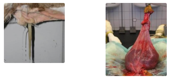
Urachal cyst/fistula
Overview of Bronchopneumonia
Prevalence in NZ
Aetiology
Viral (5)
Bacterial (6)
Risk factors
Pathogenesis
Prevalence: Relatively rare in NZ due to outdoor housing (vs. intensive dairy systems)
More common cause of death in feedlot cattle
Aetiology: MULTIFACTORIAL = Stress + virus + bacteria
Virus
Infectious bovine rhinotracheitis (BHV-1)
Parainfluenza virus (PI-3)
Bovine viral diarrhoea virus (BVDV)
Bovine respiratory syncytial virus (BRSV)
Bovine coronavirus (BCoV)
Bacteria
Mannheimia haemolytica = G- aerobe
Pasteurella multocida = G- facultative anaerobe
Histophilus somni = G- facultative anaerobe
Mycoplasma bovis = No cell wall
Trueperella pyogenes = G+ facultative anaerobe
Fusobacterium necrophorum = G- anaerobe)
Risks:
Purchase calves from auctions or markets
Mix calves from different sources (peak mortality ~16d after arrival in feedlot)
Weather conditions at arrival
Transport → Reduced mucociliary clearance and dry bronchial mucus → Suppressed immune system
Crowding
Other diseases
Climate/season: Poor ventilation, humidity, draught, cold stress, ammonia
Poor colostrum management
Pathogenesis:
Viruses and stressors damage alveolar macrophages and local ciliated epithelium
Results in suppressed immune system
Calves either heal OR become further infected
2˚ bacterial infection results in respiratory disease
Further infection with Trueperella pyogenes → abscess OR Fusobacterium necrophorum → necrosis
Results in death

Diagnosis of Bronchopneumonia
Clinical signs
7 Methods of diagnosis
Clinical Signs: DART
Depression
Anorexia
Respiratory character change (cough and dyspnoea)
Temperature elevation
± Agalactia in cows
Diagnosis: Individual and group PE →
Thoracic U/S = ID presence and severity of lung consolidation
+ve: Gold standard
Changes occur within hours
On-farm and real-time
Helps with prognosis and treatment triage
ID re-aeration of lungs = Treatment success
Lung consolidation associated with reduced ADG and lower CWT
CBC/biochemistry = Acute inflammation and FPTi
Nasal swabs = Superior for acute viral isolation
Transtracheal wash for culture and sensitivity
Radiographs with portable unit → Cranioventral lung lobe consolidation
Bronchoalveolar lavage and sterile sample
Bacterial culture
PCR
MALDI-TOF MS
Next generation sequencing
PM ± Culture of lesions
Lung U/S and presence of cough = #1 parameters to determine WHICH calf needs treatment to cure

Treating Bronchopneumonia
Treatment
Prognosis
4 Sequelae
Treatment: EARLY! Late therapy = #1 reason for treatment failure
Antibiotics for 3 - 4 days: Requires G- negative with good lung penetration
Penicillin
Florfenicol
Enrofloxacin
Tulathromycin
Cephalosporins
Oxytetracycline
NSAIDs
Prognosis: Fair to grave depending on severity and response to treatment
Response to treatment often after 12 - 24hr
Treat 2 days after recession of fever
Check U/S
Sequelae:
Chronic lung injury
Pulmonary abscess
Ear infection
Chronic, recurrent bloat (swelling of mediastinal LN)
Chronic Bronchopneumonia
2 Clinical signs
Pathogenesis
Treatment
Clinical Signs:
Initial acute bronchopneumonia (MAY or may not have been treated)
Poor growth rates and chronic weight loss despite treatment
Pathogenesis:
2˚ infection with Pasteurella multocida/Trueperella pyogenes → Fibrinosuppurative abscess → Congested OR Fusobacterium necrophorum → Necrotic tissue walled off
Difficult for antibiotics to penetrate
Extension of inflammation to pleura → Pleuropneumonia → Pleural effusion and adhesions
Common with Histophilus somni
Treatment: Cull (poor prognosis)
Rumen Drinking/Acidosis
Normal physiology of the oesophageal groove
4 Aetiologies (+ examples)
Pathogenesis
6 Clinical signs
5 Treatments
3 Methods of prevention
Normal Physiology: Suckling reflex linked to closure of the oesophageal groove
Rumen drinking = Insufficient closure of oesophageal prevent milk from bypassing the rumen
Aetiologies:
Primary suckling weakness
Difficult calving
Se deficiency
CNS problem (BVD)
Dummy calve syndrome
Prematurity (insufficient lung development)
Secondary suckling weakness due to illness
Iatrogenic = Drenching/forceful feeding of milk
Overloaded abomasum → Reflux of milk into rumen
Pathogenesis: Similar to adult rumen acidosis
Fermentation of milk in rumen
Increased lactate (D-/L-)
Drop in rumen pH
Blood acidosis
Rumenitis and bacterial translocation
Dehydration
Clinical Signs:
No/poor suckling reflex, weak and depressed
Pale faeces
Poor doer
Rough coat with alopecia
Full rumen with slushing sounds on ballottement
Teeth grinding and arched back
Treatment:
Eliminate and treat underlying cause
Flush rumen
Bicarbonate PO(15 - 30g in cold water)
IV if acidosis present
± IV dextrose for maintenance requirements (400g dextrose/40kg calf/24hr) if not good suckling reflex present
Do NOT offer milk until good suckling reflex present
± Oral antibiotics for severe rumenitis (poor prognosis) → Yoghurtised milk or rumen transfaunation
Prevention:
Gentle and patient calf husbandry (avoid negative association)
Never drench or force feed milk/MR (first colostrum is ONLY exception)
Disease monitoring

2 Ways to ID a premature calf
Incisors have not completely broken through
Very soft cartilage (rolled up ears)
Joint Ill
2 Aetiologies
5 Clinical signs
4 Methods of diagnosis
7 Treatments
Aetiologies:
Haematogenous spread (eg. navel ill or pneumonia)
Traumatic (puncture, decubitus 2˚ to contracted flexor tendons)
Clinical Signs:
Distended joints (painful as joint capsule highly innervated)
Calor, rubor, dolor (inflammation)
Increased/decreased mobility
Muscle atrophy
General symptoms: Fever, anorexia
Diagnosis: Orthopaedic exam of ALL joints →
U/S with linear transducer (5 - 7.7 MHz)
Diagnosis of arthritis, bursitis, abscess, haematoma
Normal synovial cavities in cattle DIFFICULT/IMPOSSIBLE to visualise via U/S (very small amount of normal synovial fluid)
→ Effusion that is easily visualised = Pathology
Joint tap to examine synovial fluid (do NOT joint tap if phlegmone present (inflammation of soft tissue)
Cloudy, turbid and watery
TP > 4g/dL
> 30,000 WBC/mm³
Radiography
Arthroscopy
Treatment:
Broad-spectrum AB (≥10 days and ideally 14 days)
Intra-articular antibiotics ± local anaesthesia
NSAIDs (ketoprofen or carprofen)
Joint lavage q2 days 3 - 5 times
Only if no fibrin present
Bandage
Box rest
Traumatic/open joints/multiple joints affected → Euthanasia (difficult to treat)
Abomasal Ulcers
3 Aetiologies
5 Clinical signs
PM
5 Treatments
Aetiology:
Stress/pain-related
Weather
Cl. perfringens type A???
Clinical Signs:
Melaena and occult blood
Anaemia
Acute death with perforation
Teeth-grinding
Dipping mouth into water trough
PM: Superficial, haemorrhagic and'/or perforating ulcers
Treatments:
Blood transfusion
Antacids
NSAIDs?
Surgery
Oral AB?

Abomasal Bloat
5 Risks
3 Clinical signs
Diagnosis
5 Treatments
Risks:
Feeding amount (>3L/meal)
Feeding frequency (once per day)
Milk temperature (cold milk)
Schedule
Milk quality
Clinical Signs:
Abdominal distension vs. severe colic
Dehydration
Diarrhoea
Diagnosis: Ballottement and U/S
Treatments:
Gastric tube → ± Administer vegetable oil or surfactant to pop bubbles in the frothy foam produced to remove gas
NSAIDs
Buscopan (anti-spasmodic)
Roll calf on back and relieve gas with needle (toggle principle)
Surgery
Abomasal Displacement in Calves
Aetiology
3 Treatments
Aetiology: Poor milk quality, volume and feeding interval
Treatments:
Rolling (LDA)
Abdominal surgery with full GA in left lateral recumbency (right flank approach)
Empty abomasum out as content often cause
4 Ways to prevent abomasal disease in calves
Smaller volume feeds more frequently (15 - 20% BWT/24hr over 6 feeds daily)
Ensure small teat hole to prevent rapid consumption
High milk quality (derived from milk proteins) and yoghurtised milk
Gradual transition between milk products
Feed cold milk
Atresia Ani/Colonic Aplasia
Aetiology
5 Clinical signs
2 Treatments
Aetiology: Congenital and linked to early rectal pregnancy palpation of embryo (membrane slip?)
Clinical Signs:
No anus (obvious)
No defaecation (apart from meconium)
Full abdomen
Colic
Anorexia after few days
Treatments:
Surgery
Euthanasia and PM
Bovine Neonatal Pancytopenia (BNP)/Bleeding Calf Syndrome
Signalment
Aetiology
Pathogenesis
3 Clinical signs
Treatment
Prevention
Signalment: Calves <3m
Aetiology: Associated with BVD vaccination of mothers (PregSure) and also genetic contributions?
Pathogenesis: Antibody-mediate immunopathic process
Allo-Ab directed against WBC derived from colostrum
Clinical Signs:
Generalised bleeding disorder
Fever
Death
Treatment: Symptomatic treatment usually NOT successful → Euthanasia
Prevention: Stop using this vaccine (no longer used)
Contracted Flexor Tendons
Aetiology
Signalment
4 Treatments
Complications
Aetiology: Congenital (different grades of severity)
Signalment: Large male calves predisposed
Treatment:
Physiotherapy
Hoof extensions
Casts/splints
Surgery
Complications: Decubitus and arthritis
Describe 4 challenges of NZ calves
All calves born during SHORT but INTENSE calving period → High pressure on limited staff and resources
Born in contaminated and muddy paddocks outside in cold/wet weather
→ Slippery paddocks which make it difficult to stand and suckle → Reduced colostrum intake
→ Increased energy demands to maintain body heat → Weaker calves upon arrival at calf shed
Calves raised in groups (vs. individual pens)
→ High calf-to-calf contact → Increased rate of disease transmission
→ Smaller calves forced to compete with larger, stronger calves for feed and space
More calves born than what sheds can accomodate → Calves turned onto pasture when only a few weeks old to make room for younger calves
6 Causes of calf diarrhoea (+ timing)
E. coli (enterotoxic K99 = ETEC): 0 - 5d
Rotavirus/coronavirus: 5 - 15d
Nutritional (iatrogenic): 0 - 3w
Cryptosporidiosis: 5 - 35d
Salmonella spp.: 10d - 3m
Coccidiosis (Eimeria spp.): ≥21d
Reason for clinical disease = Adverse relationship between resistance of calf and infectious pressure

3 Impacts of calf scours
#1 cause of calf death (birth → 3w) in many countries
$$$ (one case ~$150 treatment)
Labour intensive for farmers/calf rearers
5 Steps of calf scours outbreak investigation
ID scale of problem (numbers affected/died)
Verify signalment (age) and clinical signs
Dirty backside/tail
Inappetence, lethargy, hunched/arched back
Confirm vaccination status of herd → Contact MSD Animal Health if Rotavec
Assess for dehydration and acidosis (blood gas analysis)
Brix test colostrum
Blood test 12 healthy calves for FPTi
± Faecal culture/examination
ELISA TIMING important (eg. -ve result in sick calf or +ve result in healthy calf)
PM of freshly dead calves and histopathology
Small and large intestine → Fix in formalin ASAP (autolysis <30 min)
BOTH = Coronavirus
Small intestine ONLY = Rotavirus
Cryptosporidium in small intestine ONLY
Enterotoxigenic E. coli (ETEC), K99
Age
3 Clinical signs
Pathogenesis
Age: <3 - 5d
Clinical Signs:
Rapid onset of depression and recumbency with profuse scours
Rapid loss of >12% BWT in fluids
Death due to hypovolaemic shock within 12 - 24hr
Pathogenesis:
Bacteria adhere to enterocytes in small intestine
Enterotoxins produces which increases Cl- secretions
Net increase of fluid into intestinal lumen (secretory diarrhoea)
→ Dehydration, acidosis and hypersecretion
NO mucosal damage)
Rotavirus/Coronavirus
Age
3 Clinical signs
Pathogenesis
Age: 5 - 15d
Clinical Signs:
Moderately depressed
Often continue to drink milk
Scours last ~3d (± chronic with some coronavirus scours)
Pathogenesis:
Secretion of viral enterotoxin
Vasoactive agents from damaged cells → Activation of enteric nervous system
Villous atrophy and crypt cell hyperplasia in small intestine
Destruction of enterocytes in colon (coronavirus)
→ Malabsorption due to reduced SA
Cryptosporidiosis
Age
4 Clinical signs
Pathogenesis
Age: 5 - 35d
Clinical Signs:
Emaciation with persisting scours regardless of treatment
D-lactic acidosis
Death due to hypoglycaemia (even after resolution of scour) due mucosal damage → Malabsorption
Deaths more likely in cold spells and if milking restricted
Pathogenesis:
Protozoan parasite inhabits brush border of enterocytes
Villous atrophy, fusion and crypt cell hyperplasia
Prostaglandin-mediated anion secretion (Cl- or HCO3-)
Malabsorption and hypersecretion
Salmonella spp.
Age
4 Clinical signs
Age: 10d - 3m
Clinical Signs:
Fever
Depression → Recumbency and coma
Rapid weight loss
Death due to septicaemia (vs. hypovolaemic shock)
Coccidiosis
Age
PPP
Source
4 Risks
5 Clinical signs
Diagnosis
Treatment
4 Methods of prevention
Age: >21d (5 - 8w)
<12m but rare
PPP: 18 - 21d
Source: Oocysts persist in environment
Risks:
Inconsistent use of monensin → Delay or influence onset of disease
High challenge (overcomes in-feed coccidiostats)
Discontinued meal too soon after weaning
Stress (eg. weaning or weather)
Clinical Signs:
Abdominal apin
Scours
Tenesmus
Haematochezia
Severe dehydration
Diagnosis: Faecal smear or FEC
Treatment: Baycox (toltrazuril) 15mg/kg PO
Prevention:
Calf meal with coccidiostat (monensin) until ≥3w post-weaning
Strategic use of Baycox = Single drench at 4 - 5 weeks of age
Hygiene = Position water troughs to prevent faecal contamination
Steam clean calf houses annually

Nutritional (Iatrogenic) Scours
Age
4 Causes
3 Clinical signs
Prevalence
Age: <3w
Causes:
Milk contamination (malfunction of pasteuriser)
Milk replacer incorrectly mixed
Should be of high quality with actual milk proteins
Rapid diet change
Change in temperature or volume of milk
Clinical Signs:
BAR
Good appetite
Gradual weakness and emaciation if diet not corrected
Prevalence: Uncommon and overdiagnosed cause of calf scours
List 2 main problems with calf diarrhoea
Dehydration
Blood acidosis

8 Steps of managing calf scours outbreak
Stress zoonotic risk (esp. Salmonella, Crypto and E. coli)
Discuss treatment of affected calves
Discuss 3Qs of colostrum management → ID areas for improvement
Discuss biosecurity (”all-in, all-out”), ID and isolate sick calves and hygiene
Observe calf pens and bedding and ID areas for improvement
Calculate feed requirements (12 - 15% BWT in milk in first months)
Advise preventative vaccination
Report and review annually → Keep it simple and achievable
Keep records (IDs, dates and treatments)
Monitor FPT and disease diagnosis
5 Treatments for individual calf scours
Fluid therapy
Buffer
Continue milk feeding (milk withdrawal NOT recommended → No improvement of clinical outcome)
→ Malnourishment and weight loss
Milk necessary for energy and nutrients required for recovery of intestinal mucosa
High-energy ORS cannot prevent -ve energy balance in calves
± Antibiotics (routine use NOT recommended)
Indication: Systemic involvement (marked depression, anorexia and fever) with risk of bacteraemia
95% of infectious calf scours caused by rotavirus/coronavirus or Cryptosporidium
Susceptibility tests from faecal samples NOT reliable
NSAIDs (meloxicam) → Improved food intake and weight gain
ONE: DEHYDRATION
2 Causes
3 Clinical signs
%Dehydration
Volume of fluid deficit for a 40kg calf
Blood test
Treatment
Causes: Diarrhoea → Negative intestinal net fluid balance (secretion > absorption)
Lack of fluid intake
Excessive fluid loss
Clinical Signs: Tacky MM not a quantitative measure
Clinical Signs | % Dehydration | Volume in 40kg calf |
|---|---|---|
None | < 5% | ≤ 2L |
Delayed skin tent
| 5 - 7% | 3L |
Sunken eyes | 8 - 10% | 4L |
Comatose | >10% | 5L |
Blood: Increased HCT/PCV
Treatment: FLUIDS
Maintenance = 50 - 80mL/kg/d → 2L in 40kg calf
Existing deficit (%dehydration)
Ongoing losses = 5 - 10% BWT/d depending on severity
TWO: ACIDOSIS
2 Causes
Pathogenesis of D-lactic acidosis in calves
4 Clinical signs
3 Blood tests
Treatment
Causes:
Loss of buffer (bicarbonate) in faeces
Accumulation of organic acids (eg. D-lactate) in blood
Rumen drinking
Neonatal calf diarrhoea
Pathogenesis: Vicious cycle
Malabsorption results in fermentation of readily fermentable CHO in rumen/intestines
Increased production of D-lactate
Absorption of D-lactate and depletion of HCO3- buffer
Favourable environment for lactic acid-forming bacteria
Clinical Signs: NO impairment of suckling reflex
Impaired palpebral reflex
Staggering, “drunken” gait or recumbency
Somnolence
Unphysiological postures white standing/lying
D-lactic acidosis is the main cause of clinical signs
Blood:
Low/negative BE
Low blood pH/HCO3-
Increased anion gap (accumulation of unmeasured anions eg. D-lactate)
Treatment: Buffer = Bicarbonate AND fluids to increase D-lactate excretion
Bicarbonate (mmol) = BWT (kg) x Base deficit (mmol/L) x 0.6
OR “rule of thumb” Amount of bicarbonate (g) for 40kg calf = Base deficit x 2
Severity of acidosis | Clinical signs | Approximate BE | Required amount of bicarbonate (g) |
|---|---|---|---|
Mild | Standing securely | -10mmol/L | 20g |
Moderate | Standing insecurely (can be pushed over) | -20mmol/L | 40g |
Severe | Recumbent/unable to stand | -30mmol/L | 60g |
3 Methods of Fluid Therapy Administration
Indication
Dosing
Options
Oral Rehydration Solution (ORS)
Indication: Begin ASAP after scours identified BEFORE clinical signs of dehydration
Standing with suckled reflex → Bottle feed
Weak calves that cannot suckle but CAN lift head → Oesophageal tube
Weak and recumbent → AVOID (aspiration risk)
Dosing: 1 - 2L EXTRA daily in addition to milk
BUT NOT within 2 - 4hr of milk feeding (bicarbonate impairs milk clotting in abomasum)
Continue until scours stop and animal is fully rehydrated
Options:
Must include Na+, glucose, glycine/alanine, K+ and buffer
Acidotic calf → Dilute 20 - 30g sodium bicarbonate in water and drench
Citrate/propionate/acetate → Metabolised by liver to bicarbonate
Bicarbonate orally → Neutral abomasal pH → Increased risk of bacterial infection
Intravenous Fluids
Indications:
Severely depressed, recumbent, with no suckle reflex
Dehydration >8% and prolonged anorexia (>24hr)
Metabolic acidosis with increased D-lactate and L-lactate in blood
Dosing: Run first bag at shock rates (80mL/kg/hr) → 2nd bag at 50mL/kg/hr
Cephalic vein #1
Correct deficits over 4 - 6hr
Discontinue IV once calf can hold head up
Options:
250 - 750mL of 8.4% sodium bicarbonate (depending on degree of acidosis)
Acidotic, recumbent calves respond to IV sodium bicarbonate (within 1hr → Suckle)
2 - 5L of 0.9% NaCl or Hartmann’s (depending on degree of dehydration and state of patient)
Milk Feeding
Indication: ALWAYS continue milk feeding with electrolyte solution
Calves require high level of energy for size + little fat reserves if food is withheld
ORS insufficient source of energy and protein
Dosing: NEVER tube feed milk → Rumen drinker
8am: 2L milk
12pm: 2L electrolytes
4pm: 2L milk
8pm: 2L electrolytes
Provide ad lib access to electrolyte solution AND fresh water overnight → Self-rehydration
List 8 calving management factors
Environmental Management = Calving paddocks
Calving Paddock Checks
Transport to Calving Shed
Environmental Management = Calving shed
Colostrum
Vaccination
Hygiene
Well-Trained Staff
ONE: Environmental Management = Calving Paddocks
6 Recommendations
Adequate shelter belt (guarded from wind and rain)
Free draining soils (reduce mud)
Easy access for farm staff
Avoid calving cows on crop → Wet and muddy
Move springers onto new grass breaks 2x daily
Teat-spray springers
TWO: Calving Paddock Checks
3 Actions
Check ≥BID (ideally QID) to ensure colostrum ingested ≤12hr after birth AND reduce number of calves in trailer
ID and assist with calving complications
Record births
THREE: Transport of Calves to Shed
8 Recommendations
Handle calves with care to reduce stress and pain
Bend knees when lifting calves
Maintain watchful eye on dam and keep calf between you and her
Ensure all calves can lie down comfortably (prevent overloading → trampled navels)
Drive slowly to maintain stability and allow newly calved cows to follow behind
Line trailer with easy clean, non-slip material
Regularly disinfect trailer → Allow to dry in sun to reduce bacteria
Spray navel before AND after loading/unloading from trailer
FOUR: Calf Shed
11 Recommendations
Calf pens are fit for purpose and well-maintained
Divide calf pens with solid dividers to avoid direct contact between calf pens (perspex ideal as easy to clean and allows calves to see each other)
Bedding is comfortable, clean and dry
Exposed concrete, bare earth and mud are NOT acceptable bedding types
Adequate ventilation (no ammonia smell), but draft-free at calf level
High ceilings and adequate air flow
Minimum 1.5m² per calf and maximum 10 - 20 calves per pen
Access to plenty of fresh water
Adequate good quality feed (meal) → Rapid development of rumen
“All in/all out” system = Calves of similar ages kept in batches and moved together through the sheds
Keep bobby calf pens away from replacement pens
No free lying water, mud, drains or effluent near calf sheds
Isolate sick calves ASAP → Recovered calf pen NOT into normal calf pen due to prolonged shedding
Use washing station and change gloves after sick pens
FIVE: Colostrum Management
Definition
Importance
4 Advantages of good colostrum management
Describe 3 Q’s of colostrum management
7 Recommendations
Definition: Colostrum = 1st milk produced by cow post-calving which is high in Ab and nutrients
Colostrum = Gold colostrum + transition milk
Gold colostrum = 1st milking of cow post-calving
Transition milk = Milk produced over the next ~4 days (2nd - 8th milking) which contains higher level of Ab and immune cells but NOT at the level necessary for passive transfer of immunity
Useful for feeding calves between ≥ 2 - 4 days (local immunity) but NOT newborn calves
Components = IgG + WBC + growth factors + nutrients (CHO, proteins and fat)
Importance: Quality and quantity of Ab provided by colostrum within first 12hr of life determines effectiveness of calf’s immune response until calf’s immune system becomes functional
IgG is NOT transferred via the bovine placenta → Calves born with naive immune system → Insufficient immune response to bacteria/viruses typically encountered in environment
+ve: Continue even after calf becomes immunologically mature
Faster GR → Targets met earlier (less energy expended on disease)
Produce more milk in future lactation?
Little long-term effects: No difference at pregnancy testing OR lactation performance
Decreased health costs
Decreased morbidity and mortality rates (pre-weaning and post-weaning <1yr old respectively)
3 Q’s of Colostrum Management:
QUICKLY = ≤12hr → Effective passive transfer BEFORE gut closure to big IgG molecules
Calf can only absorb Ab in colostrum for SHORT time after birth
12hr post-calving → Only 5% of available Ab in colostrum absorbed
24hr post-calving → Gut closes = No further absorption of Ab
QUANTITY = 10 - 15% BWT before gut closure (4 - 6L/calf)
Pick up calves minimum 2x daily
Offer 2L fresh gold colostrum once in shed (stomach-tube if necessary)
Feed/tube again a few hours later to ensure ≥10% BWT of colostrum is received within 12hr of birth
QUALITY = High Ab and low bacterial contamination
Measured IgG with Brix Refractometer
≥22% → Acceptable for newborn calves
19 - 21% → Feed to calves 2 - 4 days of age
<19% → Feed to older calves ONLY
Measure bacterial contamination with coliform concentration (ideally < 10,000 coliforms/mL) OR measure total bacteria (ideally <100,000 bacteria/mL)
BUT contamination of NZ colostrum is extreme (millions of coliforms and billions of bacteria) → <9% of farms
#1 reason for FPTi in NZ is poor quality of colostrum (NOT picking up calves and NOT tube feeding)
Recommendations:
Dump milk from scouring cows
Clean dirty teats before cupping
Avoid pooling colostrum
Store in covered containers (freezing OR potassium sorbate)
Use colostrum ASAP after milking
Disinfect calf troughs daily
Feed calves youngest → oldest → sick calves last
Use separate equipment for sick calves OR disinfect immediately after
List 5 factors affecting colostrum quality
Colostrum quality is inversely proportional to time between calving and first milk collection
More time → Decreased Ab
Get cows in and milk ASAP post-calving
Maternal nutrition and individual variation
Avoid pooling colostrum (individual variation) → Individual selection of colostrum based on Brix refractometer
Maternal vaccinations (appropriate timing)
Bacterial contamination → Reduced ability to absorb Ab + infection
Bind IgG preventing absorption OR bind pores that allow IgG to be absorbed in the GIT
Reduce contamination by disinfecting with hot water
Time of colostrum storage (reduced IgG when being stored)
Place lid on colostrum drum and store adequately
Colostrum Storage
3 Disadvantages of pooling/mixing colostrum
3 Acceptable methods of colostrum storage
2 Unacceptable methods of colostrum storage
Storage of transition milk
-ve of Pooling Colostrum:
Dilute Ab levels → Reduced quality
Increased risk of bacterial contamination (esp. E. coli) → Reduced quality
Increased risk of disease transmission (eg. Johne’s disease and ETEC)
Acceptable Methods of Colostrum Storage:
Freezing → Reduce bacterial growth AND prolong lifespan of colostrum for up to 6 months
Thaw colostrum SLOWLY in bath of warm water (do NOT use a microwave)
Potassium sorbate (does NOT control M. bovis)
Colostrum preservatives (acid-base)
Unacceptable:
Pasteurisation (reduces bacterial contamination BUT mild decrease in IgG concentration depending on technique)
Yoghurtisation
Transition Milk Storage: CAN be mixed and stored in large containers
Ideally refrigerated at 4˚C (not always possible on farm)
Preserve with potassium sorbate = Bacterial growth inhibitor
Failure of Passive Transfer (FPTi)
Definition
5 Causes
5 Risks
Prevalence
Diagnosis
Definition: Calf does not receive adequate Ab within first 12hr of life → Calf has little immunological protection from surrounding environmental pathogens
Causes:
Inadequate staffing to meet demands of calving period (picking up calves and teaching to drink is time-consuming and requires patience)
Esp. at end of calving period (and with calves that are NOT replacement)
Staff not educated on importance of colostrum and how to measure quality, feed effectively and within correct time frame
Esp. at beginning of calving period
Poor hygiene protocols → Bacterial contamination of colostrum
Esp. at end of calving period
Farmer does not measure quality of colostrum (individual variation AND influenced by dam vaccination)
Calf does not suckle from dams in the field
Risks:
Stage of calving
Peak of calving → Higher risk of FPTi (busier)
Older dams
Younger dams (heifers) have lower risk of FPTi in their calves
Older dams have pendulous udders making suckling very difficult (conformation issues) AND produce larger volumes of colostrum → Dilute IgG
Weak, sick, small calves
Region
Otago or Southland → Increased risk of FPTi
Left on mother for 24hr → Less likely to develop FPTi vs. those tube fed
#1 reason is poor quality of colostrum being fed to picked up calves (still pick up calves and tube feed BUT give higher quality colostrum)
Prevalence: 33% (19 - 40% worldwide)
Diagnosis: Evaluate serum TP as proxy for IgG concentration in calves
Timing: Beginning and peak of calving time
Calves: 12 healthy calves 24hr - 7d old
Sample: Red top tube
Results: TP > 52g/L → Successful transfer of immunity
Calculate % of sampled calves with FPTi
Other Methods: Measure IgG in calf serum (RID or ELISA) OR measure GGT
List 12 history questions for a calf scours outbreak
Age of affected calves
Number of calves showing clinical signs
When did the outbreak start?
Any death?
Current treatment protocols
Current management protocols (sick pen)
Hygiene measures
Colostrum management protocol
Vaccination of dams for rota/corona/E. coli
Dedicated staff member for calf rearing?
Volume and frequency of feeding
Frequency of newborn calf pick up