Reversible Cell Injury III
 Call Kai
 Learn
Learn Practice Test
 Spaced Repetition
 Match
Match Flashcards
 Knowt Play
1/29

There's no tags or description
Looks like no tags are added yet.
Name | Mastery | Learn | Test | Matching | Spaced | Call with Kai | Chat |
|---|
No analytics yet
Send a link to your students to track their progress
30 Terms
Secondary amyloidosis
Amyloidosis that is caused by complication of underlying disease
chronic inflammation
example of secondary amyloidosis
deep brown, yellow
In detection of amyloid grossly, Lugol’s iodine stains amyloid ______ while normal tissue is color _____
H&E, homogenous pink, smudgy extracellular material
In using light microscopy as amyloid detection, what stain should we use? and what is it gonna look like?
Polarizing microscopy
What type of microscopy is used to detect amyloid that uses Congo Red stained section and results in green birefringence?
fine non-branching fibrils about 7.5-10nm in diameter
In electron microscopy, you’ll see _____ in amyloid positive tissue sample.
Beta-pleated sheet structure of fibrils
X-ray diffraction
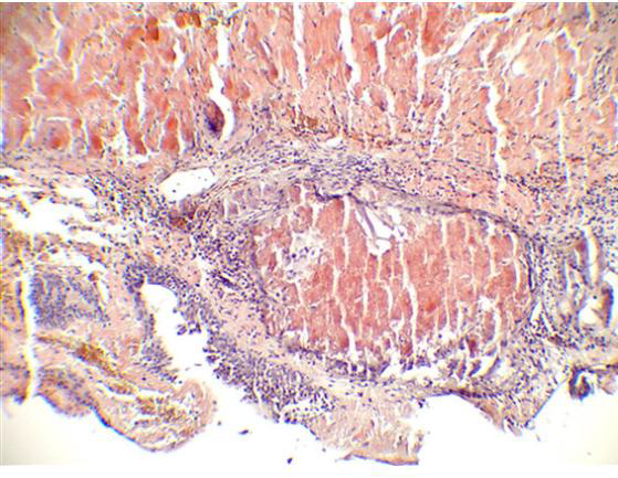
bladder amyloidosis in congo red staining
what this?
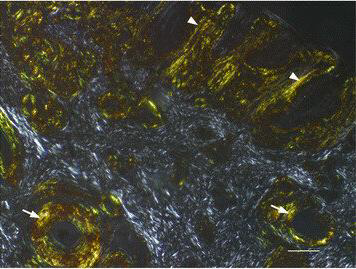
Nasal amyloid display in apple green birefringence in polarized light
what this?
fibrinoid degeneration/fibrinous degeneration/fibrinoid necrosis
Cellular accumulation characterized as poorly defined, deeply acidophilic, homogenous refractile deposits
connective tissue, blood vessel walls
fibrinoid degeneration occurs in
fibrin
some fibrinoid degeneration staining reactions resembles
phosphotungstic acid haematoxylin (PTAH)
Masson’s trichome
Martius Scarlet Blue
spacial stains of fibrinoid degeneration
glycogen degeneration
Cellular accumulation characterized as excessive accumulation of glycogen in cells following defective carbohydrate metabolism
parenchymal cells, liver cells
Glycogen degeneration if seen in _______ particularly in ____
atrophy from malnutrition
vacuolar degeneration
associated changes with glycogen degeneration
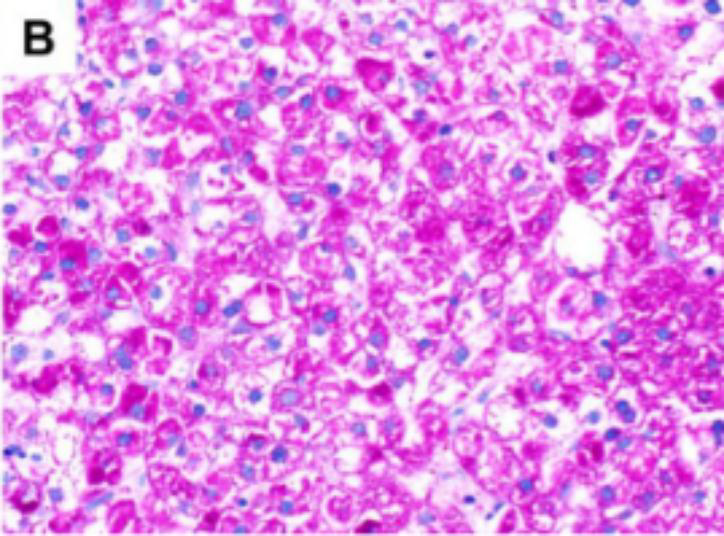
PAS on frozen section
special stains of glycogen degeneration
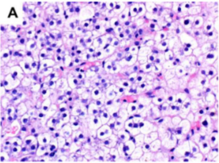
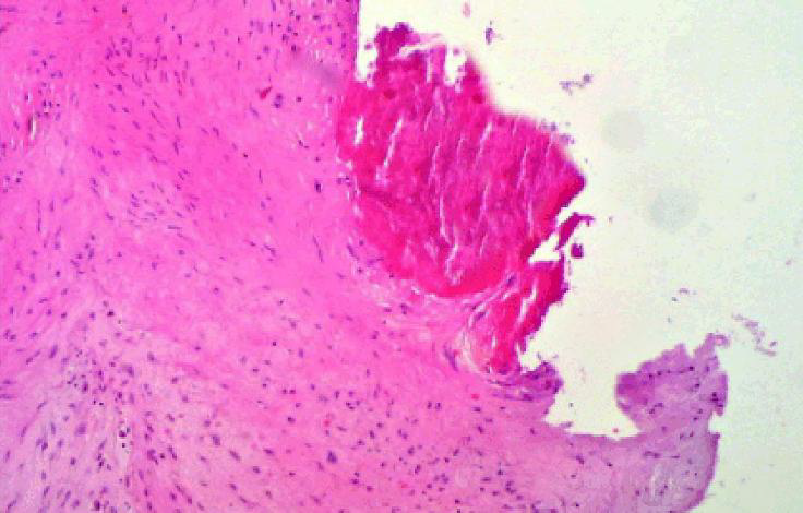
mucopolysaccharidosis
Cellular accumulation of mucopolysaccharide , also known as lysosomal storage disease
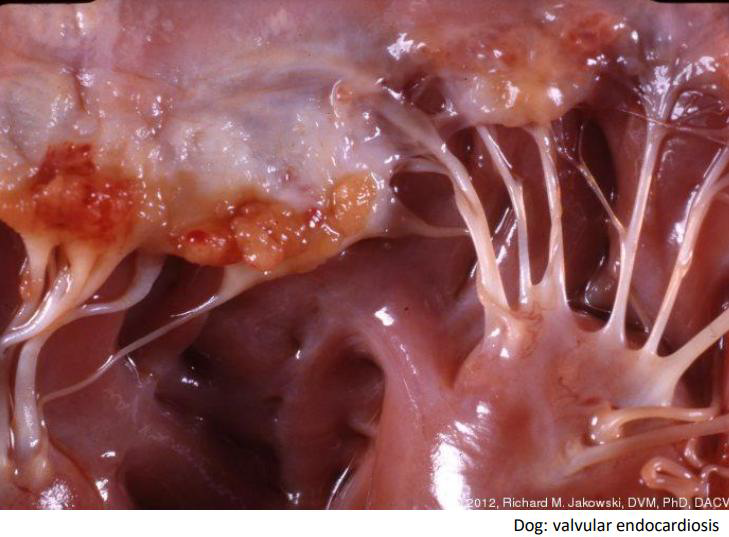
lysosomal enzyme system that breaks glycosaminoglycans
Basic defect of mucopolysaccharidosis is the absence or loss of critical _________ usually from genetic disorders
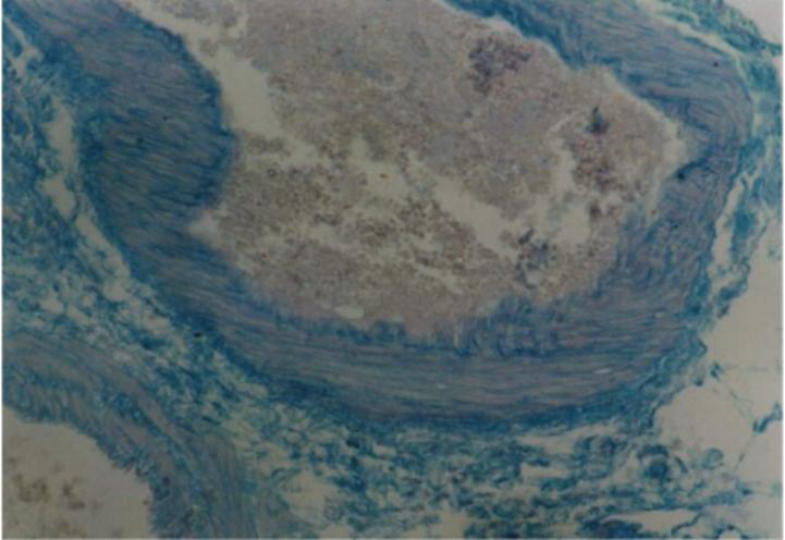
myxoid degeneration
myxomatous degeneration
mucoid or mucinous degeneration
Old terms of Mucopolysaccharidosis
Toluidine blue in resin section
Stain of mucopolysaccharidosis
endogenous
exogenous
What are the pigment sources of pathological pigmentation?
hemoglobin derivatives (hemosiderin, bilirubin)
melanin
lipofuscins
under endogenous pigment source
pneumoconiosis
anthracosis
under exogenous pathological pigment source
okie dokie yo
study this diagram pleae
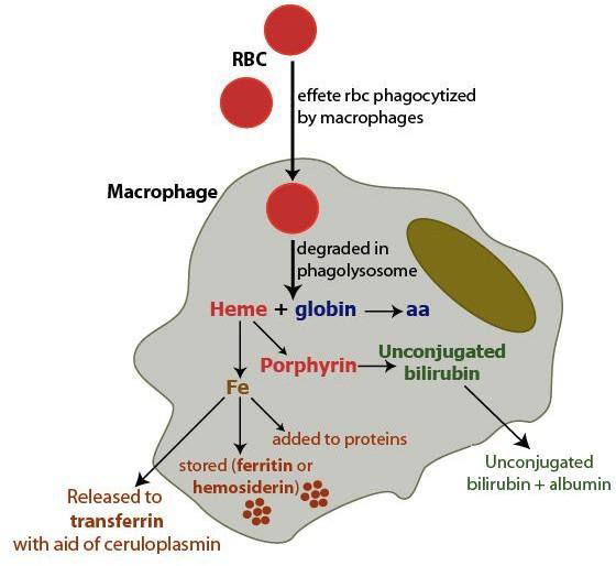
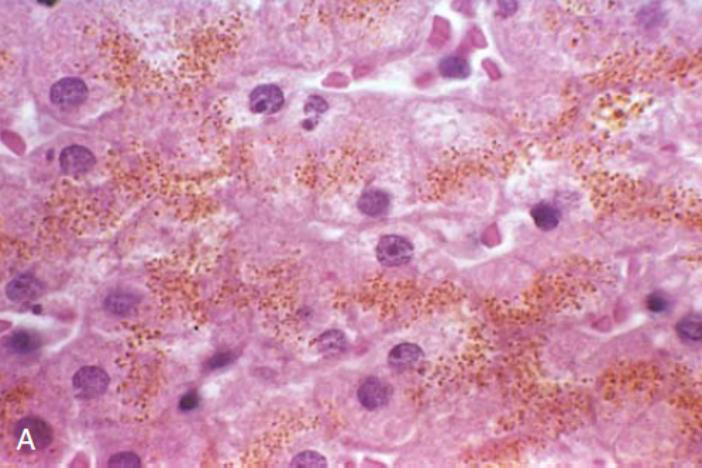
hemosiderin
Local deposits on tissues denote local breakdown of red blood cells
hemorrhages and tissue trauma
sample of hemosiderin
ferritin
storage form of iron that is usually seen in sections of spleen and in bone marrow
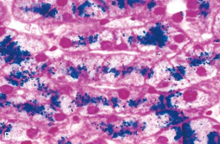
Perls’ prussian blue test
detections of hemosiderin
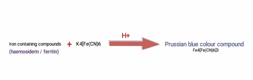
ferrocyanide
In Perls’ Prussian Blue Test, ferritin reacts with _____ in the stain to form insoluble Prussian blue dye in situ