Penny chapter 10 GI and ABD Wall, HERNIA
1/59
There's no tags or description
Looks like no tags are added yet.
Name | Mastery | Learn | Test | Matching | Spaced | Call with Kai | Chat |
|---|
No analytics yet
Send a link to your students to track their progress
60 Terms
b. 3 mm.
The measurement identified by the arrows in
Figure 10-20 should not exceed:
a. 2 mm.
b. 3 mm.
c. 3.5 mm.
d. 6 mm.
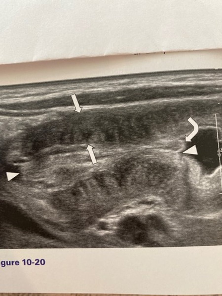
d. 2 to 6 weeks of age
What is the most common age range at which the abnormality occurs in Figure 10-20?
a. 5 to 10 years of age
b. 1 to 4 weeks of age
c. 3 months to 3 years of age
d. 2 to 6 weeks of age
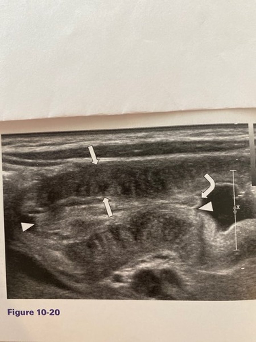
What is the sonographic sign noted in Figure
10-20?
a. Doughnut sign
b. Pyloric sign
c. Cervix sign
d. Cinnamon-bun sign
c. Cervix sign
What would be the least likely clinical finding associated with Figure 10-20?
a. Weight gain
b. Olive sign
c. Projectile vomiting
d. Dehydration
a. Weight gain
d. 6 mm.
The patient in Figure 10-21 presented with right lower quadrant pain and nausea. The anteroposterior measurement of this structure in this image should not exceed:
a. 4 mm.
b. 3 mm.
c. 10 mm.
d. 6 mm.
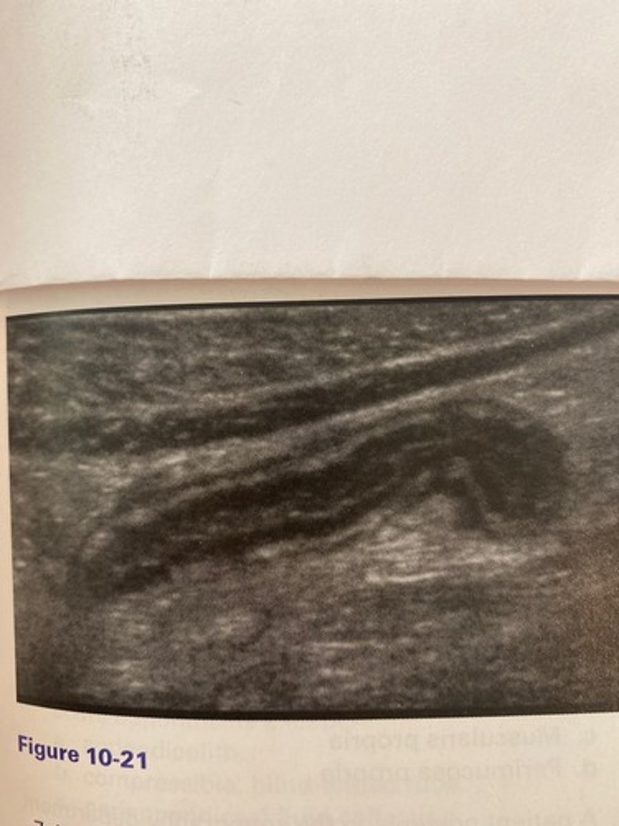
b. Leukocytosis
Which of the following would be another common clinical finding for the patient in Figure 10-21?
a. Thyroid in the belly sign
b. Leukocytosis
c. Kernicterus
d. Hypernatremia
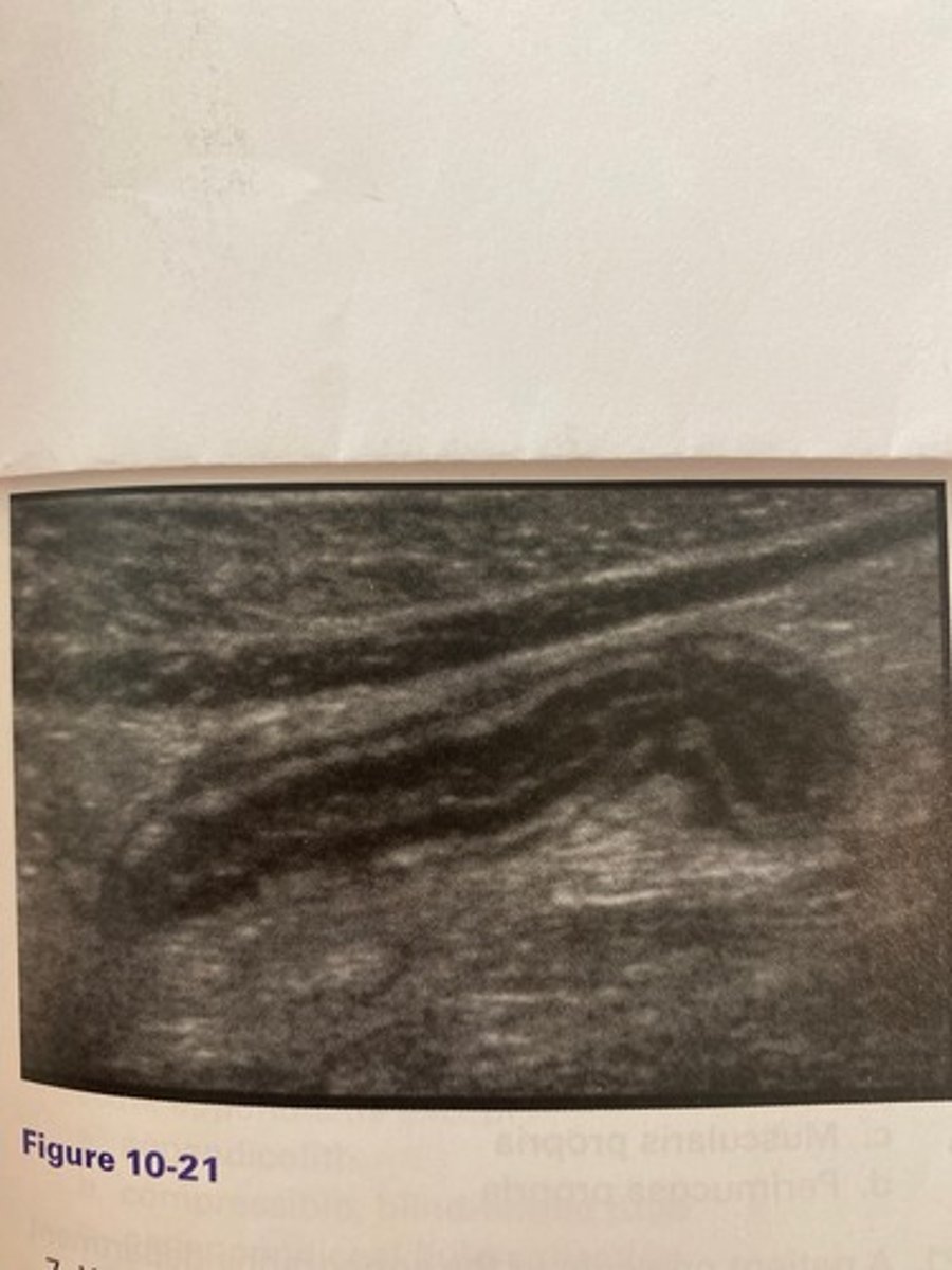
What is the thyroid in the belly sign?
a. Anechoic fluid surrounding an inflamed bowel segment
b. Hypochoic material adiacent to a distended pyloric stenosis
c. Enlargement of the distal colon in the presence of diverticulitis
d. Hyperechoic edematous connective tissue surrounding an inflamed appendix
d. Hyperechoic edematous connective tissue surrounding an inflamed appendix
c. Stomach
What does the large white arrow in Figure 10-22 indicate?
a. Esophagus
b. Transverse colon
c. Stomach
d. Pyloric stenosis

What is another name for the right colic flexure?
a. Splenic flexure
b. Hepatic flexure
c. Ascending flexure
d. Descending flexure
b. Hepatic flexure
What is the term that denotes twisting of the bowel?
a. Intussusception
b. Reflux
c. Volvulus
d. lleus
c. Volvulus
What is the most common type of ventral hernia?
a. Paraumbilical hernia
b. Spigelian hernia
c. Incisional hernia
d. Linea alba hernia
a. Paraumbilical hernia
Where is the most common location of endometriosis in the abdominal wall?
a. Paraumbilical
b. Cesarean section scar
c. Rectus sheath
d. Mesentery
b. Cesarean section scar
Which of the following would be among the most commonly encountered metastatic diseases of the bowel?
a. Brain cancer
b. Diverticulitis
c. Splenic carcinoma
d. Melanoma
d. Melanoma
What bowel segment is most often affected by diverticulitis and what is the most common location?
a. Sigmoid colon in the left lower quadrant
b. Cecum in the right lower quadrant
c. Ascending colon in the right upper quadrant
d. Splenic flexure in the left upper quadrant
a. Sigmoid colon in the left lower quadrant
Which of the following statements is true of a rectus sheath hematoma?
a. It may occur during defecation.
b. It is best seen with the Valsalva technique.
c. It is often located adiacent to the proximal duodenum.
d. It presents with general abdominal pain that eventually shifts to the left upper quadrant.
a. It may occur during defecation.
Which of the following is typically found in older patients and may lead to a bowel blockage?
a. Lactobezoar
b. Trichobezoar
c. Mycobezoar
d. Phytobezoar
d. Phytobezoar
The diameter of intussuscepted bowel will exceed:
a. 1 cm.
b. 5 mm.
c. 9 cm.
d. 3 cm.
d. 3 cm.
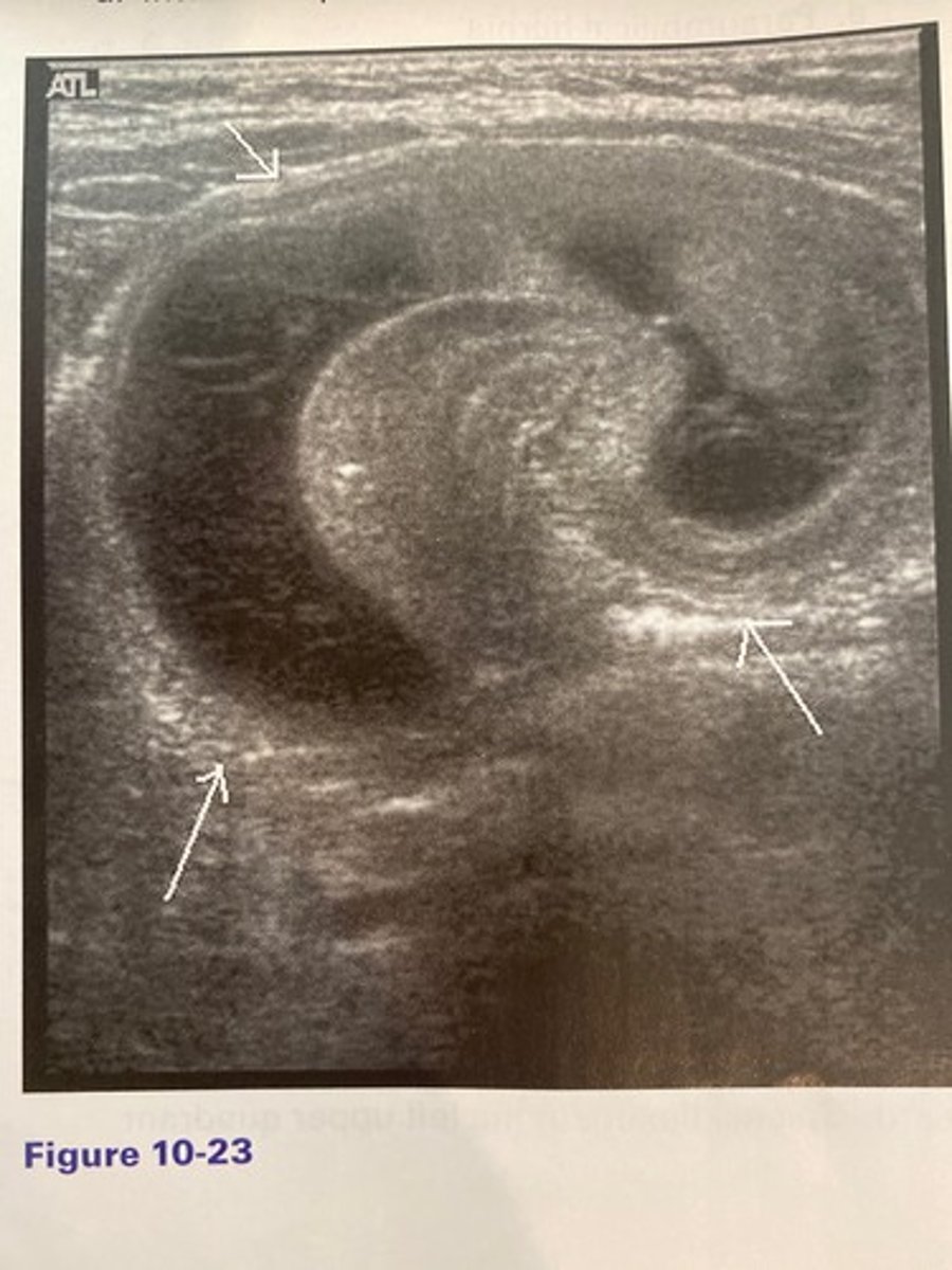
d. Intussusception
The finding in Figure 10-23 was discovered in the right lower quadrant of an 18-month-old patient with a history of intermittent, severe abdominal pain, and vomiting. What is the most likely diagnosis?
a. Appendicitis
b. Colitis
c. Mechanical obstruction
d. Intussusception
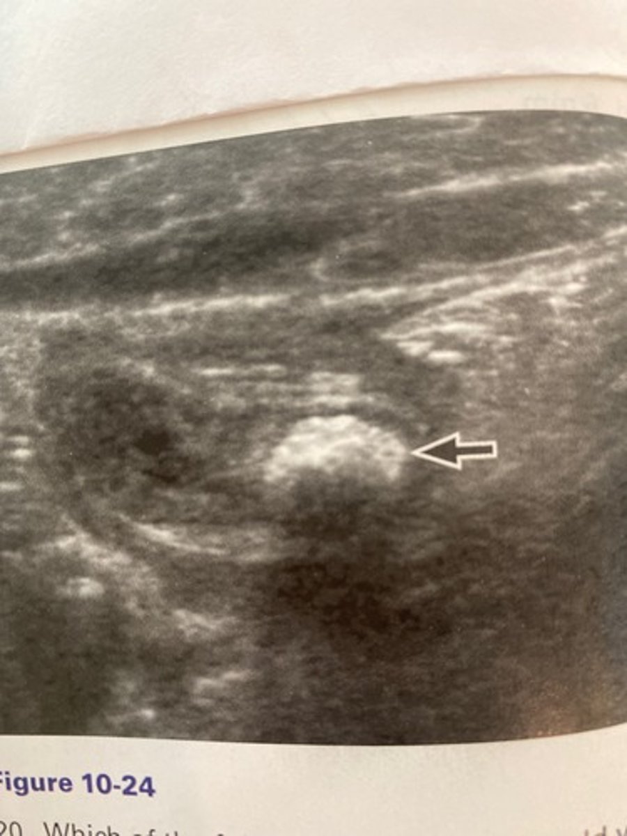
a. Appendicolith
What does the arrow demonstrate in the patien in Figure 10-24 who complained of focal right lower quadrant pain?
a. Appendicolith
b. Thyroid in the belly
c. Olive sign
d. McBurney sign
Which of the following gut layers would yield an echogenic pattern?
a. Deep mucosa
b. Superficial mucosa
c. Muscularis propria
d. Perimucosa propria
b. Superficial mucosa
A patient presents to the sonography department with bilious vomiting. While investigating the pediatric patient for pyloric stenosis, you note that while the pyloric sphincter appears normal, the SMA is abnormally located to the right of the SMV. What is the most likely diagnosis?
a. Pylorospasm
b. Intussusception
c. Crohn disease
d. Midgut malrotation
d. Midgut malrotation
What anatomic structure may be noted as a bull's-eye structure anterior to the abdominal aorta and posterior to the left lobe of the liver in the sagittal scan plane?
a. Pyloric sphincter
b. Duodenal antrum
c. Gastrosophageal junction
d. Distal jejunum
c. Gastrosophageal junction
Which of the following is not a layer of gut identified with sonography?
a. Visceral
b. Serosa
c. Submucosa
d. Mucosa
a. Visceral
All of the following are true of normal intestinal findings with sonography except:
a. normal bowel does not compress.
b. normal bowel should have observable peristalsis.
c. colon wall should measure less than 9 mm.
d. normal bowel has little to no color Doppler signals.
a. normal bowel does not compress.
Upon sonographic evaluation of the right lower quadrant in a patient complaining of focal abdominal pain in that area, you visualize a hyperemic blind-ended, tubular structure that contains a shadowing focus. What is the most likely etiology of the shadowing focus?
a. Ureteral stone
b. Appendicolith
c. Gallstone
d. Herniated omentum
b. Appendicolith
All of the following are sonographic criteria in the diagnosis of pyloric stenosis except:
a. wall of the pylorus is focally thinned.
b. length of the pylorus measures more than
17 mm.
c. doughnut appearance in transverse.
d. cervix appearance in longitudinal.
a. wall of the pylorus is focally thinned.
All of the following are sonographic findings of acute appendicitis except:
a. appendicolith.
b. compressible, blind-ended tube.
c. periappendiceal fluid collection.
d. hyperemic flow.
b. compressible, blind-ended tube.
Clinical findings of acute appendicitis include all of the following except:
a. leukocytosis.
b. right lower quadrant pain.
c. constipation.
d. rebound tenderness.
c. constipation.
All of the following are common clinical findings in infants who present with pyloric stenosis except:
a. weight loss.
b. dehydration.
c. olive sign.
d. first-born female.
d. first-born female.
Pseudomyxoma peritonei can result from:
a. intussusception.
b. pyloric stenosis.
c. Crohn disease.
d. appendix cancer.
d. appendix cancer.
A patient presents to the sonography department with a painful, superficial abdominal mass located within a prior cesarean scar. What clinical feature would be most consistent with scar endometriosis?
a. Hematuria
b. Chronic headaches
c. Cyclical pain
d. Bloody diarrhea
c. Cyclical pain
What abnormality associates red currant jelly stools?
a. Diverticulosis
b. Appendicitis
c. Intussusception
d. Pyloric stenosis
c. Intussusception
Other abnormalities that can present much like pyloric stenosis include all of the following except:
a. midgut malrotation.
b. pylorospasm.
c. gastroesophageal reflux disease.
d. intussusception.
d. intussusception.
Which of the following would be the most likely clinical feature of colitis?
a. Inguinal herniation of the bowel
b. Right shoulder pain
c. Watery diarrhea
d. Midline hematoma
c. Watery diarrhea
Gastric cancer is most often in the form of:
a. cystadenocarcinoma.
b. adenocarcinoma.
c. rhabdomyocarcinoma.
d. angiosarcoma.
b. adenocarcinoma.
Pediatric patients could suffer from bowel obstructions that are caused by a buildup of ingested hair. The mass associated with this type of obstruction is termed a:
a. phytobezoar.
b. lactobezoar.
c. trichobezoar.
d. permabezoar.
c. trichobezoar.
An autoimmune disease characterized by periods of inflammation of the gastrointestinal tract describes:
a. Crohn disease.
b. intussusception.
c. pyloric stenosis.
d. Meckel diverticulitis.
a. Crohn disease.
The telescoping of one segment of bowel into another is referred to as:
a. volvulus.
b. Crohn disease.
c. intussusception.
d. pyloric stenosis.
c. intussusception.
Which of the following types of obstruction refers to the bowel being physically blocked by something?
a. Mechanical
b. Nonmechanical
c. Obstreperous
d. Bezoarine
a. Mechanical
Which of the following would be useful to employ during a sonographic evaluation of a suspected abdominal wall hernia?
a. Upright positioning
b. Prone positioning
c. Graded compression
d. Valsalva
d. Valsalva
The situation when bowel protrudes into the groin is referred to as a(n):
a. inguinal hernia.
b. linea alba hernia.
c. umbilical hernia.
d. spigelian hernia.
a. inguinal hernia.
The situation when bowel protrudes into a weakened area in the lower one-fourth of the rectus muscle is referred to as a(n):
a. inguinal hernia.
b. linea alba hernia.
c. umbilical hernia.
d. spigelian hernia.
d. spigelian hernia.
The area of pain and rebound tenderness with acute appendicitis is most likely at:
a. Meckel point.
b. McBurney point.
c. Murphy point.
d. Olive point.
b. McBurney point.
Which of the following best describes the location of McBurney point?
a. Left lateral to the umbilicus and medial to the left iliac crest
b. Halfway between the anterior superior iliac spine and the umbilicus
c. Midway between the umbilicus and the symphysis pubis
d. Medial to the superior iliac spine
b. Halfway between the anterior superior iliac spine and the umbilicus
The olive sign is best described as:
a. the palpation of the inflamed appendix with rebound tenderness
b. an area of pain halfway between the anterior superior iliac spine and the umbilicus
c. an enlarged palpable pyloric sphincter
d. the sonographic appearance of pyloric stenosis
c. an enlarged palpable pyloric sphincter
Rebound tenderness is associated with:
a. appendicitis.
b. intussusception.
c. diverticulitis.
d. gastric carcinoma.
a. appendicitis.
47. The most common location of the vermiform appendix is in the area of the:
a. jejunum.
b. descending colon.
c. cecum.
d. sigmoid colon.
c. cecum.
48. Which of the following is the development of small outpouchings within the sigmoid colon?
a. Diverticulitis
b. Crohn disease
c. Diverticulosis
d. Midgut malrotation
c. Diverticulosis
Which of the following is not associated with a rectus sheath hematoma?
a. Palpable abdominal mass
b. Increased hematocrit
c. Childbirth
d. Sneezing
b. Increased hematocrit
Which of the following is not a sonographic finding consistent with Crohn disease?
a. Bowel wall thickening
b. Noncompressible bowel that has a target
appearance
c. Increased peristalsis
d. Hyperemic wall
c. Increased peristalsis
All of the following are common clinical findings in infants who present with intussusception except:
a. vomiting.
b. first-born male infant.
c. red currant jelly stools.
d. leukocytosis.
b. first-born male infant.
The sonographic finding of fluid-filled, distended loops of bowel is consistent with:
a. Meckel diverticulum.
b. diverticulitis.
c. gastrosophageal reflux disease.
d. intestinal obstruction.
d. intestinal obstruction.
Traditionally, treatment for intussusception is by means of:
a. surgery.
b. external manipulation.
c. compression sonography.
d. therapeutic enema.
d. therapeutic enema.
The most common cause of intestinal obstruction in children less than 2 years of age is:
a. intussusception.
b. midgut malrotation.
c. pyloric stenosis.
d. acute appendicitis.
a. intussusception.
What abnormality may be diagnosed by observing fluid mixed with gas bubbles traveling from the stomach to the esophagus with
sonography?
a. Pylorospasm
b. Pyloric stenosis
c. Gastrosophageal reflux disease
d. Midgut malrotation
c. Gastrosophageal reflux disease
In what position is the infant often placed for better sonographic visualization of the pyloric sphincter?
a. Right lateral decubitus
b. Left lateral decubitus
c. Prone
d. Upright
a. Right lateral decubitus
An adult patient presents to the sonography department with left lower guadrant pain, fever, and bouts of both constipation and diarrhea.
Which of the following would be the most likely etiology?
a. Diverticulitis
b. Intussusception
c. Midgut malrotation
d. Appendicitis
a. Diverticulitis
What are the diagnostic criteria for pyloric stenosis?
a. 17 mm in thickness and 2 mm in length
b. 17 mm in thickness and 3 mm in length
c. 3 mm in thickness and 10 mm in length
d. 3 mm in thickness and 17 mm in length
d. 3 mm in thickness and 17 mm in length
Clinical findings of a patient with Crohn disease include all of following except:
a. palpable abdominal mass.
b. rectal bleeding.
c. abdominal pain.
d. weight loss.
a. palpable abdominal mass.
Which of the following would be most likely a cause of colitis?
a. Gastroesophageal reflux disease
b. Antibiotic therapy
c. Dehydration
d. Rectus sheath hematoma
b. Antibiotic therapy