Anatomy 2200 - Midterm
1/99
There's no tags or description
Looks like no tags are added yet.
Name | Mastery | Learn | Test | Matching | Spaced | Call with Kai |
|---|
No analytics yet
Send a link to your students to track their progress
100 Terms
Spinal nerve
mixed nerve carrying motor and sensory information between spinal cord and body
Segments of the spine
cervical (8 nerves, 7 vertebrae), thoracic, lumbar, sacral, coccygeal
Spinal nerve components
spinal cord - dorsal/ventral rootlets - dorsal/ventral roots - spinal nerve - dorsal/ventral ramus
Dorsal vs. ventral roots
dorsal - sensory - posterior
ventral - motor - anterior
Paresthesia
compression of nerve - abnormal sensation (burning, prickling, tingling, numbness)
Paresis
partial or incomplete paralysis from damage or compression of nerve
Brachial plexus
network of nerves that supply upper limb - begins at neck and ends in axilla (armpit)
Composition of brachial plexus
Roots (C5, C6, C7, C8, T1)
Trunk (anterior and posterior)
Divisions (lateral, posterior, medial)
Cords
Branches (musculocutaneous, median, ulnar, axillary, radial)
Nerves of the brachial plexus
musculocutaneous (C5, C6, C7 - arm flexors - lateral forearm sensory)
axillary (C5, C6 - shoulder - shoulder sensory)
median (C5, C6, C7, C8, T1 - forearm + hand flexors - lateral palm, 1-3 digits, 1/2 of 4th digit)
radial (C5, C6, C7, C8, T1 - arm + forearm extensors - posterior arm, forearm, lateral hand)
ulnar (C8, T1 - forearm + hand flexors - medial hand, 1/2 of 4th digit, 5th digit)
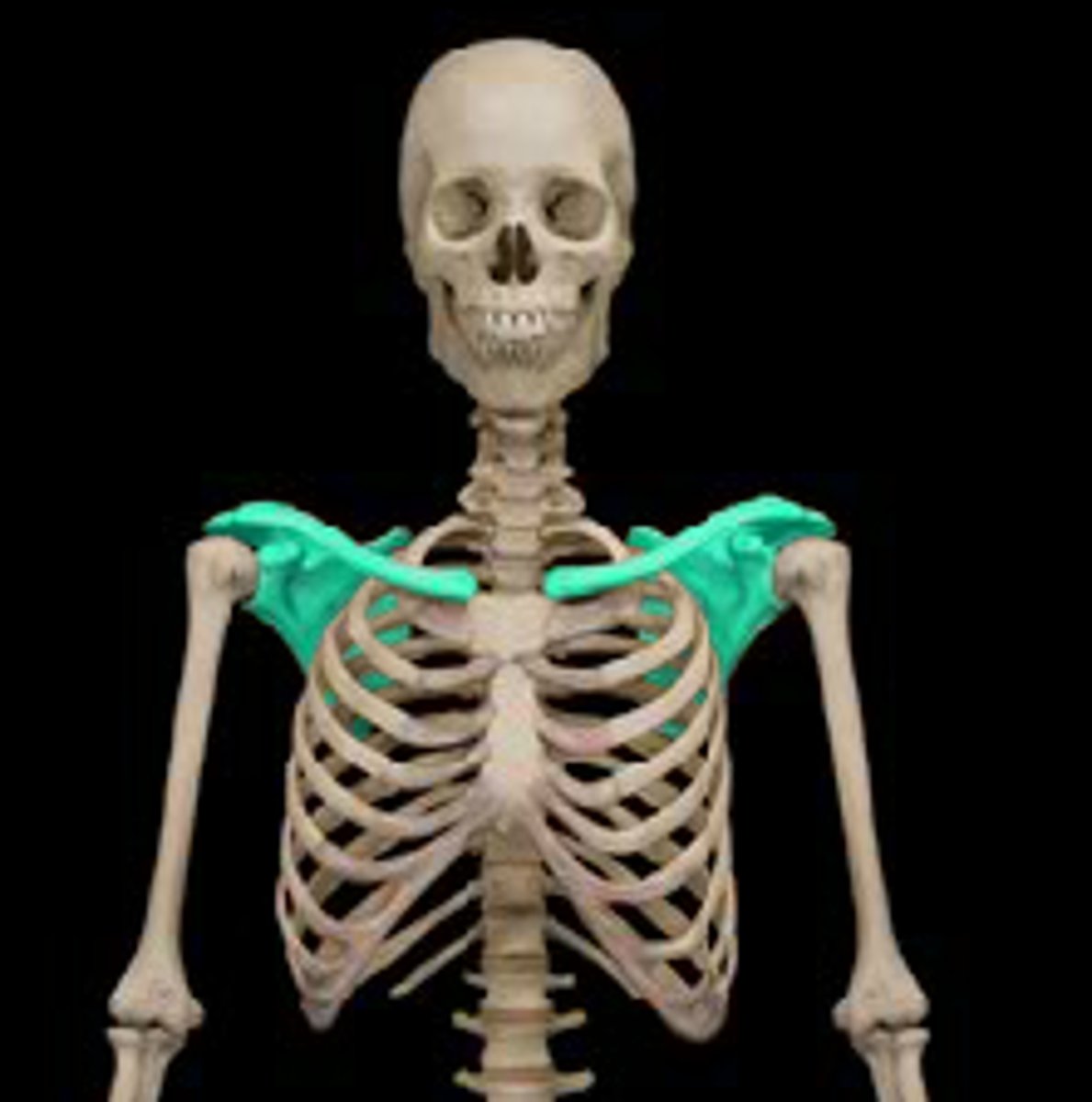
What is the pectoral girdle and its function?
clavicle and scapula - attachment of upper limb to skeleton and movement flexibility
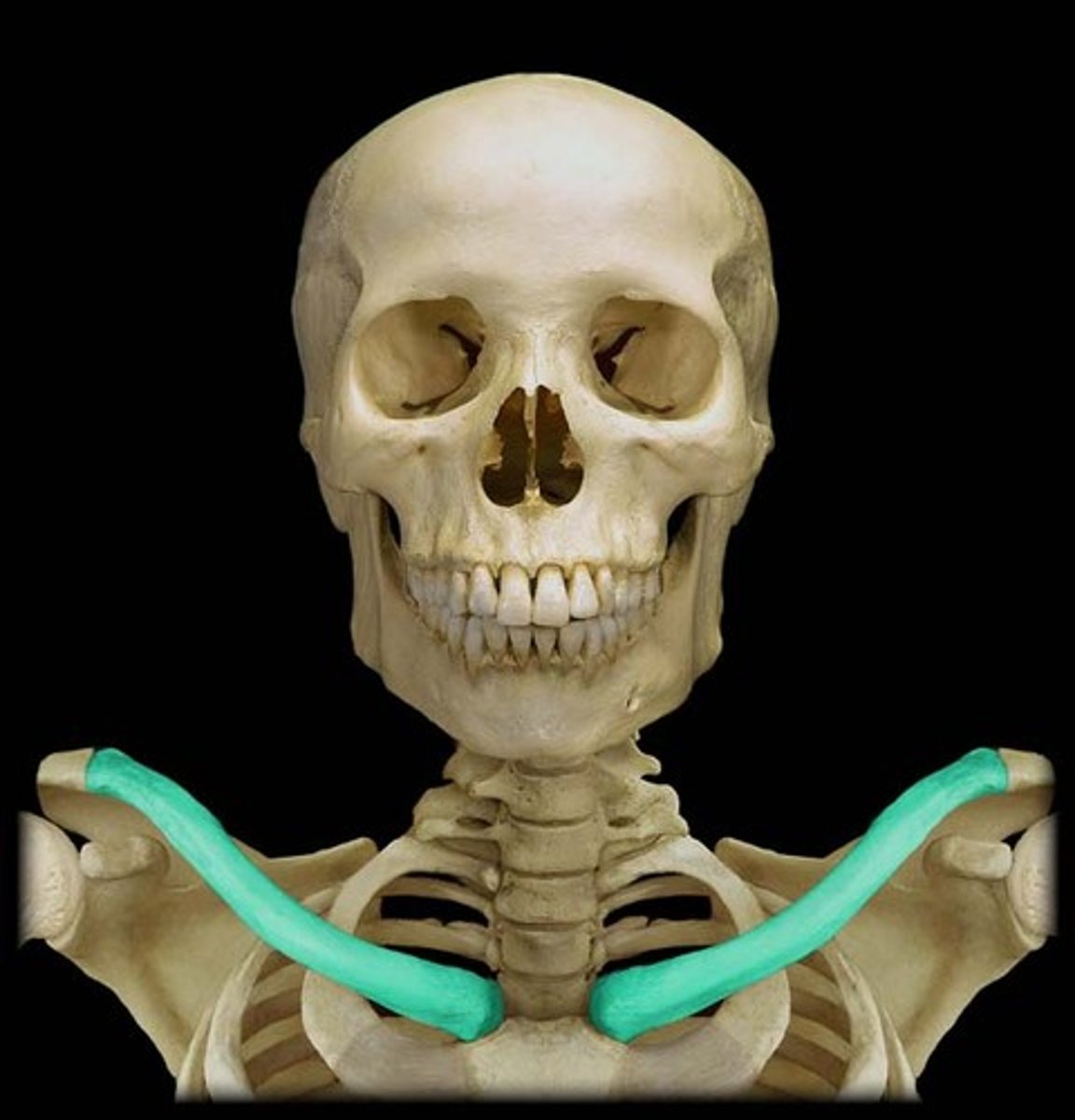
Describe clavicle and its function
"s" shaped bone - sternal end (medial, attach to sternum) - acromial end (lateral, attach to acromion process)
muscle attachment, "braces" - holds scapula and arms out laterally from trunk
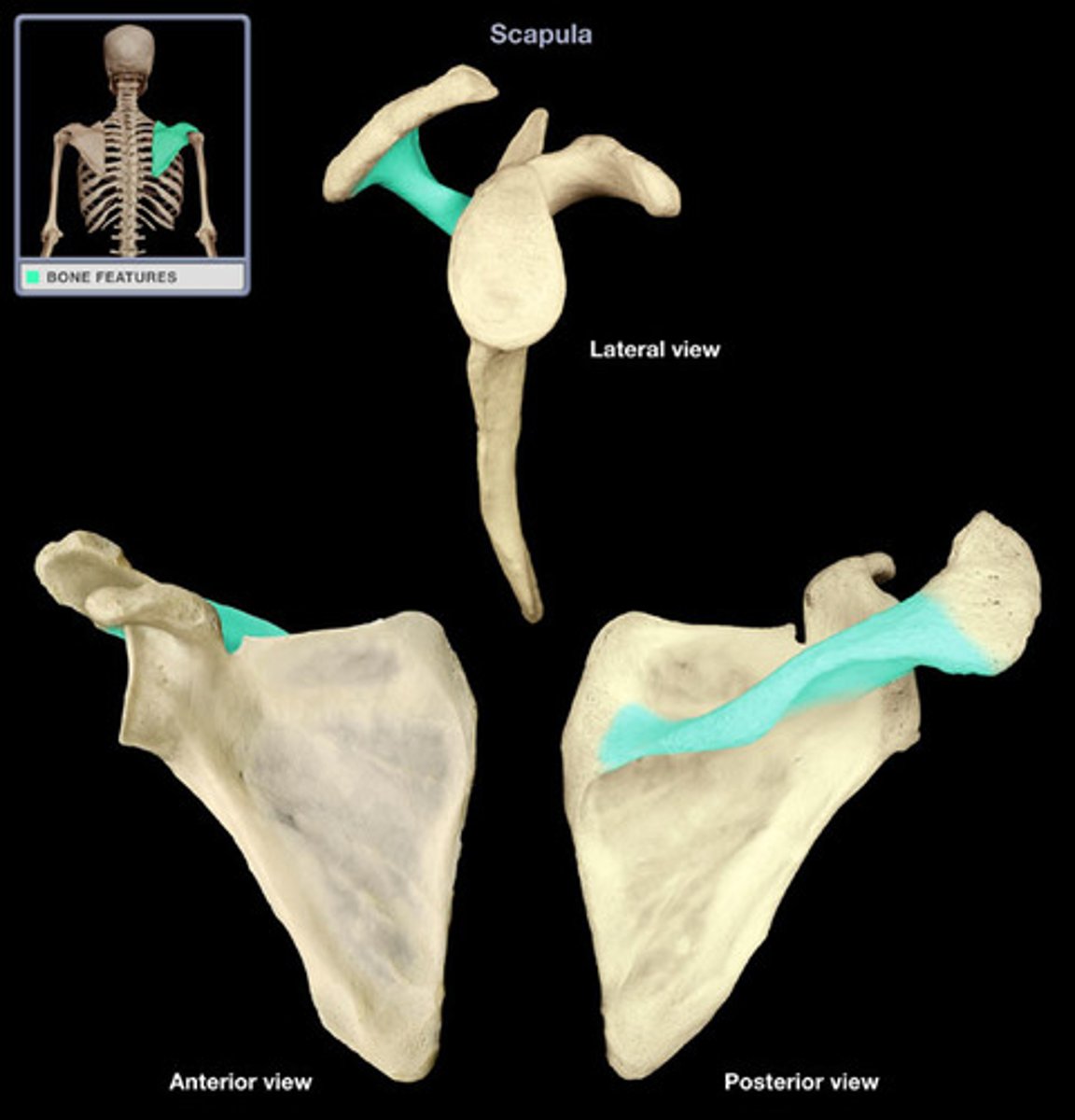
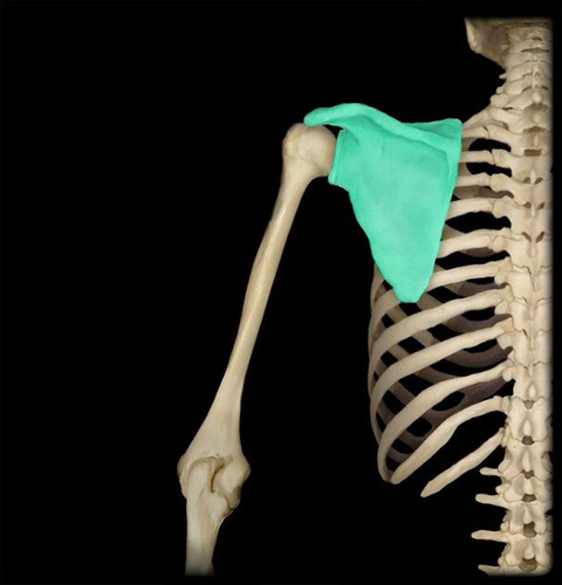
Label scapula
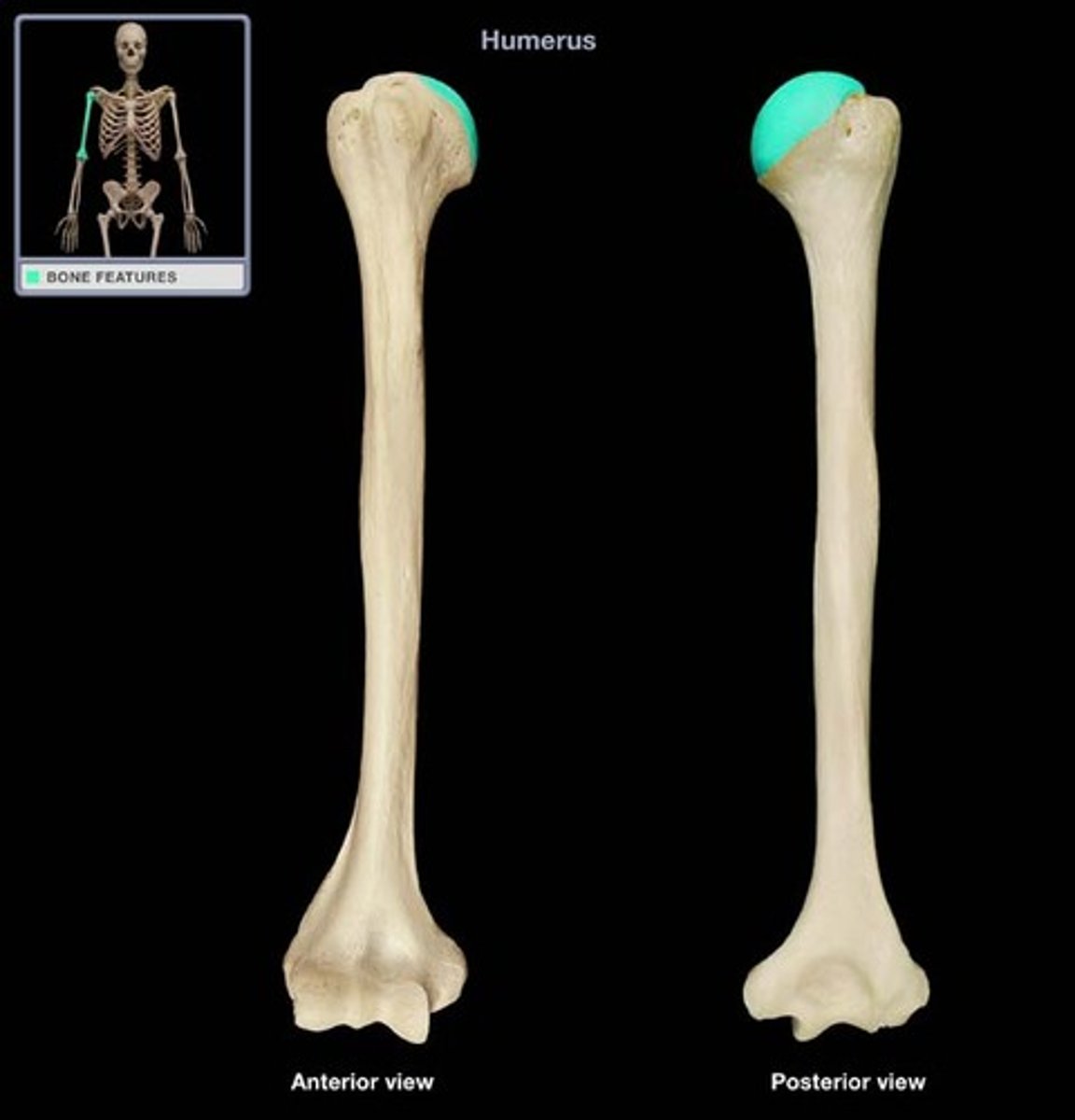
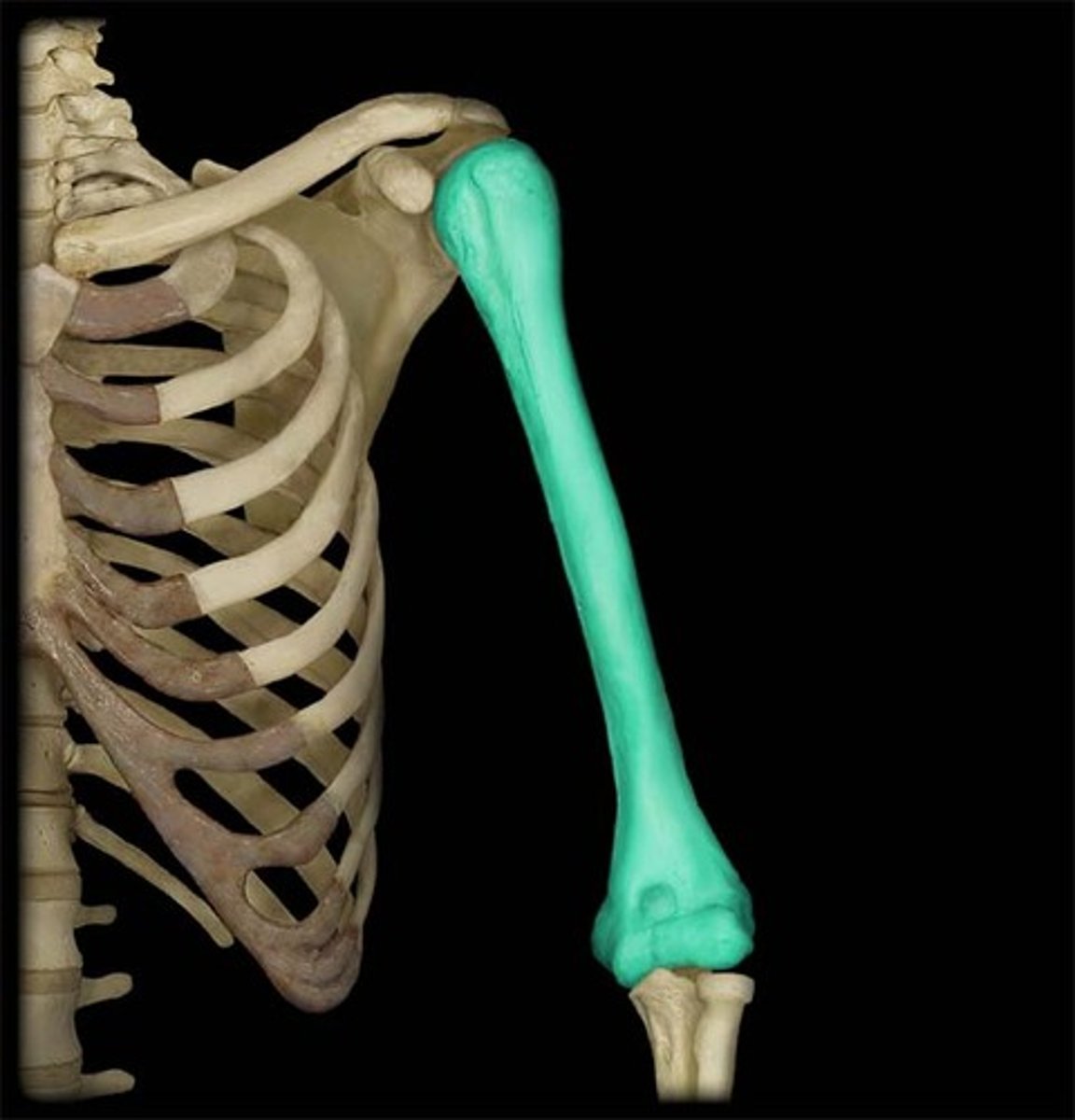
Label humerus, what does it do
articulates with glenoid cavity
Muscles can be divided into:
prime mover/agonist: major action
antagonist: opposes action
synergist: assist muscle action
Posterior muscles of the scapula
levator scapulae, trapezius, rhomboids (major + minor)
Levator scapulae
Origin: upper neck, C1-C4
insertion: superior portion of medial border
action: elevates + rotates scapula
innervation: dorsal scapular + cervical nerves
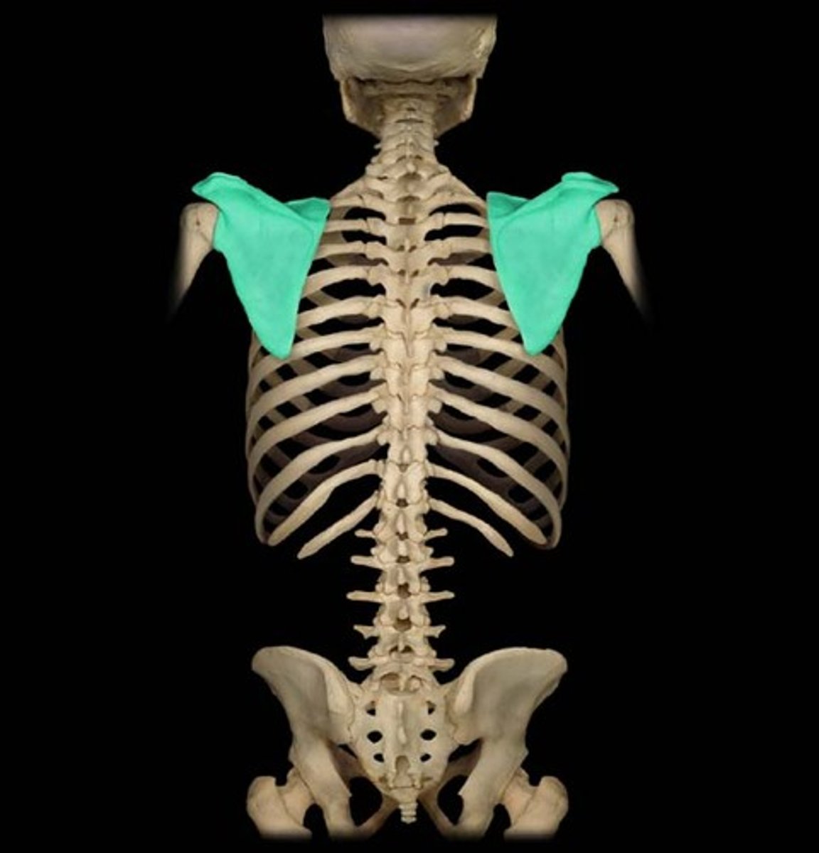
Trapezius
origin: along spine C7-T12
insertion: clavicle, acromion, spine of scapula
action: elevate, rotate, retract scapula
innervation: accessory nerve
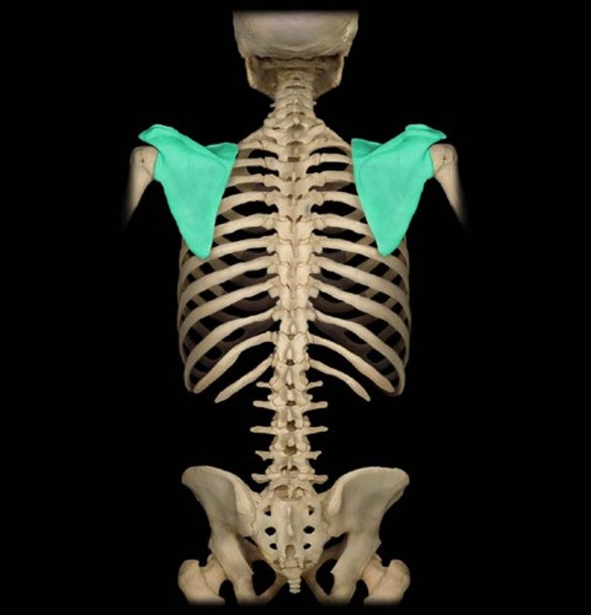
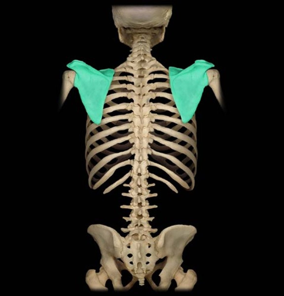
Rhomboids
origin: major (T2-T5) + nuchal ligament; minor (C7-T1)
insertion: medial border
action: retract scapula towards spine (medially), rotate
innervation: dorsal scapular nerve
Anterior muscles of the scapula
pectoralis minor, serratus anterior
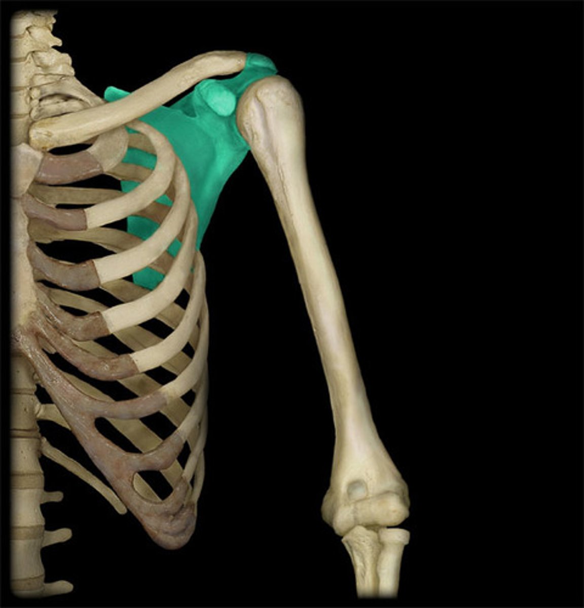
Pectoralis minor
origin: upper ribs (3-5)
insertion: coracoid process
action: stabilize scapula, pull inferiorly and anteriorly
innervation: medial pectoral nerve
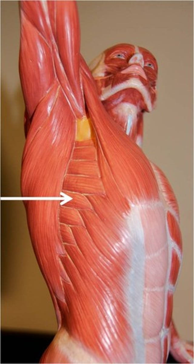
serratus anterior
origin: fleshy slips of upper ribs
insertion: medial border
action: protracts (punching), stabilize, upward rotation
innervation: long thoracic nerve
Rotator cuff muscles: what are they? what do they do?
subscapularis, suprascapularis, infraspinatus, teres major
encircle shoulder like a cuff, help anchor head of humerus into glenoid cavity
subscapularis
rotates arm medially, inserts into lesser tubercule, fills subscapular fossa, subscapular nerve
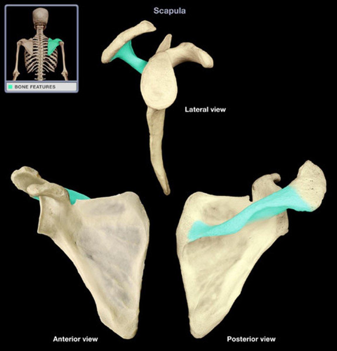
suprascapularis
abducts arm, inserts into greater tubercule, fills supraspinous fossa, suprascapular nerve
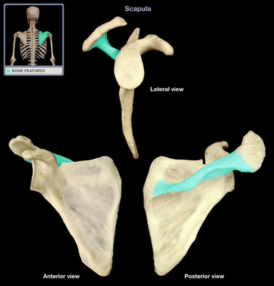
infraspinatus
rotates arm laterally, inserts into greater tubercule, fills infraspinous fossa, suprascapular nerve
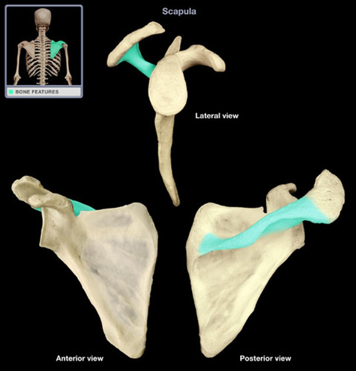
teres minor
rotates arm laterally, inserts into greater tubercule, fills infraspinous fossa, axillary nerve
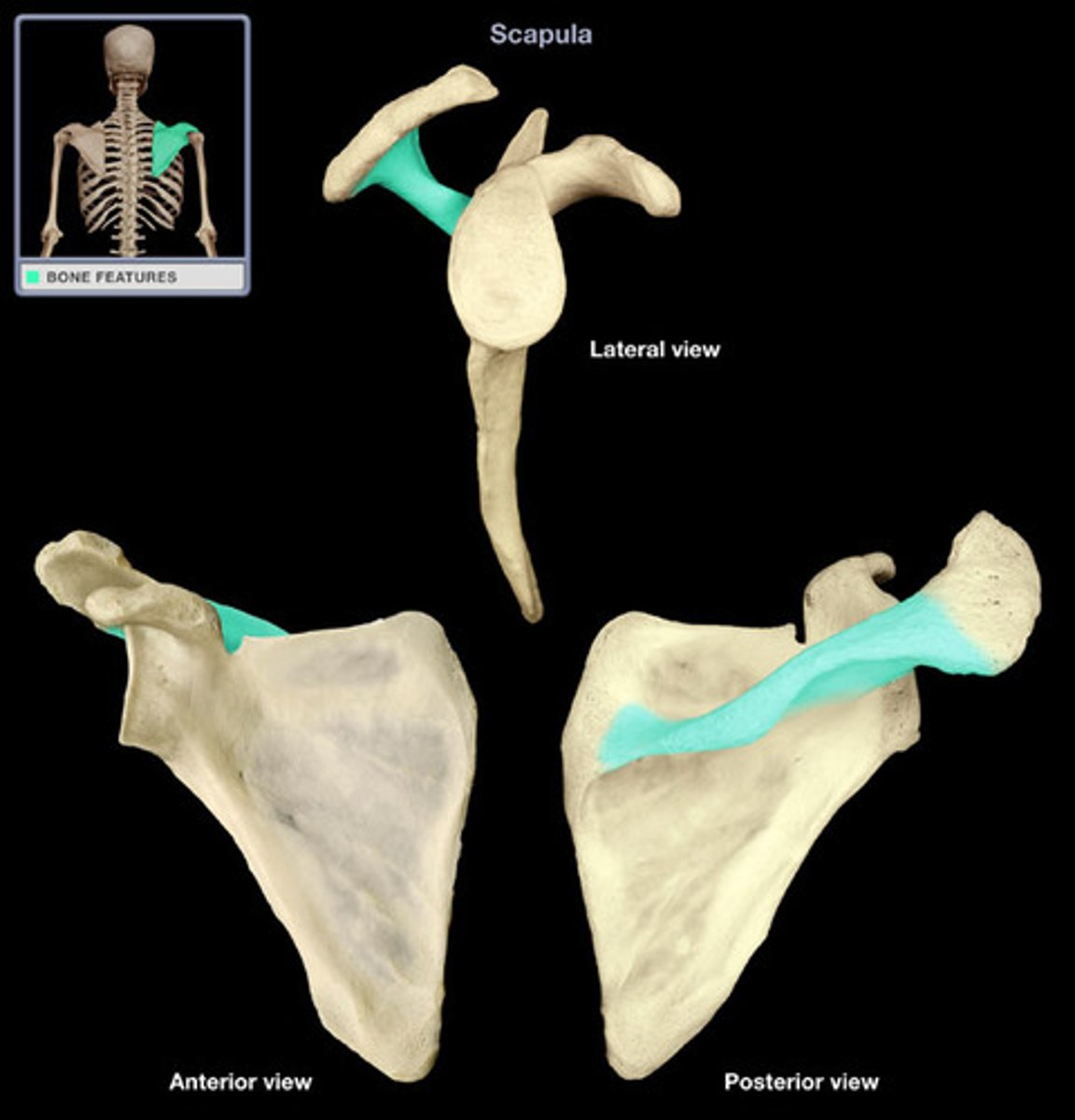
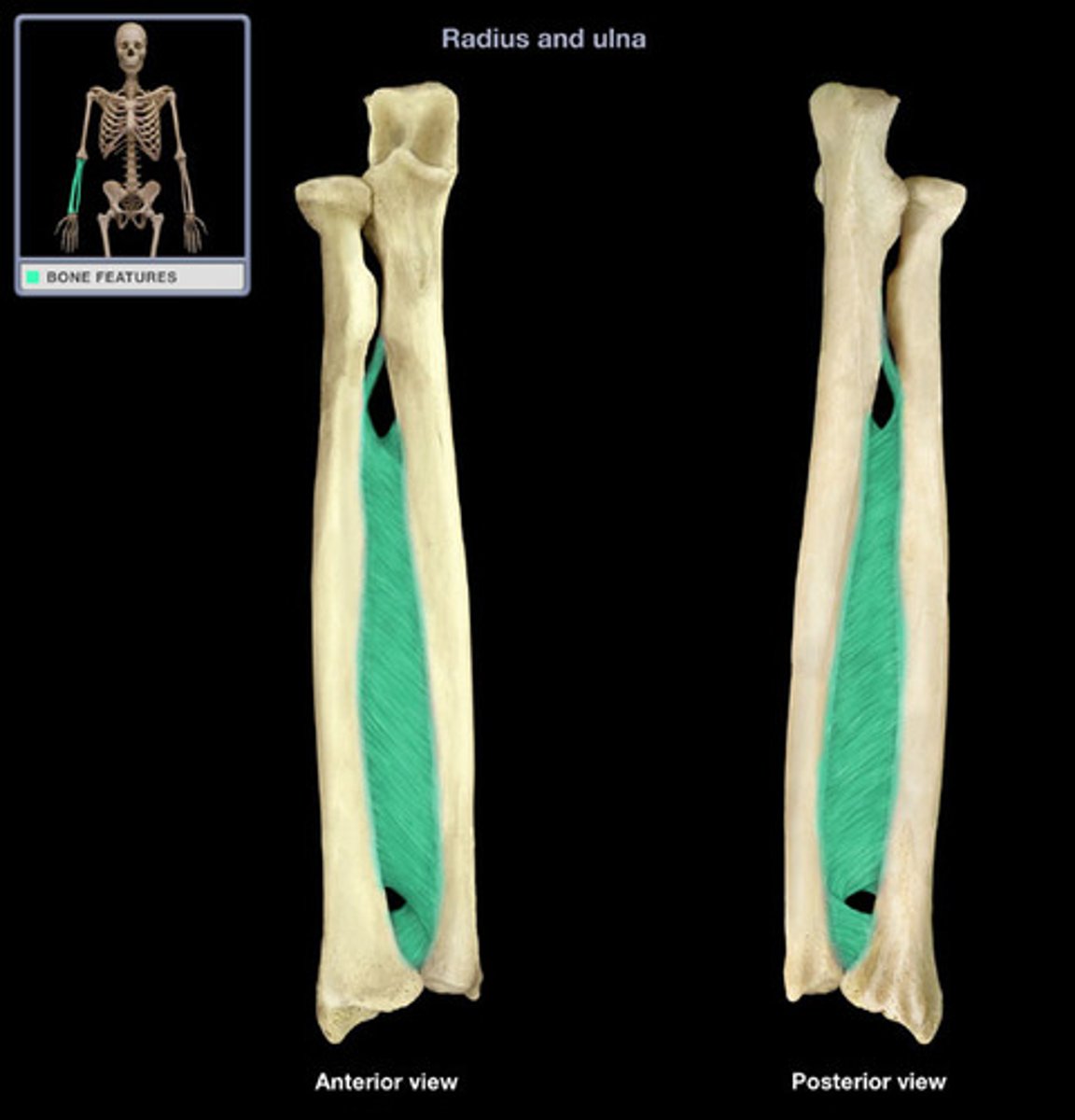
interosseous membrane
separation between muscles at front of forearm and muscles at back of forearm
What type of joint is the elbow?
synovial hinge joint (uniaxial motions - flexion + extension)
Positions in a flexed elbow
head of radius tucked into radial fossa, coronoid process tucked in coronoid fossa
Positions in an extended elbow
olecranon process fits into olecranon fossa
Label elbow joint
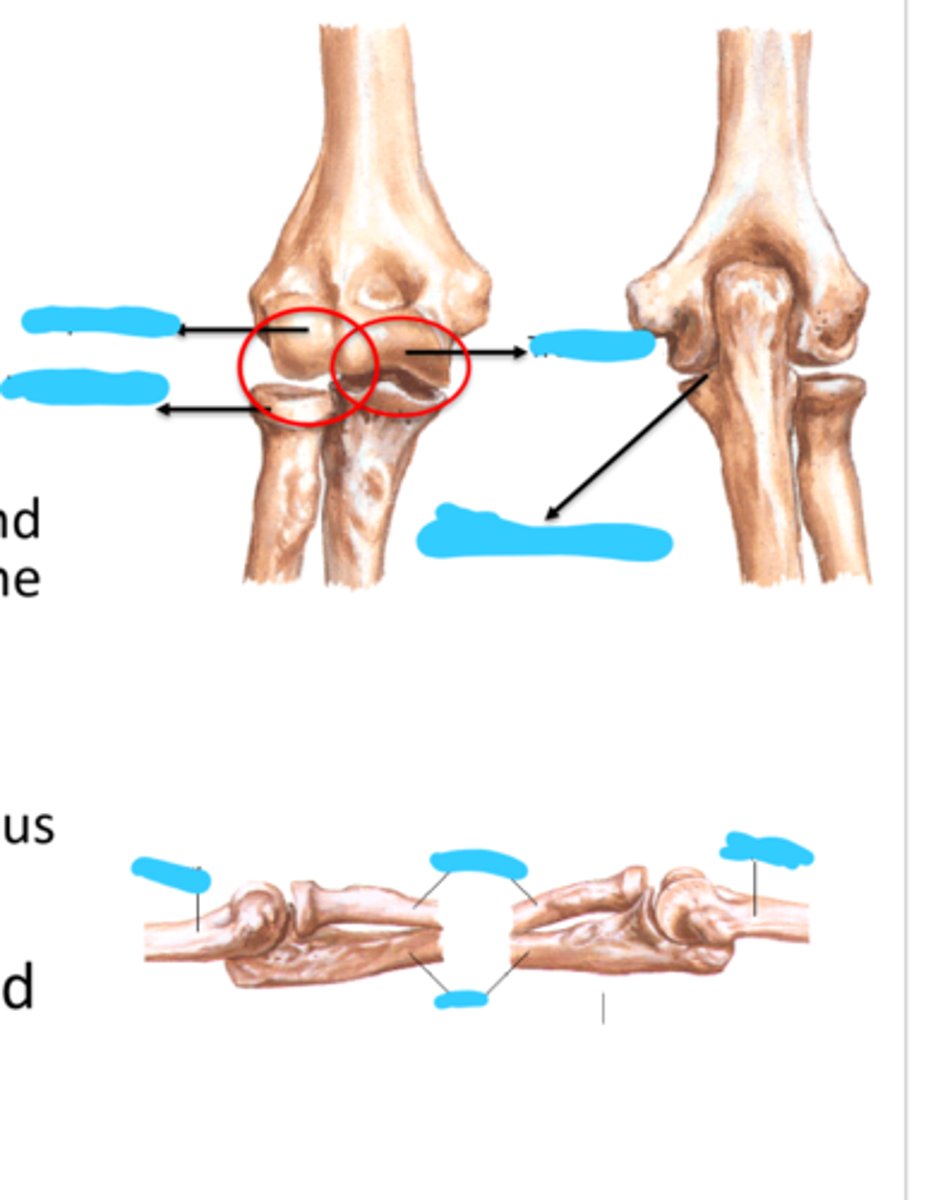
Proximal radio-ulnar joint
allows for supination and pronation
pronation: radius radiates over ulna, ends up more medial
Anterior muscles of the shoulder and arm
pectoralis major, coracobrachialis, deltoid
pectoralis major
origin: sternal end of clavicle, sternum and cartilage of ribs 1-6
insertion: lateral edge of intertubercule groove
action: arm flexion, medial rotation, arm adduction
innervation: medial + lateral pectoral nerve
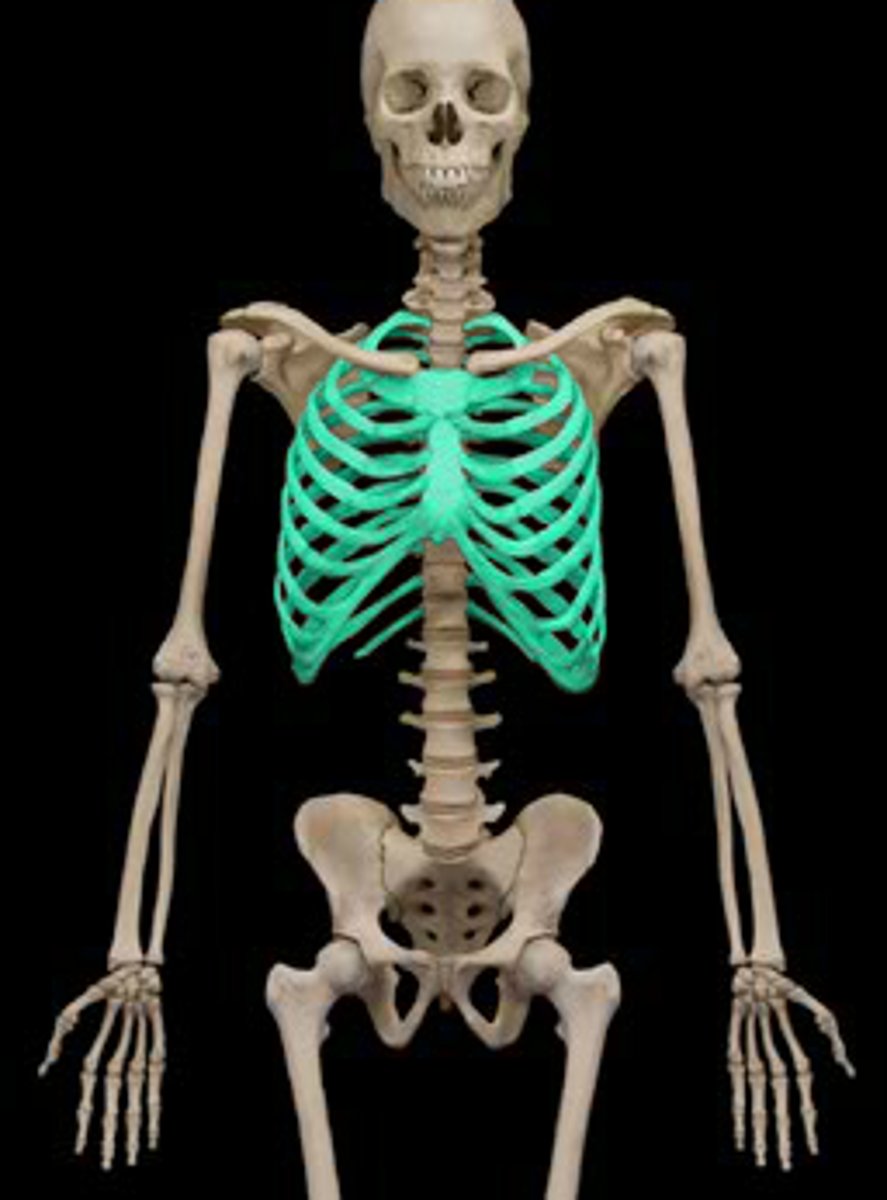
coracobrachialis
origin: coracoid process
insertion: midshaft of humerus medially
action: arm flexion, adduction
innervation: musculocutaneous nerve (pierces it)
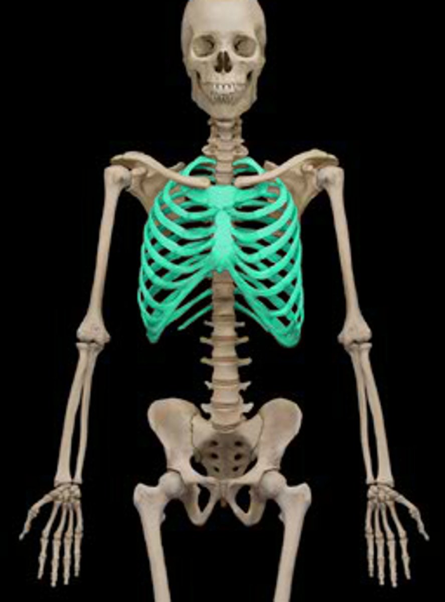
deltoid
origin: clavicle, acromion, spine scapula (posteriorly)
insertion: deltoid tuberosity
action: fiber dependent
- all = abduction
- anterior fibers = flexion + medial rotation
- posterior fibers = extension + lateral rotation
innervation: axillary nerve
Posterior muscles of shoulder and arm
latissimus dorsi, teres major
latissimus dorsi
origin: thoracolumbar fascia, iliac crest, T7-T12, lower ribs
insertion: intertubercule groove floor (ant)
action: arm extension, adduction, medial rotation
innervation: thoracodorsal nerve
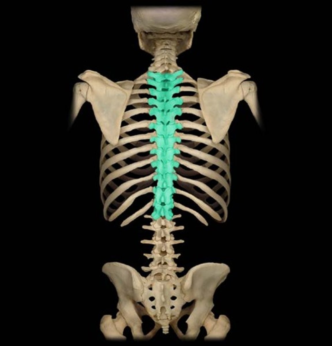
teres major
origin: inferior angle of scapula
insertion: medial edge intertubercule groove (ant)
action: arm extension, adduction, medial rotation (lats little helper)
innervation: lower subscapular nerve
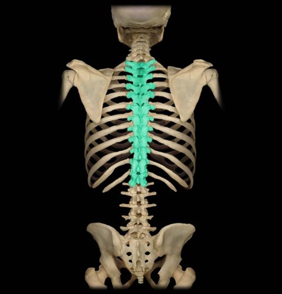
Muscular compartments of the arm
posterior: triceps, extensors, innervated by radial nerve
anterior: biceps + brachialis, flexors, innervated by musculocutaneous nerve
biceps brachii
origin: long head = supraglenoid tubercule of scapula; short head = coracoid process
insertion: radial tuberosity
action: supination, elbow flexion
innervation: musculocutaneous nerve
bicep injuries
proximal tendon rupture - aka popeye deformity, young ppl in baseball, weightlifting - old people from wear and tear
distal tendon rupture - severe hyperextension of elbow forced
brachialis
origin: anterior surface of distal humerus
insertion: coronoid process of ulna
action: primary elbow flexion
innervation: musculocutaneous nerve
triceps brachii
origin: long head - infraglenoid tubercule of scapula; lateral head - post. shaft of humerus above radial groove; medial head - post. shaft of humerus below radial groove (deep)
insertion: olecranon of ulna
action: extension of forearm; long head - extension and adduction
innervation: radial nerve
Nerves of the arm compartments
radial nerve - C5-T1, posterior compartment, runs in radial groove
musculocutaneous - C5-C7, anterior compartment, pierces coracobrachialis
Label radial notch and ulnar notch, radio-ulnar joints, radial styloid process, ulnar styloid process
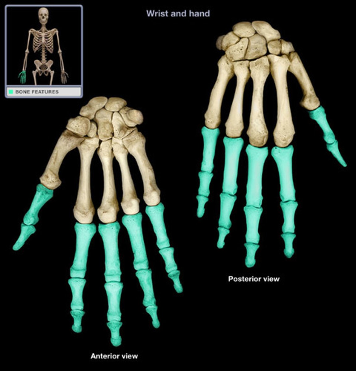
Label the bones of hand
flexor retinaculum
holds contents of wrist in place
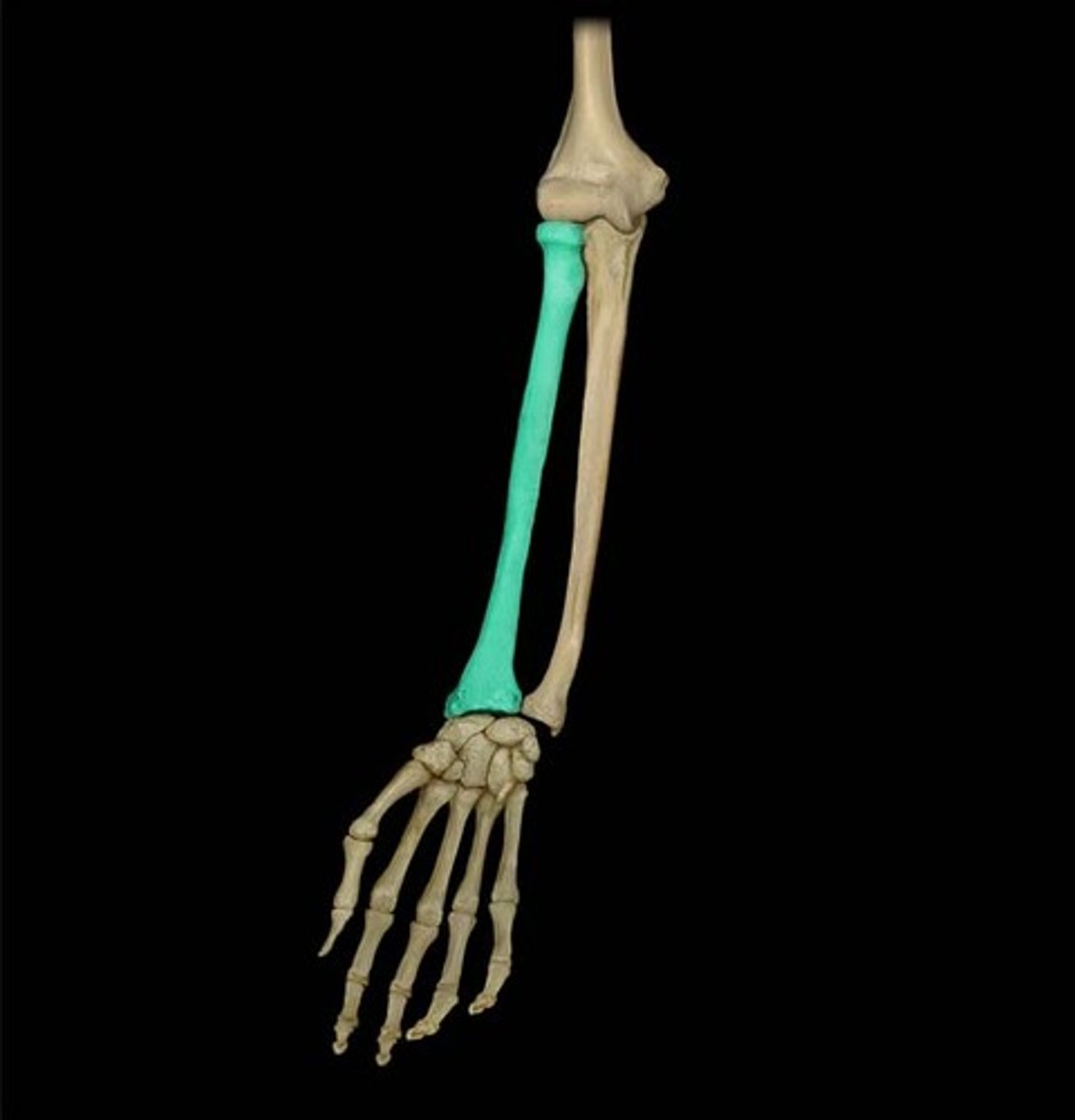
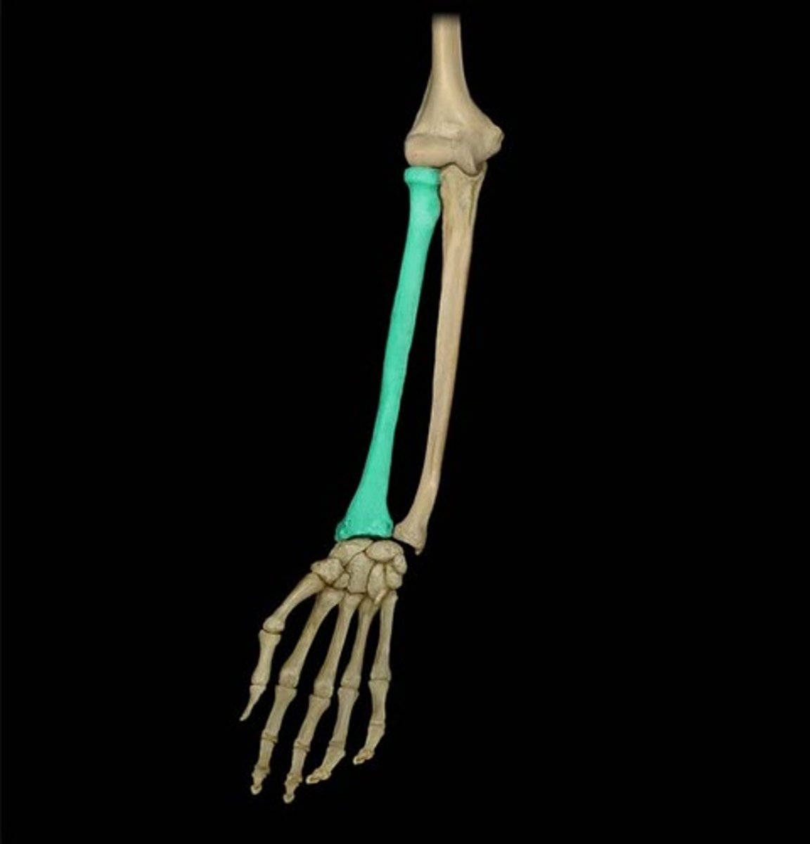
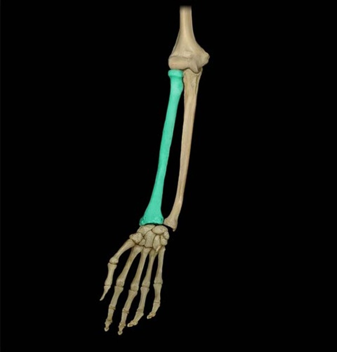
Describe the anterior forearm
3 layers: superficial, middle, deep
actions: flexion (elbow, wrist, fingers), pronation
innervation: median nerve (2 exceptions)
anterior compartment superficial muscles
pronator teres, flexor carpi radialis, palmaris longus, flexor carpi ulnaris
pronator teres
origin: medial epicondyle of humerus + coronoid process of ulna
insertion: distal lateral radius
action: weak elbow flexion, pronation
innervation: median nerve
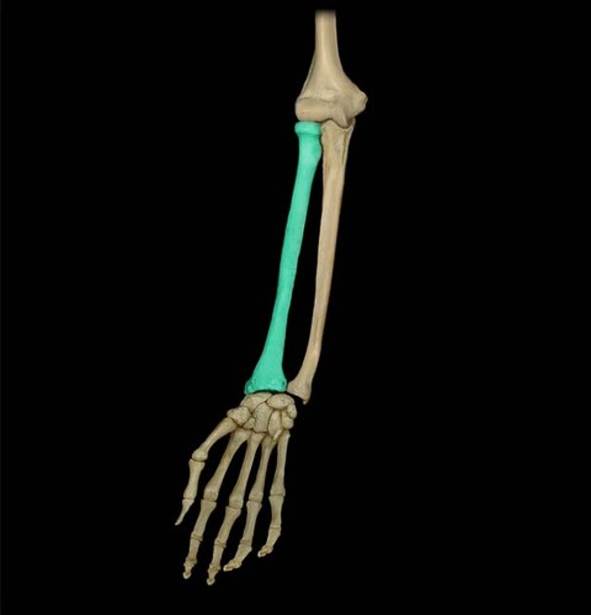
flexor carpi radialis
origin: medial epi
insertion: base of 2nd metacarpal
action: wrist flexion, hand abduction
innervation: median nerve
palmaris longus
origin: medial epi
insertion: palmaris aponeurosis
action: wrist flexion, tighten aponeurosis
innervation: median
flexor carpi ulnaris
origin: medial epi and olecranon
insertion: base of metacarpal 5, carpal bones
action: wrist flexion, hand abduction
innervation: ulnar nerve
anterior forearm deep muscles
flexor pollicus longus, flexor digitorum profundus, pronator quadratus
flexor pollicus longus
origin: anterior radius, inteross. membrane
insertion: distal thumb
action: flexes thumb, weak wrist flexion
innervation: median nerve
flexor digitorum profundus
origin: ulna
insertion: distal palmar phalanges of II, III, IV, V
action: wrist flexion, finger flexion of 2, 3, 4, 5 (MCP, PIP, DIP)
innervation: 2, 3 - median; 4, 5 - ulnar
pronator quadratus
origin: distal ulna
insertion: distal radius
action: pronation
innervation: median
Brachioradialis
origin: distal humerus
insertion: radial styloid process
action: elbow flexion
innervation: radial nerve
posterior forearm superficial muscles
extensor carpi radialis longus, extensor carpi radialis brevis, extensor digitorum, extensor carpi ulnaris
extensor carpi radialis longus
origin: lateral supracondylar ridge humerus
insertion: base 2nd metacarpal
action: extend + abduct hand at wrist
innervation: radial nerve
extensor carpi radialis brevis
origin: lateral epi humerus
insertion: base 3rd metacarpal
action: extend + abduct hand at wrist
innervation: radial nerve (posterior interosseous)
extensor digitorum
origin: lateral epi of humerus
insertion: extensor expansions of 4 fingers
action: extends 4 fingers at MCP joint + extend wrist
innervation: radial nerve (post. inter.)
extensor carpi ulnaris
origin: lateral epi
insertion: base 5th finger
action: extends + adducts hand at wrist
innervation: radial nerve (post. inter)
posterior forearm deep muscles
supinator, abductor pollicis longus, extensor indicis, extensor pollicus longus, extensor pollicis brevis
supinator
origin: lateral epi, ulna
insert: lateral, post, ant. surfaces of proximal 1/3 radius
action: supination
innervation: radial nerve
extensor indicis
origin: post. surface ulna
insert: extensor expansion of index finger
action: extend index finger, extend hand
inn: radial nerve
abductor pollicis longus
origin: post surface ulna, radius
insert: base of thumb
action: abducts thumb, extends it
innervation: radial nerve
extensor pollicus longus
origin: ulna
insertion: base of distal part of thumb
action: extend distal part of thumb
innervation: radial nerve
extensor pollicis brevis
origin: radius
insertion: base proximal part of thumb
action: extend proximal part of thumb
inn: radial nerve
anatomical snuff box
indent in thumb from ext. poll. longus + brevis tendons
access point for radial artery, cutaneous branch of radial nerve, scaphoid bone
extrinsic vs intrinsic muscles
extrinsic: muscle bellies located in forearm not in palm
intrinsic: muscle bellies located in palm
groups of intrinsic muscles
thenar: fleshy part on palm below thumb
hypothenar: fleshy part below pinky
palmar muscles: lumbricals + interossesous (3 PAD, 4 DAB)
thenar muscles
abductor pollicis brevis, flexor pollicis brevis, opponens pollicis (median nerve)
adductor pollicis (ulnar nerve)
hypothenar muscles
abductor digiti minimi, flexor digiti minimi brevis, opponens digiti minimi (ulnar nerve)
palmar muscles - interosseous
3 palmar interossi: adducts fingers toward middle finger
4 dorsal interossi: abducts fingers away from middle finger
palmar muscles - lumbricals
allow flexion at MCP while maintaining extension at IP - 4 in each hand
1,2 = median nerve ; 3,4 = ulnar nerve
wiring of upper limb
musc. n. -> arm (anterior compartment) -> forearm (cutaneous/sensory)
median n. -> forearm (anterior compartment, pronators/flexors except 1 1/2) -> hand (3 lateral thenars + 2 lateral lumbricals)
ulnar n. -> forearm (flex. carpi ulnaris, medial 1/2 flex. digitorum profundus) -> hand (all remaining intrinsic hand muscles)
radial n. -> arm (post. compartment + brachioradialis) -> forearm (muscles of post. compartment) -> hand (cutaneous/sensory)
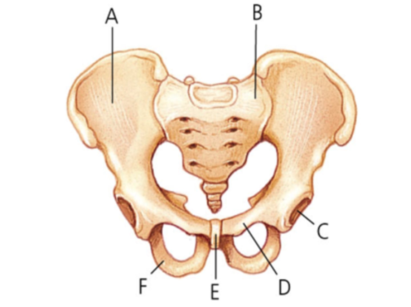
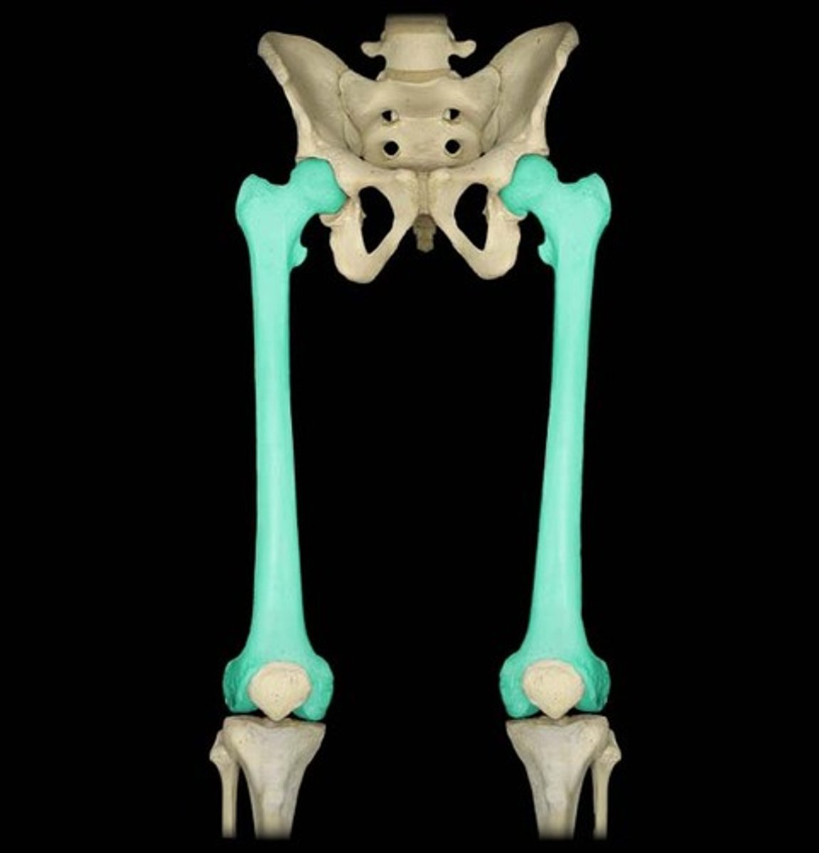
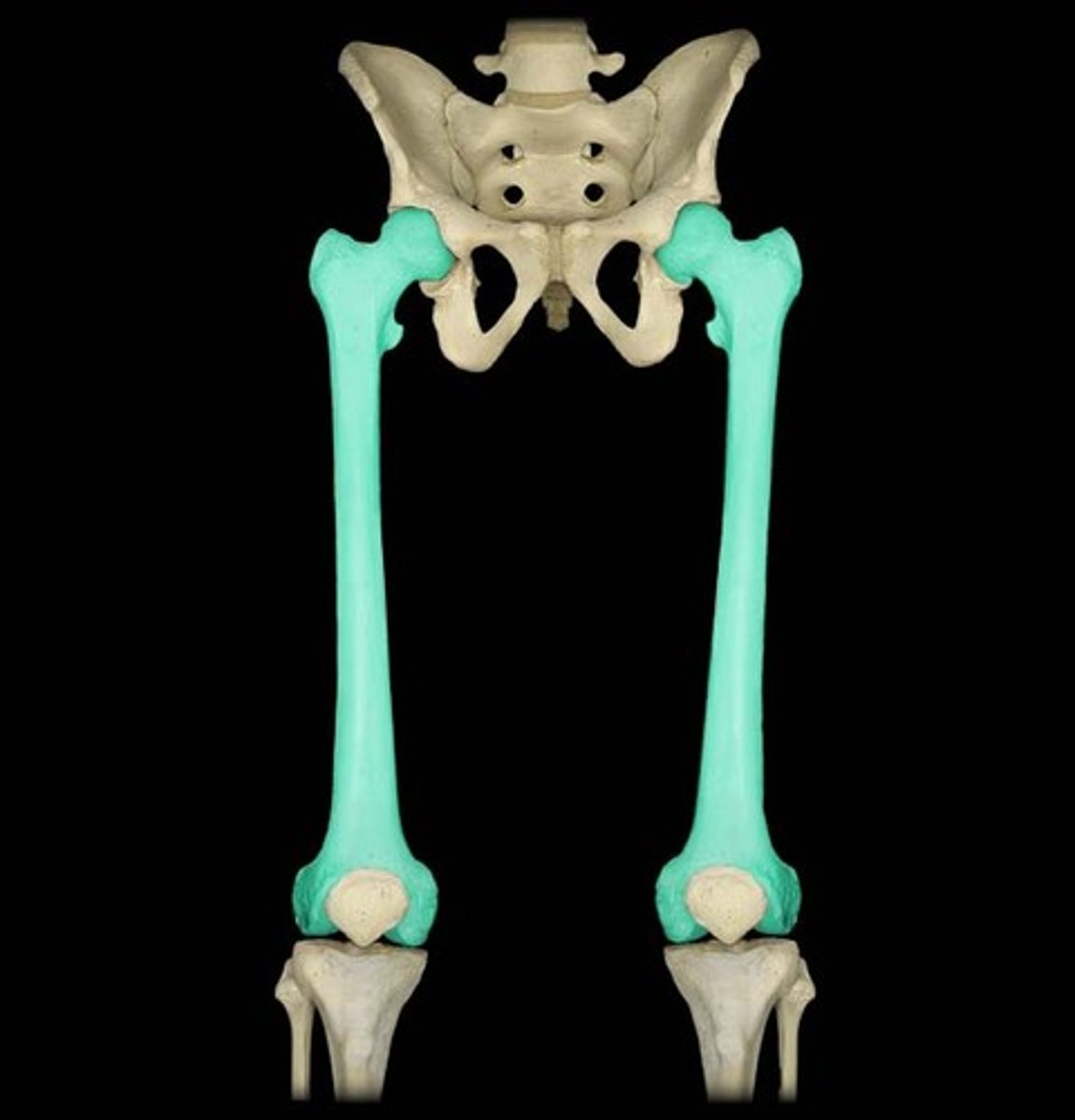
Label pelvic girdle
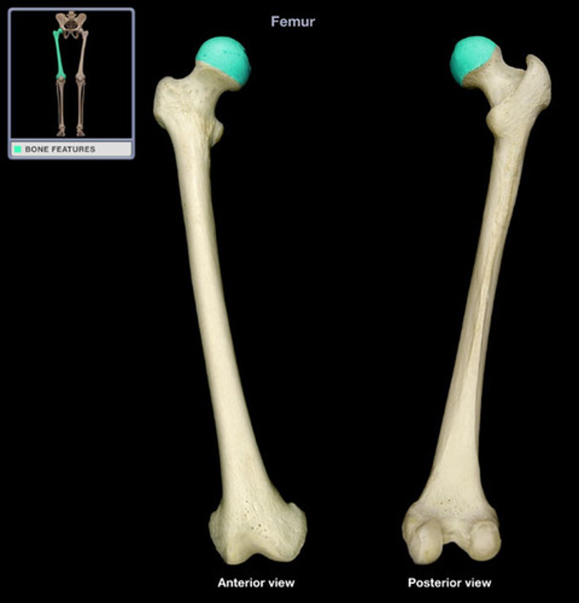
Label the femur
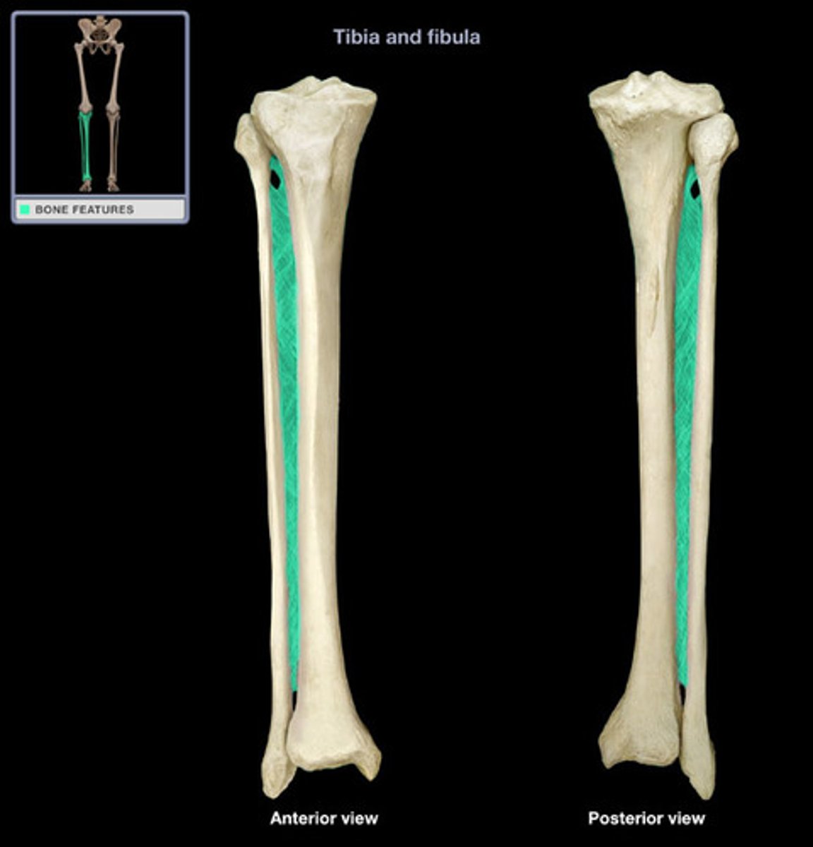
Label the tibia and fibula
Compartments of the thigh
anterior: hip flexors, knee extensors
posterior: hip extensors, knee flexors
medial: adductors
Anterior muscles of the thigh
iliopsoas, sartorius, 4 quadriceps muscles (rectus femoris, vastus medialis, vastus lateralis, vastus intermedius
Iliospoas
origin: iliacus; iliac fossa + ala
insert: lesser trochanter
action: prime mover thigh flexion + flexing trunk
inn: femoral nerve
sartorius
origin: ASIS
insertion: medial tibia
action: flex thigh at hip mainly
innervation: femoral nerve
rectus femoris
origin: ASIS, superior acetabulum
insert: patella and tibial tuberosity
act: extend knee and flex thigh at hip
innervation: femoral nerve
vastus medialis
origin: linea aspera, medial supracondyle
insertion: patella and tibial tuberosity
act: exend knee
inn: femoral nerve
vastus lateralis
origin: linea aspera, greater trochanter
insertion: patella and tibial tuberosity
act: exend knee
inn: femoral nerve
vastus intermedius
origin: anterior + lateral surfaces of shaft of femur
insertion: patella and tibial tuberosity
act: exend knee
inn: femoral nerve
Patellofemoral Syndrome
pain in knees - rectus femoris pulls patella laterally, vastus medius pulls patella medially to straighten it out - if muscle is weak then patella doesn't track straight -> pain
more common in women because women have wider hips (wider pull)
medial muscles of the thigh
adductor longus, pectineus, gracilis, adductor brevis, adductor magnus
adductor longus
superficial
origin: pubis
insertion: linea aspera
action: adduction and flex thigh
innervation: obturator nerve
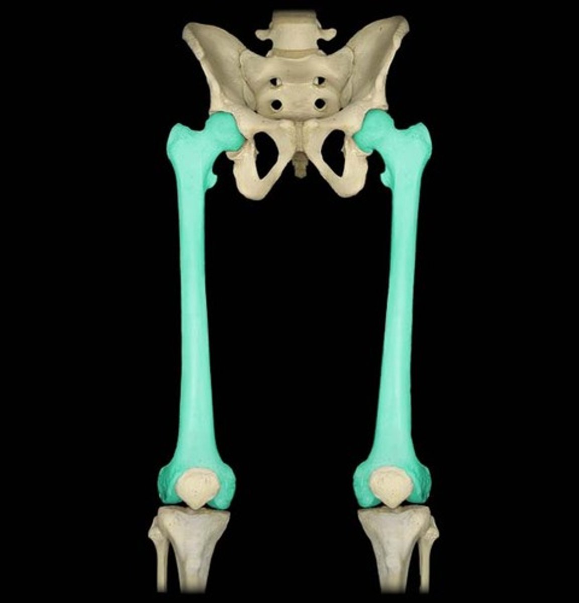
pectineus
superficial
origin: pectineal line of pubis
insert: lesser trochanter to linea aspera
act: adducts and flex thigh
inn: femoral and sometimes obturator
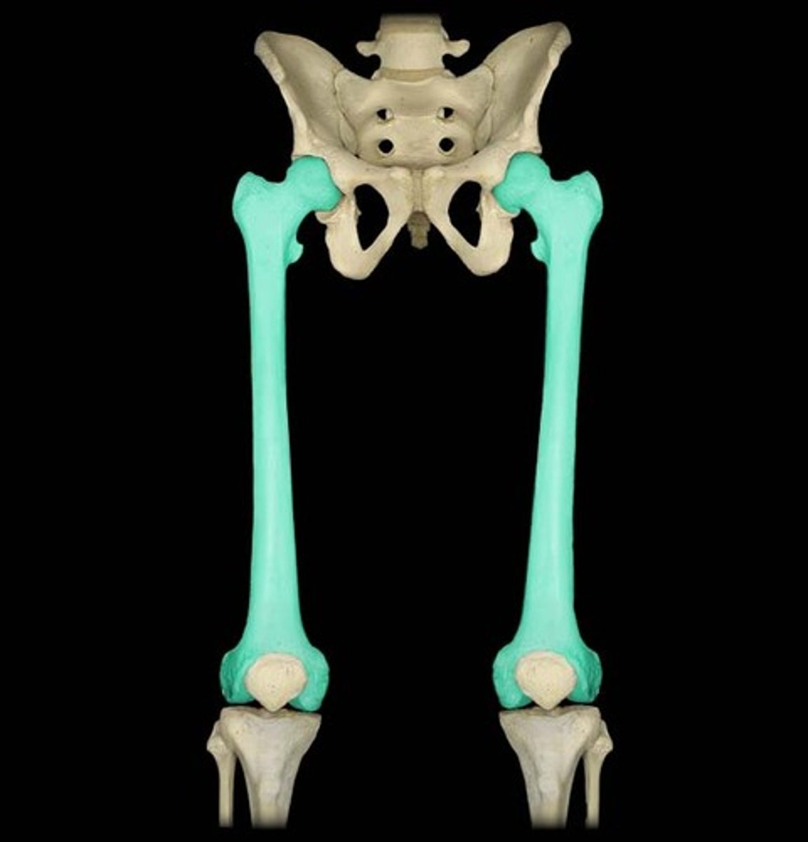
gracilis
origin: pubis
insertion: tibia
act: adduct thigh, flex leg at knee, med rotation of leg
inn: obturator nerve
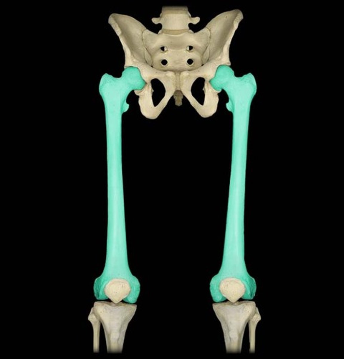
adductor brevis
deep
origin: inferior ramus, pubis
insertion: linea aspera
act: adducts thigh
inn: obturator nerve
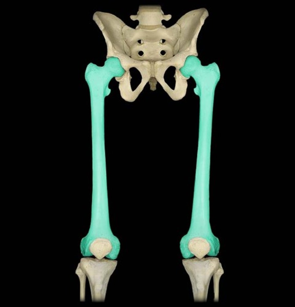
adductor magnus
origin: ischial tuberosity
insert: linea aspera, down femur
action: ant fibers (adduct, med rotation and flex thigh); post fibers (assist hammys in extension)
inn: sciatic nerve + obturator
tensor fasciae latae
sandwiched in IT band
origin: ASIS, anterior iliac crest
insert: IT band
act: flex hip, keep IT band tight to stabilize knee
inn: superior gluteal nerve
gluteus maximus
origin: dorsal ilium, sacrum and coccyx
insertion: gluteal tuberosity of femur, IT band
action: major thigh extensor, lat rotation + abduction
inn: inferior gluteal nerve
gluteus medius
origin: lateral surface of ilium
insertion: greater trochanter
action: abducts, med rotates thigh, steady pelvis when walking
inn: superior gluteal nerve