Clin Med- Cardiology
 Call Kai
 Learn
Learn Practice Test
 Spaced Repetition
 Match
Match Flashcards
 Knowt Play
1/180

There's no tags or description
Looks like no tags are added yet.
Name | Mastery | Learn | Test | Matching | Spaced | Call with Kai | Chat |
|---|
No analytics yet
Send a link to your students to track their progress
181 Terms
Dyslipidemia
Abnormal levels of lipids (fats) in the blood that are divided on the basis of density
HDL: good, more the better, (transport; most dense)
LDL: bad, want less (less dense; triglyceride component)
VLDL: bad, want less (least dense; triglyceride transport)
Total cholesterol: HDL+LDL+VLDL
Lipoprotein (a): key role in development of atherosclerosis (LDL-C)
Niacin
dyslipidemia medication that should be discontinued in all patients
HDL
Good cholesterol
>40 mg/dL in men, >50 mg/dL in women
Low HDL: insulin resistance, obesity, smoking, high carbs, drugs, decreased physical activity
High HDL, low risk of coronary heart disease
LDL
bad cholesterol
Low LDL will decrease rate of MI, stroke, angina and need for CABG
Primary prevention: decrease risk with reduction of LDL-C
Secondary prevention: prevents future additional occurrences
Triglycerides in the blood should be
<150 mg/dL
diet and exercise (lifestyle) are key
Secondary causes of dyslipidemia
Alcohol use, diabetes, pancreatic and liver disease, hyper/hypothyroidism, drugs
Signs and symptoms of dyslipidemia
Most are non-specific and caught on routine lab screening
>1000 mg/dL in triglyceride/VLDL- eruptive xanthomas (red-yellow papules on buttocks)
High LDL- tendinous xanthomas (Achilles, patella, back of hand)
Lipidemia retinalis (cream colored blood vessels in the fundus)
Screening for dyslipidemia should start
at 20 years old and continue once a year
Dyslipidemia Dx
Labs (lipid panel/profile)
Dyslipidemia Tx
Raise HDLs, improve diet, increase exercise (lifestyle changes)
If does not improve, go med route
Statins (atorvastatin, fluvastatin), Ezetimibe (Zetia), PCSK9 inhibitors (Repatha, Praluent), Omega-3-fatty acids, Bempedoic acid (nexletol)
Pericarditis
Inflammation of the pericardial lining
Acute: 2/4 criteria (chest pain, pericardial friction rub, ST changes, new or worsening effusion) and symptoms less than 4-6 weeks
Incessant: Longer than 4-6 weeks, none at 3 months, no interruption
Recurrent: longer than 4-6 weeks, has interruptions
Chronic: lasting longer than 3 months, no breaks
Acute pericarditis
less than 2 weeks, inflammation of the pericardium
Risks: idiopathic, infection, surgery, CT disease, radiation, pericardial injury. myxedema
Infectious
Viral: coxsackie, echo, influenza, EBV, varicella, hepatitis, mumps, HIV, COVID
Males <50 yo
Bacterial: rare (TB)
Uremic, neoplastic, post MI (Dressler syndrome), radiation, CT disease, drug induced
S/S of Acute Pericarditis
chest pain (significant, continuous, sharp), dyspnea, fever, pericardial friction rub
TB: TB symptoms
Bacteria: inflammatory, toxic, critically ill (septic)
Uremic: w/ or w/o symptoms, absent fever
Neoplastic: Often painless, increase risk of pericardial effusion
Dx of acute pericarditis
Viral: clinical diagnosis based on criteria, leukocytosis, echo is normal
TB: TB testing
Bacterial: pericardiocentesis
Uremic: “shaggy pericardium”- irregular and thickened pericardium on ECHO or CT
ECG: shows ST waves elevated in all ECG leads
CXR: cardiac enlargement, masses (neoplastic)
Neoplastic: cytologic examination
Post MI: increased ESR, large pericardial effusion
Myxedema: hypothyroidism, cholesterol crystals
Acute Pericarditis Tx
Tx of underlying cause
NSAIDs
Colchicine
Cardiac Tamponade
Pressure that affects pericardial filling, cardiac perfusion, and cardiac output
Triad
Jugular venous distention (JVD)
Hypertension
Muffled heart sounds
DO NOT CONFUSE WITH TENSION PNEUMO
Tx of Cardiac Tamponade
Restriction in activity (3 mo)
ASA, NSAID
Colchicine + NSAID (3 mo) for prevention (cyclophosphamide or azathioprine if not tolerated)
Underlying cause
TB: TB drug therapy
Radiation: symptomatic tx
Uremic: dialysis (tamponade common)
Neoplastic (poor prognosis): drain effusion, chemo, tetracycline
Dressler Syndrome (post MI pericarditis)
recurrence of pain with pleural-pericardial features, the rub is audible and repolarization changes on ECG may be confused with ischemia
prior MI (weeks or months ago), large effusions (uncommon)
S/S: pain, fever, malaise, leukocytosis
DX: S/S and Hx
TX: spontaneous resolution
Post MI: ASA and colchicine for 3 months, no NSAIDS or Steroids
Refractory: colchicine (6 mo), Indomethacin doses, corticosteroids
Pericardial Effusion
accumulation of fluid in the pericardial sac
gradual= stretch and accommodate
quickly= cardiac tamponade
Transudate= L heart failure, overhydration, hypoproteinemia
Exudate= renal failure, malignancies, infection
1st affected= right atrium and ventricle
Pericardial Effusion S/S
underlying cause, dull chest pain, dyspnea, muffled heart sounds, poorly palpated apical pulse, pulsus paradoxus
Pericardial Effusion Dx
CXR: “water bottle heart”- enlarged flask shaped cardiac silhouette
Echo: visualize fluid around heart
Pericardial effusion tx
Underlying cause
NSAIDs (pain relief)
Large effusion/cardiac tamponade (pericardiocentesis)
Recurrent/Refractory effusion (pericardial window)
Pericardial tamponade + Constrictive pericarditis
Effusive-Constrictive pericarditis
Constrictive pericarditis (over time)
leads to thickened, fibrotic, adjacent pericardium that restricts diastolic filling and produces chronically elevated venous pressure
Risks: immune mediated/neoplastic pericarditis (increases risk after bacterial pericarditis)
Constrictive pericarditis S/S
S/S: dyspnea, fatigue, weakness (principle)
chronic edema, increase JV pressure, Kussmaul sign, pericardial knock, pulsus paradoxus, A-fib
Constrictive pericarditis Dx and Tx
X-ray: normal, some cardiomegaly or peripheral calcification
ECHO: septal bounce (best test choice)
CT/MRI": pericardial thickening
Cardiac cath: confirmatory but invasive and expensive
Tx: aimed at specific etiology, anti-inflammatories, diuretics, pericardiectomy if dieuresis fails
Metabolic syndrome
cluster of conditions that increase risk of heart disease, stroke, and type 2 diabetes
Metabolic syndrome criteria (must have at least 3)
Waist line: >40 inch for men, >35 inch in women
Hypertension: >130/85 or on anti-hypertensive medications
High blood sugar: >100 or on meds to control blood sugar
High triglycerides: >150 or on meds to control
Low HDL: <40 for men, <50 for women
Risk factor of metabolic syndrome
Insulin resistance, obesity (abdominal fat), physical inactivity, poor diet, aging, genetics
Consequence of metabolic syndrome
increased cardiovascular disease risk, increased risk for type 2 diabetes, sleep apnea and osteoarthritis
Prevention and treatment of metabolic syndrome
Lifestyle change: diet, physical activity, weight loss, quit smoking/alcohol, stress and sleep management
Treatment for metabolic syndrome
Medication for blood pressure, cholesterol, and blood sugar
Claudication
cramping discomfort, weakness, tired sensation caused by lower extremity exercise and relieved with rest
Severely stenotic
pain continuous and severe from constant inadequate tissue perfusion
Peripheral artery disease (PAD) is also known as
Peripheral vascular disease (PVD), peripheral vascular occlusive (PVOD), peripheral arterial occlusive disease
Peripheral artery disease (PAD)
Most commonly atherosclerotic disease of the arteries that perfuse the limbs an occlusion can reduce or completely obstruct blood flow to distal tissues (arterial insufficiency) it chronic or acute
occlusion of 50% of arterial lumen (75% of vessel diameter), produces significant reduction in blood flow
Decreases blood flow causes tissue hypoxia and ischemia (severe: tissue necrosis w/ gangrene)
Plaques develop @ bifurcations, lower extremities affected more
Atherosclerosis: chronic, slow process
Risks for peripheral artery disease
Smoking, hypercholesterolemia, diabetes mellitus, hypertension, family hx, male sex
S/S and PE findings for peripheral artery disease
Intermittent claudication: calves MC, thighs and buttocks, severing claudication (in worsening cases)
SEVERE: rest pain, severely stenotic artery (foot) (pain at rest requires surgical intervention)
Aortic iliac: Leriche’s Syndrome- erectile dysfunction
Intestinal Angina: visceral artery insufficiency- postprandial pain (fear of eating and weight loss), pain out of proportion to PE
Amaurosis Fugax: transient monocular blindness (ophthalmic branch of ICA)
PE: diminished/absent peripheral pulses, bruits (severely stenotic- absent), pallor on elevation, erythema on dependency, poor capillary refill, digital cyanosis, tropic changes (hair loss), painful ischemic changes (gangrenous ulcers)
DX of Peripheral Artery Disease
Ankle-Brachial Ratio/Index (ABI/AAI)- normal is 1.0-1.1, <0.9 is PAD, <0.5 is critical PAD
Most common first thing done (non-invasive)
USN: arterial duplex scanning
Done 1st after ABI
CTA/MRA: precise location and extent of vascular occlusion with contrast
Gold standard test
TX of peripheral artery disease (PAD)
Reduce cardiovascular risk (smoking cessation, lipid/BP control, weight loss, antiplatelet)
Exercise program (daily walking and tracking point of claudication)
Meds (after initial lifestyle changes)
Aspirin
High dose statin (atorvastatin)
± Rivaroxaban (DOAC)
± Cliostazol (symptom relief of claudication)
SEVERE: vascular reconstruction or angioplasty and stenting, arterial grafts, thromboendarectomy (repairing diseased arteries), amputation in the case of gangrene
F/U 3-6 months with stable disease and consult vascular surgery
Complications: secondary infection (cellulitis, tissue necrosis, ischemic ulcers
Aneurysm
Localized dilation or out pouching of a vessel wall or cardiac chamber
aorta is susceptible
Atherosclerosis contributes
Genetic conditions predispose to aneurysm
Acute arterial insufficiency
Usually occurs acutely as a result of thrombosis or embolism (MCC- emboli from heart)
emboli from A-fib/flutter, mitral stenosis, transmural infarction
10x more likely in lower extremity than upper (most common superficial femoral artery)
Acute occlusion= severe ischemia (no collateral circulation)
Must catch within 4-6 hours to minimize the risks of limb loss
Acute arterial insufficiency S/S
Acute onset of severe pain, loss of pulses, ischemic changes (cool limbs and cyanosis), unilateral, can present as stroke
5 P’s
Pain: constant and aggravated by movement
Pallor: occurs initially, followed by cyanosis
Pulselessness: w/ cold limb
Paresthesia: caused by anoxia to peripheral nerves
Paralysis: necrosis of muscles and nerves
Dx of acute arterial insufficiency
Hx (time of onset, previous heart disease, atherosclerotic PVD, hypotension)
5 P’s (HALLMARK)
Finding distal to occlusion
Paresthesia and paralysis are late findings
MRA/CTA: GOLD STANDARD, if no time straight to OR
Tx of acute arterial insufficiency
Consult vascular surgery for operative intervention
Immediate anticoagulation with heparin
Cardiac embolism: PERMANENT oral anticoagulation
Complications if not treated: limb loss, organ failure, compartment syndrome, reperfusion syndrome
Abdominal aortic aneurysms (AAA)
Localized dilation or out pouching of abdominal aorta, can be apart of aging, 90% are below renal artery
Healthy young man 2 cm
Aneurysm present when diameter exceeds 3 cm
Rarely rupture until >5 cm
Male to female ration 4:1, occurs in 2% of men over 55 yo
Involve the aortic bifurcation and often the common iliac arteries
AAA S/S
Asymptomatic: found incidentally on CT/ultrasound or palpated
Symptomatic: mild/severe mid-abdominal discomfort often radiating to the lower back
RUPTURE- SURGICAL EMERGENCY
Rupture causes severe pain, palpable abdominal mass, hypotension
Free rupture into peritoneal sac: death
AAA DX and TX
Dx: abdominal ultrasounds, aneurysm around 5 cm perform a CT
F/U every 2 yrs< 4cm, 6 mo if 5 cm Aneurysm present
Screen 65-74 yo men who were previous or current smokers, no enlargement do not repeat
Tx: Elective repair if >5.5cm or rapid expansion >0.5 cm in 6 mo
Pain, tenderness, impending rupture= urgent repair
Open Sx repair= ruptured
Endovascular repair = noncomplicated, unruptured
Thoracic aortic aneurysms
Happens due to atherosclerosis, CT disorders (Marfan), less than 10% are thoracic
S/S: most asymptomatic, may have substernal or neck pain, can cause SVC syndrome
Dx: CTA scan (SCAN OF CHOICE), could be followed by cardiac catheter and ECHO to see where arteries are
Tx: > 6cm consider repair
Ascending aorta/proximal aortic arch: open procedure
Descending thoracic aorta: endovascular grafting
Aortic dissection
Spontaneous initimal tear develops, blood dissects into the media of the aorta and blood flows into the lining and not the lumen, HTN is a key finding, often confused for an MI
If there is an absence of HTN, abnormalities of smooth muscle, elastic tissue, or collagen are present
Type A: arch proximal to the left subclavian artery
Type B: proximal descending thoracic aorta beyond the left subclavian artery
Aortic dissection risks and S/S
Risks: pregnancy, bicuspid aortic valve or coarctation
Occludes major aortic branches: cause ischemia of brain, intestine, kidney, and extremities
S/S: severe sudden onset of chest pain, radiates down back-anterior chest-and neck, HTN, syncope or paralysis of lower extremities, new murmur, peripheral pulses diminisheD
Aortic Dissection Dx and Tx
Dx
EKG (to eliminate MI confusion)- can show LVH might be clear
CT Chest and Abdomen (TEST OF CHOICE): line (dissection)
Tx
Bring down BP: Beta blockers (labetelol or Esmolol), CCB if doesn’t work (nicardipine or nitroprusside IV)
Prior to definitive Dx even being known
Sx
Type A: URGENT Sx repair
Type B: early thoracic stent repair if signs of malperfusion
Uncomplicated B: BP control, urgent Sx not required, monitor with yearly CT
Varicose veins
Dilated tortuous superficial veins in the legs due to high venous pressure
Risks: prolonged standing, pregnancy, heavy lifting, hereditary
S/S: dull aching heaviness of legs, worse w/ standing, itching over the ankles or large varicosities
older patients bleed with minimal trauma
Dx: Hx and PE
Tx:
Non-surgical: medical grade compression stockings
Scleortherapy: sclerosing agents to veins
Surgical: thermal ablation or vein stripping
Superficial Venous Thrombophlebitis
Red, painful in duration along a superficial veins, MC @ the site of an IV Line
Risks: Pregnancy, trauma, varicose veins, systemic hyper-coagulable states, IV lines
Monitor site for SEPTIC phlebitis (erythema, fever, fatigue, leukocytosis)
Dx: Hx and PE
Tx:
Symptomatic: analgesics (Tylenol) and warm compresses
Septic thrombophlebitis: remove lines/catheters, IV abx (VANC and CEFTRIAXONE)
Chronic venous insufficiency
Inadequate venous return over a long period of time
Risks: prior DVT, leg trauma/surgery, obesity
S/S: progressive pitting edema, itching-dull-aching after standing, taut shiny skin at ankle, brownish discoloration of the skin, ulcerations above the ankles, frequent cellulitis
Dx: Hx and PE
Tx: prevention of primary causes, graduated compression stockings, meticulous wound care in cellulitis
Superior Vena Cava Syndrome (SVCS)
Occlusion of the superior vena cava leading to venous distention in the upper extremities and head (“swollen head”)
Risks: MCC is malignancy, histoplasmosis, TB, CF, benign masses, invasive procedures
S/S: headache, fuzziness, dizziness, dilated facial veins, facial flushing, edema of face-neck-arms, cyanotic appearance of skin
Bending over and lying down exacerbates the symptoms
Dx: PE signs, CT of the chest (BEST IMAGING)- shows site of occlusion
Tx: elevate the head, balloon angioplasty/stent of the occlusion, Tx of the underlying cause, treatment of the neoplasm
Lymphedema
Progressive disease that results in accumulation of interstitial fluid and fibroadipose tissue
Primary form: congenital abnormalities of distal lymphatics (often unilateral)
Secondary form: inflammatory or traumatic lymphatic obstruction
Trauma, external beam radiation tx, lymph node removal, inflammatory disorders, obesity, FILARISIS (WORM)
Lymphedema S/S, Dx, and Tx
S/S: insidious onset of symptoms, limb hypertrophy, thickened and fibrotic skin, pitting edema initially- slowly becomes less pitting overtime, complain of limb pain, decrease ROM
DX: clinical, USN Doppler
TX: underlying cause, refer to specialty wound care, elastic compression stockings or lymphedema wraps, meticulous skin care to avoid cellulitis
Infection= use CEPHALEXIN
Lymphangitis (Lymph/fluid vessels infection)/ Lyphadenitis (lymph node infection)
Bacterial infection (hemolytic strep or staph aureus) that feeds bacteria into lymphatics
Red streak extending towards involved nodes
Infection can progress rapidly, may lead to septicemia and death
S/S: throbbing pain and warmth in areas of cellulitis, indurated cellulitis with spreading streaks, malaise, fever, chills, sweating, tachycardia
streaking can be faint and easily missed in people of color
Dx: Labs (leukocytosis), blood cultures (+ if septicemia)
Tx: heat, elevation, immobilization, analgesia (Tylenol), meticulous wound care
ABX: Staph and strep CEPHALEXIN, AUGMENTIN (MRSA: BACTRIM)
Raynaud’s Phenomenon
Vasospastic attacks of small arteries of the fingers (sometimes the toes too)- vasoconstrict and decrease blood flow
1. Primary: young women, course is more benign
2. More severe, causing digital ulceration or gangrene
S/S: change in skin color and sensation, attacks precipitated by cold-stress-vibrations, pallor-cold sensation- numbness, sluggish blood flow and ischemia (cyanosis), chronic attacks (thin/brittle nails and skin), asymptomatic between attacks
Dx: Hx and S/S
Tx: Avoid cold to the extremities, keep body warm, NO SMOKING, avoid sympathomietric drugs
Meds: CCB- nifedipine, amlodipine
Sx: only in refractory cases
Thromboangitis Obliterans (Buerger Disease)
Segmental, inflammatory and thrombitic disease, occurs in distal arteries (sometimes veins) of extremities, usually plantar and digital vessels of the foot and lower leg (It is arteritis, inflammation of the arterial wall)
Risks: male smokers, middle-aged
S/S: lesions on toes and patient <40 yo, rest pain is frequent, progresses to tissue loss and amputation
MRA/CTA: distal tree obliteration
TX: Stop smoking (curative), if pt doesn’t stop smoking amputation to the extremities will be the outcome
Giant cell arteritis (GCA) or Temporal arteritis
Chronic-inflammatory disease involving large and medium sized arteries, can be widespread causing stenosis and aneurysm of affected vessels
Risks: Age (almost never before 50, usually 70-79), MC in women, caucasians (Scandinavian), Polymyalgia Rheumatica
S/S: fever, fatigue, weight-loss, headache (scalp tenderness), jaw claudication, ocular involvement (amaruosis fugax)
Dx: Labs- increased ESR, CRP, TEMPORAL ARTERY BIOPSY
Tx: 1 mo prednisone (do steroid taper)
Supraventricular arrhythmias
Originate ABOVE the cardiac ventricles
narrow QRS complex, less serious because ventricle activity is preserved
S/S
Fast heart rate: palpitations, chest pain, nausea, diaphoresis
Slow heart rate: dizziness, syncope
Sinus Bradycardia
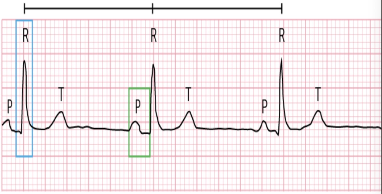
Sinus rhythm w/ HR <60 bpm, regular rhythm slow-rate, P-waves followed by narrow QRS complexes, Can be a normal finding in healthy individuals, athletes, or during sleep
Risks: Medications (Beta-blockers, CCB, Digoxin), athletes/healthy individual, sleep (benign)
S/S: dizziness, weakness, fatigue, SOB, chest pain, syncope (fainting)
Dx: confirm and exclude other causes of bradycardia, ELECTROLYTES and THYROID FXN TESTS
Tx: not always needed
Symptomatic: Atropine (acute and symptomatic), pacemaker (chronic)
Sinus tachycardia
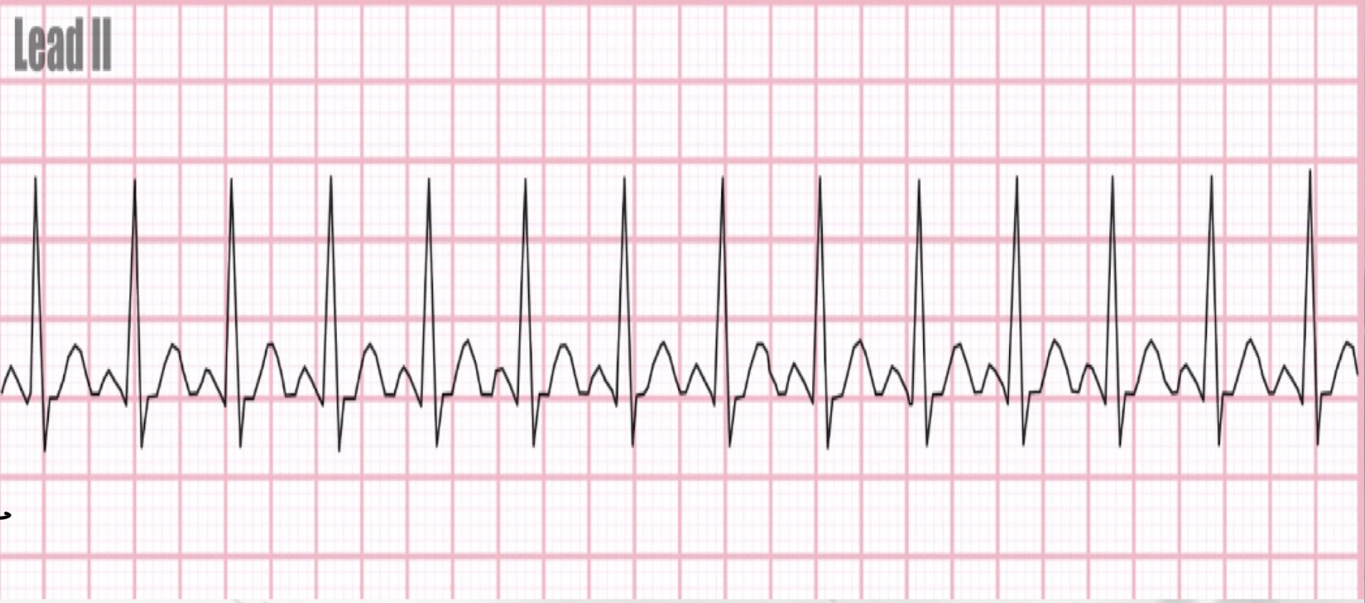
Heart rate >100 bpm, typically between 100-160 bpm originating from SA nodes, normal rhythm-fast rate
Risks: exercise/exertion, stress-anxiety, fever, hyperthermia, hypoxia, anemia, stimulants
Tx: Underlying conditions (pain, fever dehydration), avoid stimulants, address stress and anxiety problems, adequate hydration and sleep
Meds: Beta-Blockers (metoprolol, labetolol), or CCB (verapamil, diltiazem) to control heart rate
Premature atrial contractions (PACs)
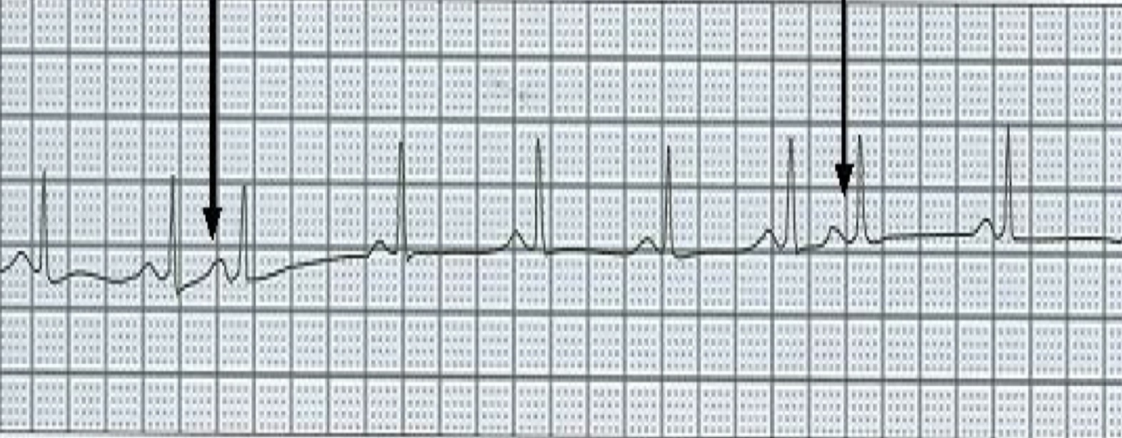
Early beat originating from an ectopic pacemaker site in the atria, early beat interrupts the regularity of sinus rhythm, increase automacity of pacemaker cells
Same ectopic site= regular irregular rhythm
Different ectopic site= irregular, regular rhythm
Risks: caffeine, nicotine, cocaine, amphetamines, thyroid disease, post-MI, stress/anxiety, hypoxia, CHF, Pulm dz
Tx
Infrequent: no treatment
Frequent: underlying cause
Multifocal atrial tachycardia and wandering atrial pacemaker (WAP)
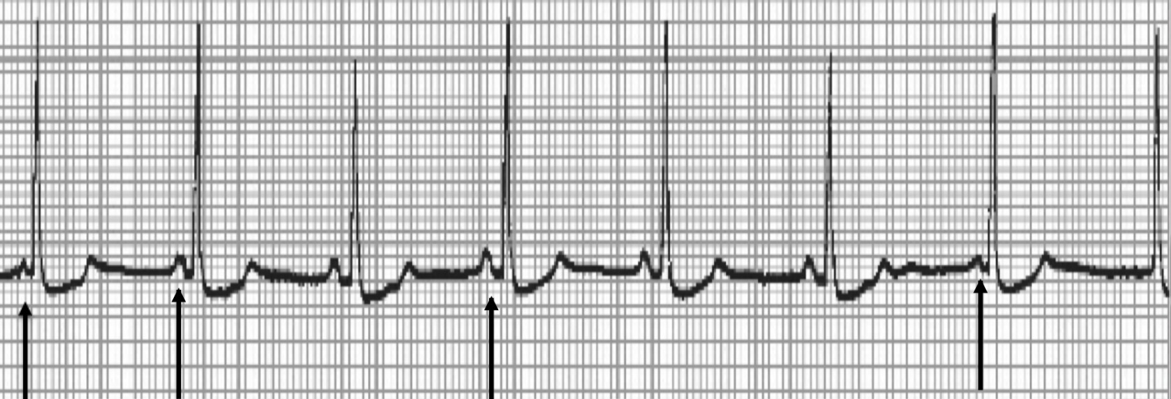
Occurs when the pacemaker site shifts back and forth b/t sinus node and ectopic atrial sites; P-waves vary in size, shape or direction across the rhythm strip as the pacemaker “wanders” b/t multiple sites
3 different P-wave morphologies
MAT: HR>100
WAP: HR<100
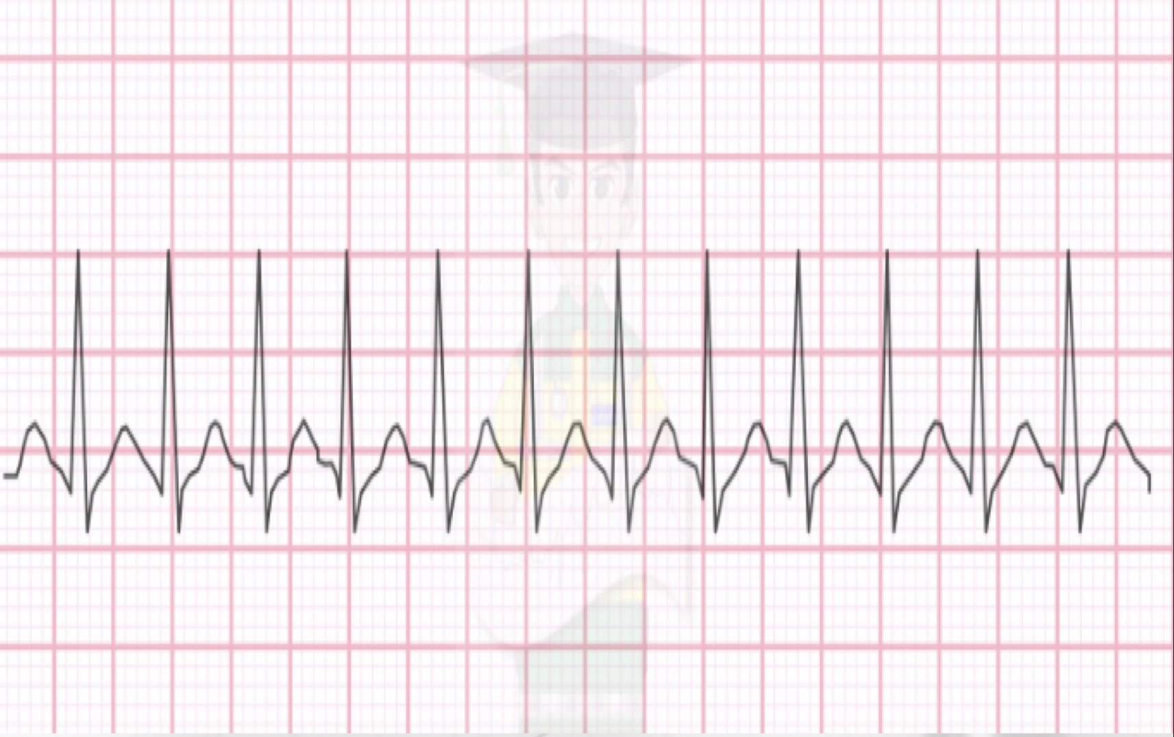
Supraventricular Tachycardia (SVT)
Often occurs in patients without structural heart disease, episodes start and end abruptly (seconds to hours)
Rate 140-240 bpm, regular rhythm (not sinus)
Loss of normal contour, P-wave differs in contour from sinus beats and is often buried in the T-wave (no sinus)
Normal QRS complex
S/S: palpitations and anxiety
Tx: depends on pt stability
Unstable (hypotension, AMS, HF): cardioversion (synchronized electric shock)
Stable (least to most invasive)
Vagal maneuvers (carotid massage, Valsalva)
Bolus of adenosine IV →If that doesn’t work do another bolus of adenosine→ If ineffective do another bolus of adenosine
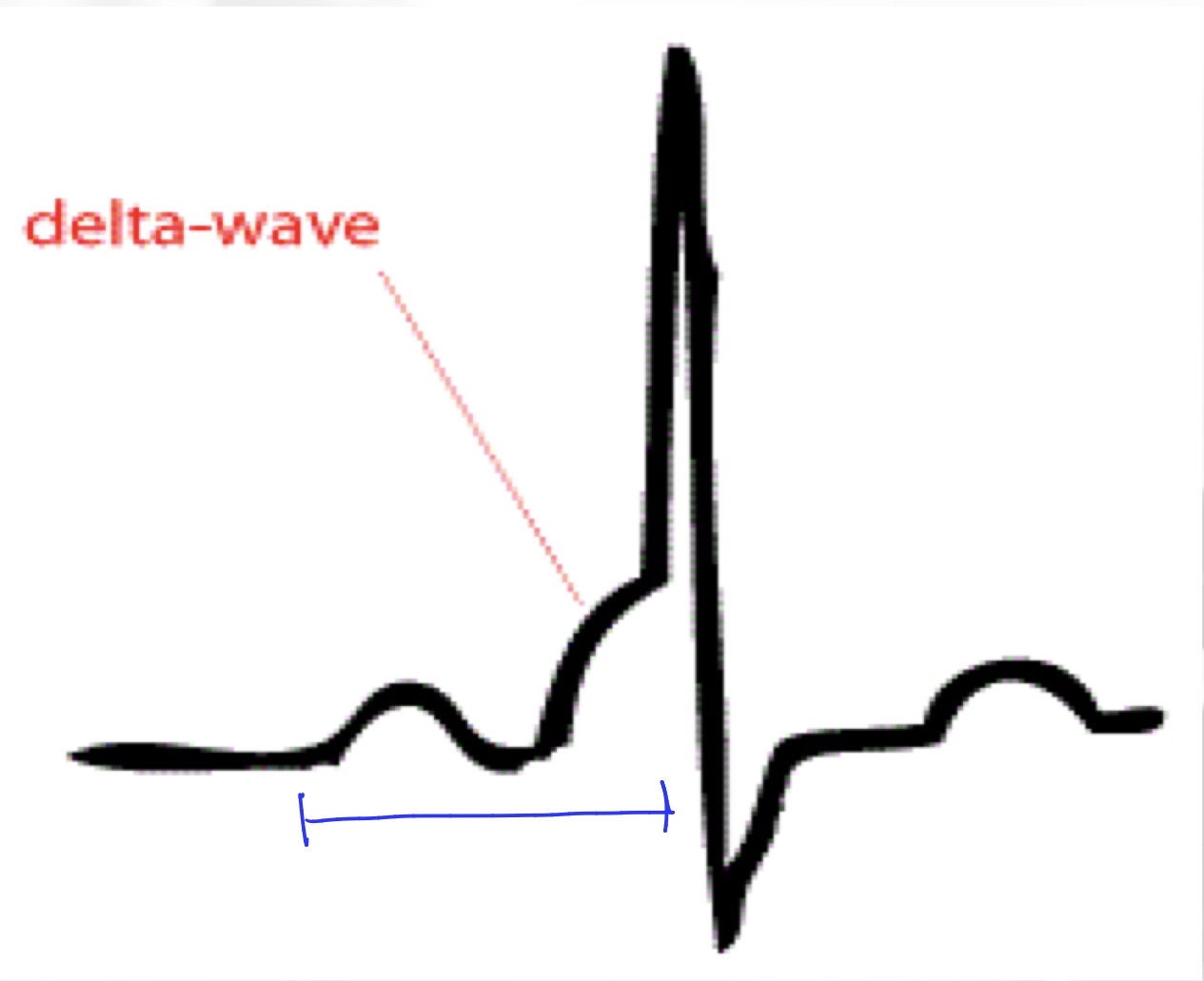
Wolff-Parkinson White (WPW)
Accessory pathway between atria and ventricles causes tachycardia
short PR interval, HALLMARK DELTA WAVE
Risks: sudden cardiac death
S/S: palpitations, dizziness, dyspnea, chest pain, episodes of syncope
Tx: Accessory pathway ablation (radio frequency energy)
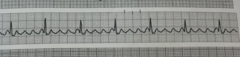
Atrial flutter
Originates in ectopic pacemaker site in the atria discharging atria impulses at a rate of 250-400 bpm; ventricular contraction rate typically regular and ranges from 75-150 bpm, blood pools and the atria cannot drain
rapid atrial impulses create a V-shaped waveform with a sawtooth pattern
TX:
Unstable: Cardioversion (synchronized electric shock)
Stable: Rate control (B-blocker or CCB) or Rhythm control (amiodarone or cardioversion with med failure), catheter ablation (PREFERRED LONG TERM MANAGEMENT)
Anticoagulation (decrease stroke incidence)
Onset <48 hrs can convert rhythm
Onset >48 hrs begin anticoag or schedule TEE for clot evaluation before cardiovert
Long term anticoag with a DOAC
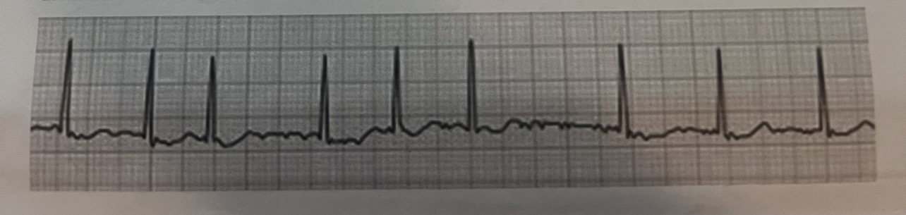
Atrial Fibrillation (A-Fib)
MC arrhythmia requiring treatment and can be secondary to many medical conditions; atria contracts 300-500 contractions in a minute
atrial pathology: increase atrial pressure, atrial dilation (disrupts underlying conduction path)
Atrial rate >300 bpm, irregular rhythm, variable ventricular rate, absence of P-waves, irregularly regular, varying R-R interval
Risks: enveloping blood clots, stroke, 60 and older
S/S: dizziness, palpitations, fatigue, mild dyspnea, chest pain
Tx: rate control, rhythm control, stroke prevention (anticoagulation)
Unstable: Cardioversion (synchronized cardioversion)→ TEE or anticoag first
DO a CHADS-VAS risk score (stroke risk >2 in men, >3 in women)
Stable: 1ST LINE- Rate control and anticoag for long term maintenance
Rate: Beta blocker (metoprolol, atenolol), CCBs (diltiazemn, verapamil), amiodarone
Anticoag: DOAC (apixaban, rivaroxaban)
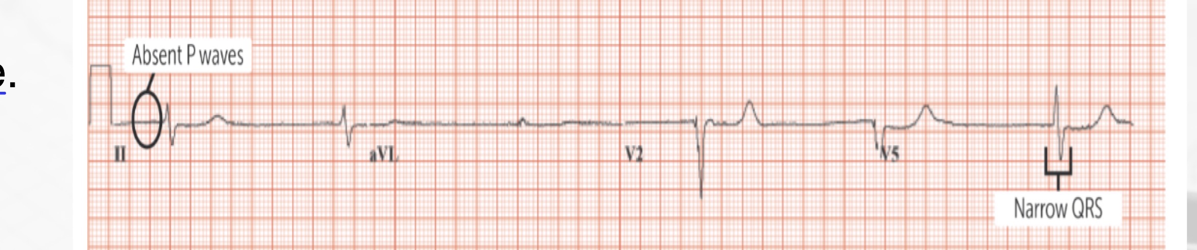
Junctional escape rhythm
Junctional arrhythmia the heart rate is low at 40-60 bpm, SA node fails to initiate impulse so a secondary pacemaker within AV junction takes over
P-waves typically absent/buried
Bradycardia
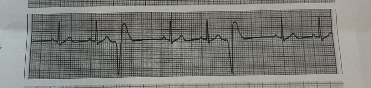
Premature ventricular contractions (PVCs)
Early ectopic impulse that originates in ventricular tissue either in the right or left ventricle, premature wide morphology
QRS complex is wide and morphology is different b/c of underlying rhythm
Pause follows the PVC, usually compensatory
Risks: MC in those w/ heart disease
Tx: not indicated if asymptomatic
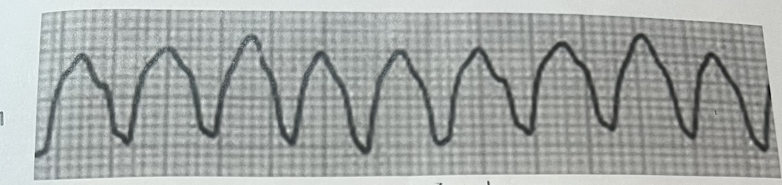
Ventricular Tachycardia (V-tachycardia) aka monomorphic tachycardia
Originates from an ectopic site in the ventricles discharging impulses at a rate of 140-250 beats per minute (complication of MI or cardiomyopathy)
3 or more premature beats
Tombstone appearance
Tx
Unstable: cardioversion (w/pulse)→ if recurs give amiodarone and shock again
Stable: amiodarone, if unsuccessful sedate patient and perform cardioversion as indicated
Long term: B-blocker or CCB
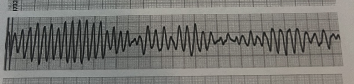
Torsades De Pointes→ Polymorphic Vtach
Prolonged QT interval, “twisting of the points”- twist around isometric line, QRS complexes are wide and have varying heights, widths, and shapes; seen anywheres from 200-300 bpm
Risks: severe hypokalemia, severe HYPOMAGNESEMIA or prolonged QT interval
Tx: O2 therapy, UNSYNCHRONIZED CARDIOVERSION and magnesium maintenance infusion (1st line)
Also correct electrolyte inbalance and d/c QT prolonging drugs
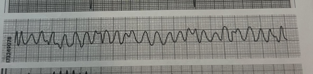
Ventricular fibrillation
No regularity, no complexes or waves present are able to be analyzed (no pulse), death is imminent unless rhythm is treated immediately
1. Defibrilate at highest energy level (unsuccessful do IV lines and intubate)
2. Administer epinephrine and perform 5 cycles of CPR
3. Continue drug therapy, CPR and defibrillation attempts
Once pt is revived, tx the underlying cause
Heart Block
Rhythms where there is a delay or block in the conduction of impulses from the atria to the ventricles, 3 classes
1. First degree AV block: all conducted just delayed
2. Second Degree AV block
A. Mobitz I: Progressive PR interval with a dropped beat
B: Mobitz II: Fixed PR interval with a dropped beat
3. Third degree AV block: non conducted, firing completely on their own
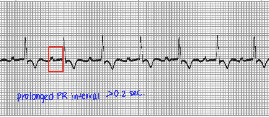
First degree AV block
Electrical impulse leaves the sinus node and travels normally to AV node where it is delayed longer than normal before conducted to the ventricles
prolonged QR interval (longer than 0.2 seconds)
Mildest form, typically requires no specific treatment
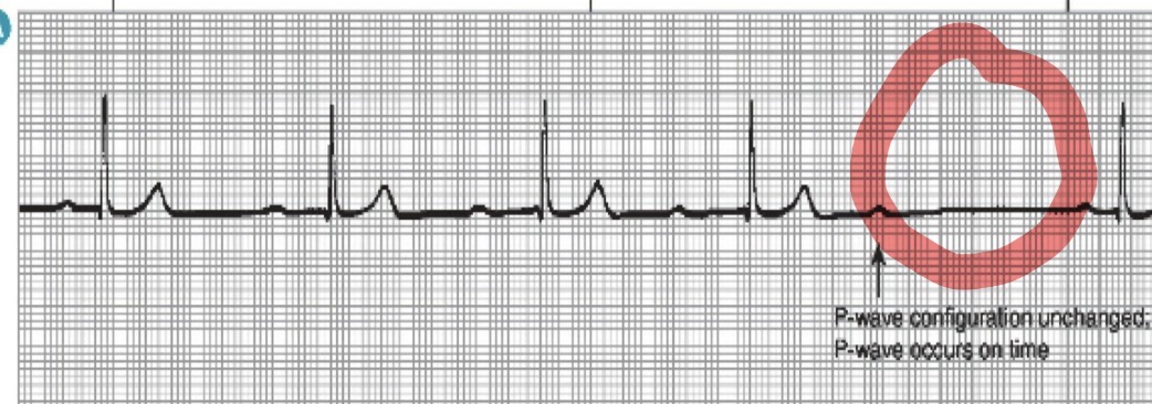
Second degree heart block Mobitz Type I (Wenckebach)
Initial impulse leaves the sinus nose and travels through the AV node, is conducted through the ventricles but each successive impulse has increasing difficulty passing through the AV node until finally an impulse does conduct to the ventricles
PR intervals longer and longer until dropped QRS
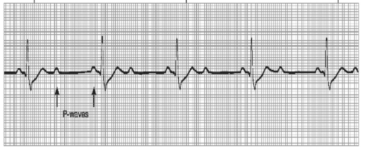
Second Degree Heart block Mobitz Type II
More than one P-wave to each QRS complex
PR is normal or prolonged but will be consistent
Can progress suddenly to 3rd degree
TX: PERMANENT PACING if it does not resolve
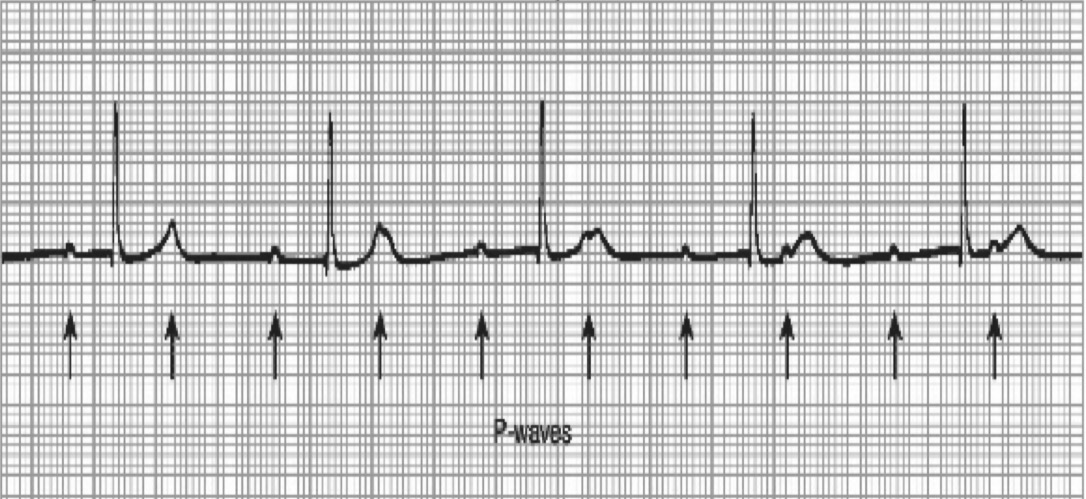
Third Degree Heart Block
Impulses fail from atria to ventricles, complete heart block, beat totally separate of eachother
TX: permanent pacemaker
Non-modifiable risk of HTN
Age
Family hx/genetics
Male sex
Modifiable risks of HTN
Obesity
Diet
High sodium
Smoking
Alcohol
Sleep apnea
Metabolic disorders
Emphasis on lifestyle factors (sodium, alcohol, stress, weight, heart healthy habits)
Essential/Primary HTN
HTN without an identifiable secondary cause
Secondary HTN
Due to a specific underlying disease
Hypertension measurements and risks
Goal: <130/80 mmHg
Normal: less than 120/80
Elevated 120-129 mmHg and <80 mmHg
Stage I Hypertension: 130-139 mmHg or 80-89 mmHg
Stage II: >140 mmHg or >90 mmHg
Risks: Coronary artery disease, heart failure, A-Fib, stroke, dementia, CKD, heart attack
Hypertension S/S
Often asymptomatic, if present can present as
Headache
Dizziness
Visual disturbances
Palpitations/chest discomfort
Nosebleeds
Hypertension Dx
Dx
PREVENTTM risk calculator to evaluate risk for heart attack, stroke, and heart failure
Ratio of urine albumin/creatine (recommended for all pts with HTN)
Screening for primary aldosteronism (plasma aldosterone/renin)→ secondary causes
Confirmed with 2+ readings on >2 visits and home BP monitoring
Hypertension Tx
1. First line: lifestyle modification
2. Medication- initiate if
Stage 1 HTN with CVD, CKD, diabetes, Prevent risk >7.5%
BP >130/80 mmHg after 3-6 mo of lifestyle change
Stage 2 HTN (>140/90 mmHg): start 2 drugs (single pill combo preferred)
ACE/ARB+CCB (MC)
Tx of HTN with medications (after lifestyle modification ineffective or other indication)
First Line Medications
Thiazide diuretics (HCTZ, chlorthalidione)- Mild HTN
DHP-CCBs (amlodipine, nifedipine)
ACE/ARBs (protects CKD)
Second Line Medications
Beta-blockers
Vasodilators (hydralazine)
Central Alpha agonists (Clonidine)
ACE in HTN
First line in pts with HTN with a comorbidity such as diabetes and chronic kidney disease (CKD)
ACE and ARB are kidney protective
CCB in HTN (DHPs)- amlodipine, nifedipine, nicardipine
Black patients with uncomplicated HTN (first line)
Could also use HCTZ which is a thiazide diuretic
Alpha blocker in HTN (prazosin terazosin, doxazosin)
HTN and benign prosthetic hyperplasia, smooth muscle relaxer (tx both conditions)
Tx hypertension and relaxes smooth muscle
Beta-Blocker for Pt with recent MI and HTN
Patients discharged for MI, all patients with recent MI, and also treats HTN (decreases O2 demand to the heart)
Thiazide diuretics in HTN
Tx mild, uncomplicated HTN
Decreases urinary Ca++ excretion (GOOD for use in kidney stones)
Hypertensive Urgency
Chronic uncontrolled HTN or medication nonadherence, slow vascular adaptation, NO ORGAN DAMAGE
>180 or >120 without organ damage
Risk: non-compliance with meds, abrupt withdrawal of B-blocker or clonidine, pain/anxiety/stress
Dx: BP measurement and evaluation for organ damage
Tx: Outpatient or observation unit
Usually always a PO med- ACE/ARB, CCB (DHP), BB
Med adherence, diet, and at home BP monitoring
Hypertensive emergency
Sudden rise in systemic vascular resistance, endothelial injury, fibrinoid necrosis of arterioles, ischemia, organ dysfunction
usually >180, >120 WITH ORGAN DAMAGE
Risks: med non-adherence, renal disease, drugs, post-op, aortic dissection
S/S: organ damage, headaches, confusion, HF, aortic dissection, acute kidney injury, retinal hemorrhages, stroke symptoms
DX: confirm BP, Labs, Imaging (for signs of organ damage)
TX: Need ICU and continuous monitoring
IV Meds (sodium nitroprusside, B-Blocker, CCB), NO ACE
Cardiovascular disease, heart disease, coronary heart disease
1. Cardiovascular disease- The Big Umbrella: all types of diseases that affect the heart or blood vessels
Coronary Heart Disease: CAD and other conditions that affect coronary arteries like spasms and inflammation
Coronary Artery Disease: Narrowing or blocking of coronary arteries caused by build-up of plaque, MC coronary heart disease and presents a MI
Coronary heart disease
Risks:
Nonmodifiable: age (Men >45, women >55), family hx, Males, Race (Hispanic, south asain, Indian)
Modifiable: Hyperlipidemia (hypercholesterolemia), HTN, smoking, DM, obesity, sedentary, diet/alc, psychosocial, METABOLIC SYNDROME
S/S: Always in a state of inflammation (high-sensitivity CRP inflammatory marker)
Atherosclerosis: CHD results from endothelial cell dysfunction, dyslipidemia and inflammation
1. Stage 1: endothelial dys leads to formation of a fatty streak
2. Stage 2: LDL enters the intima and becomes oxidized, attracts macrophages
3. Stage 3: plaque matures to fibrous cap, vessel of lumen narrows
Plaques can remain stable or progressive or narrowing of arteries or can rupture
Risks for rupture: high lipid content, high concentration of macropahage, thin fibrous cap (signs of young, immature plaques)
Can be asymptomatic; angina, arrhythmia, MI, possible sudden death
Myocardial ischemia pain
Dull, achin pressure, heaviness, tightness, squeezing
Location: retrosternal or left precordial
Referred: jaw, shoulder, arm, upper abdomen, back
Ischemic S/S first felt with exercise and stress, progresses to at rest and unstable angina
S/S: SOB, dizziness, nausea, diaphoresis, doom
NOT reproduced with palpation, DEPRESSION in women, 1/3 will have no chest pain