HSC 214: taste and smell, eye, ear
1/77
There's no tags or description
Looks like no tags are added yet.
Name | Mastery | Learn | Test | Matching | Spaced | Call with Kai |
|---|
No analytics yet
Send a link to your students to track their progress
78 Terms
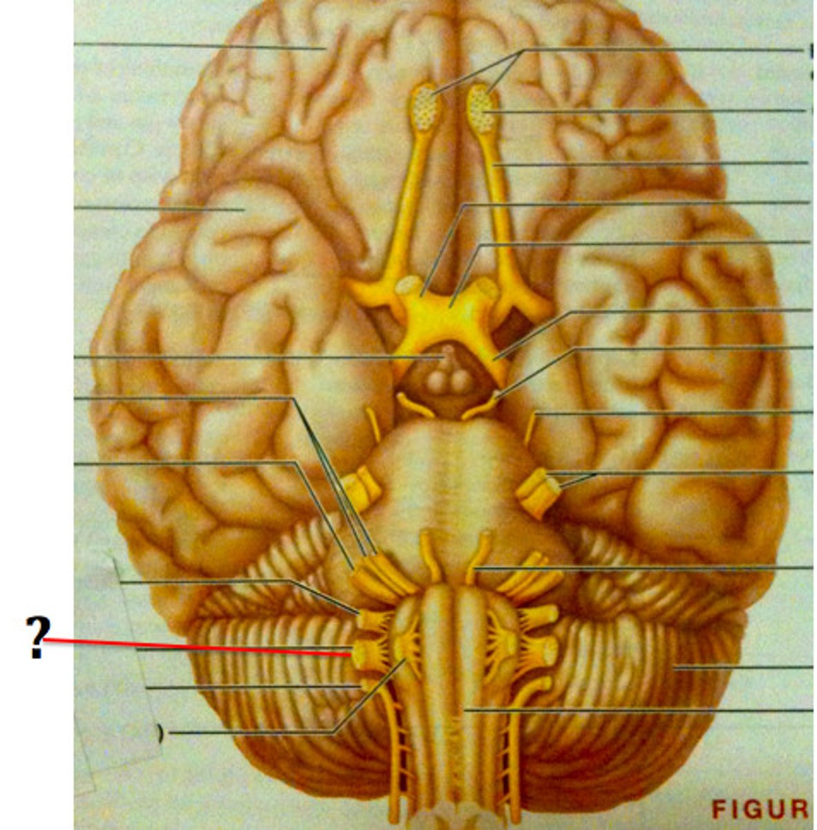
glossopharyngeal nerve
cranial nerve arising from the medulla oblongata translating the special sense of taste, posterior third of tongue
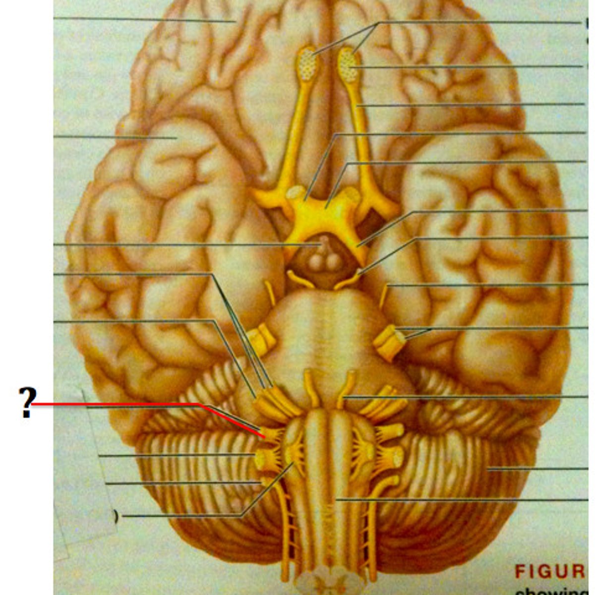
facial nerve
cranial nerve arising from the medulla oblongata translating the special sense of taste and motor innervation to the face, anterior two thirds of tongue
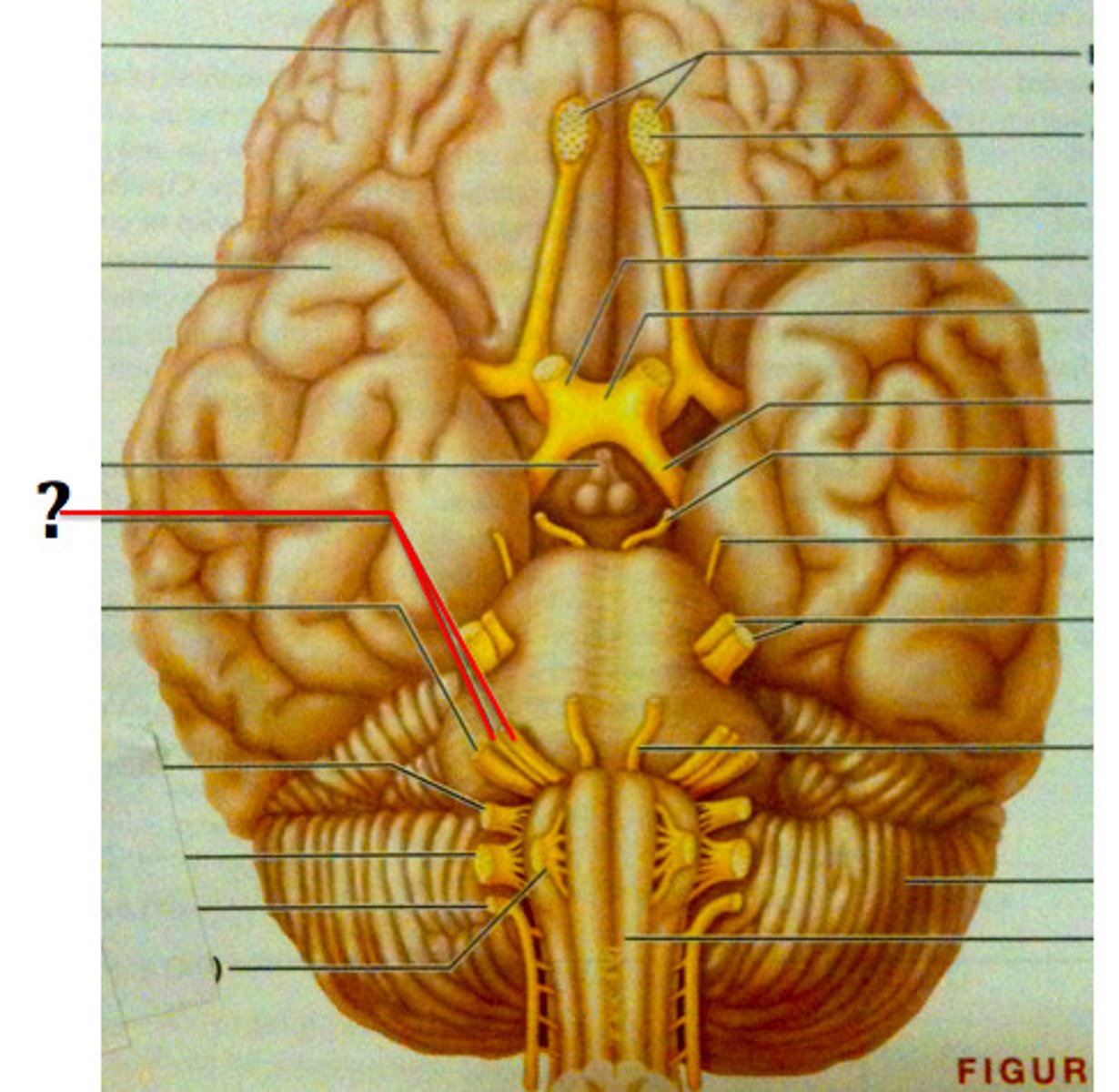
vagus nerve
cranial nerve arising from the medulla oblongata translating the special sense of taste and motor innervation to the parotid gland, to the epiglottis
olfactory nerve
cranial nerve arising from the olfactory bulb translating the special sense of smell, nasal cavity
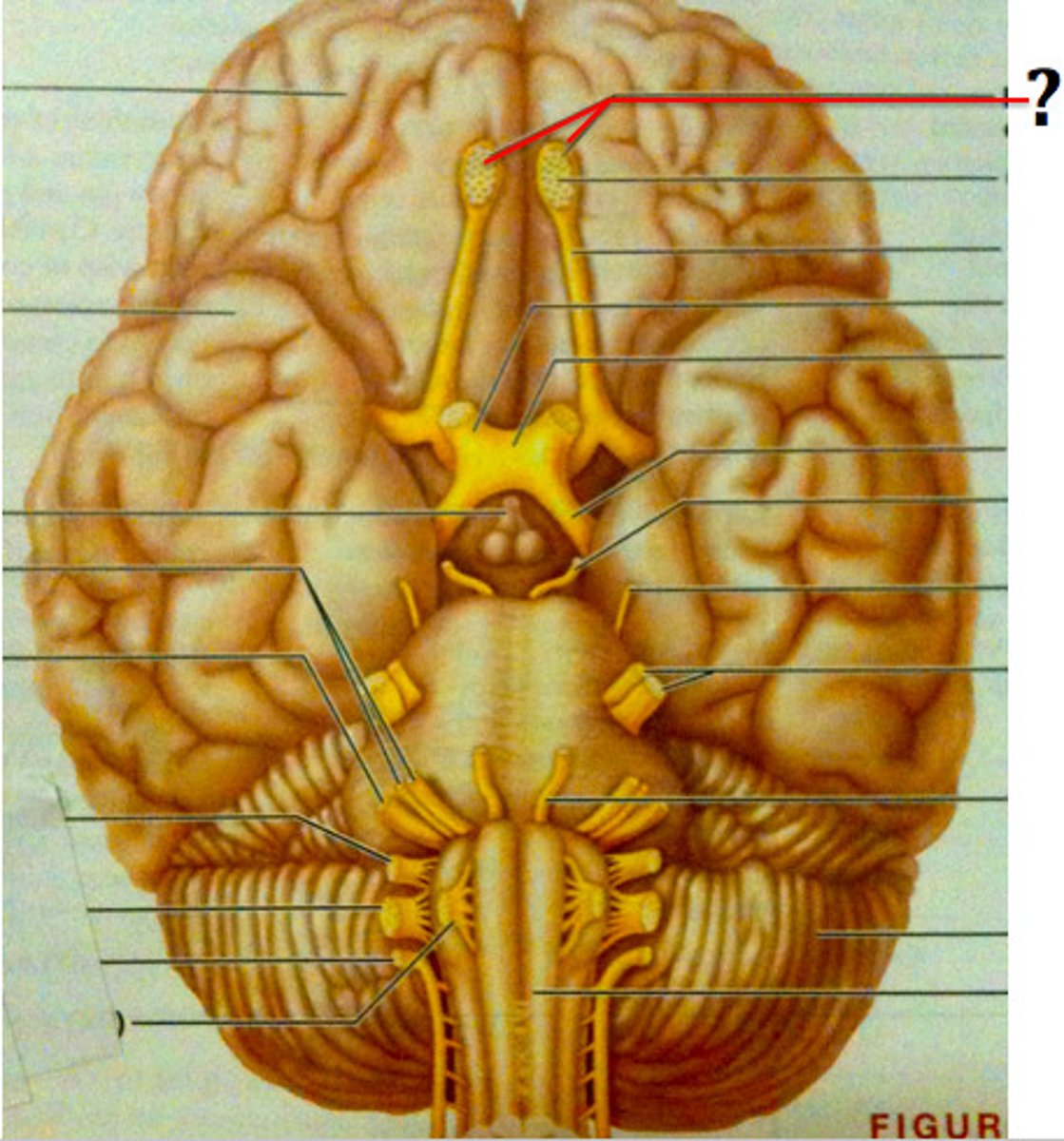
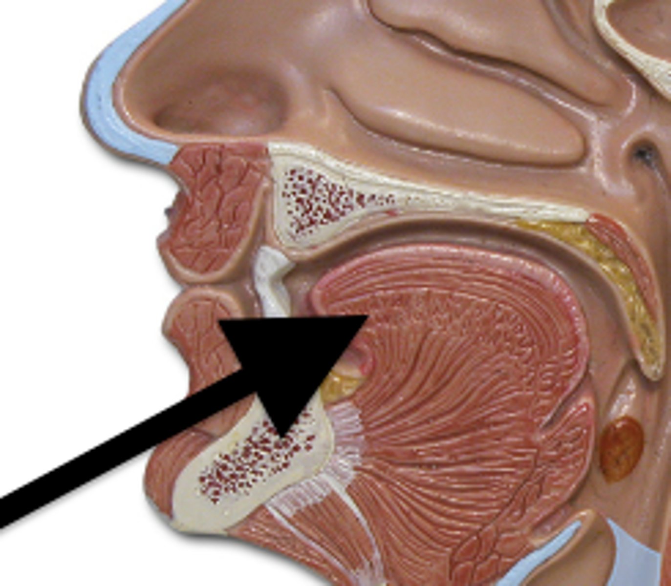
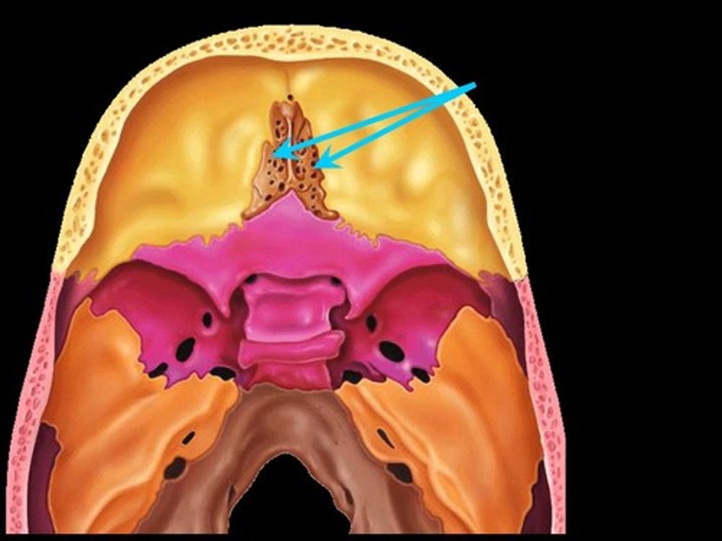
olfactory bulb
enlarged bundle of nervous tissue sitting in the cribriform plate
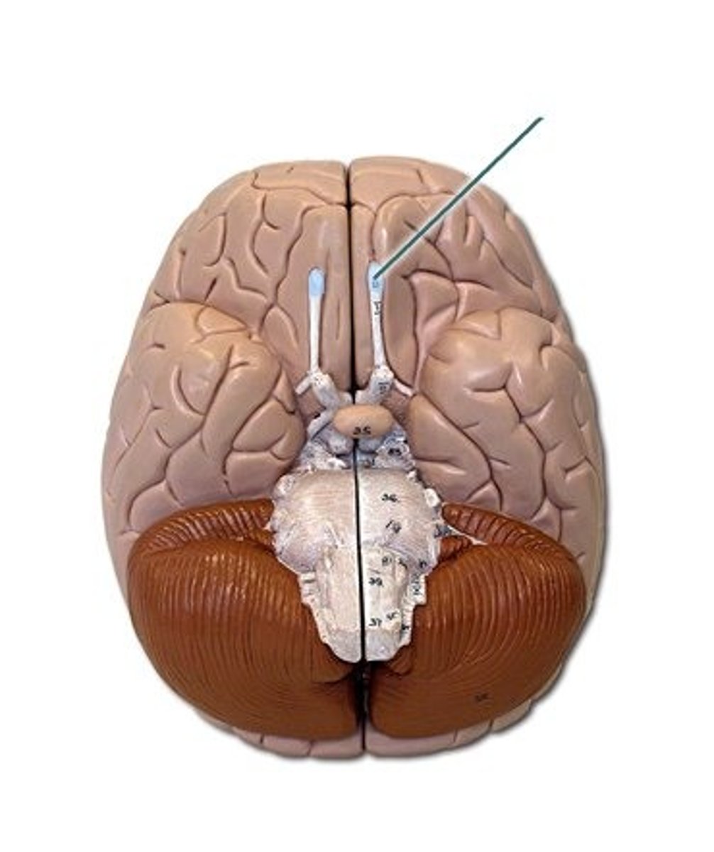
olfactory tract
bundles of axons from the olfactory bulb to the temporal lobe

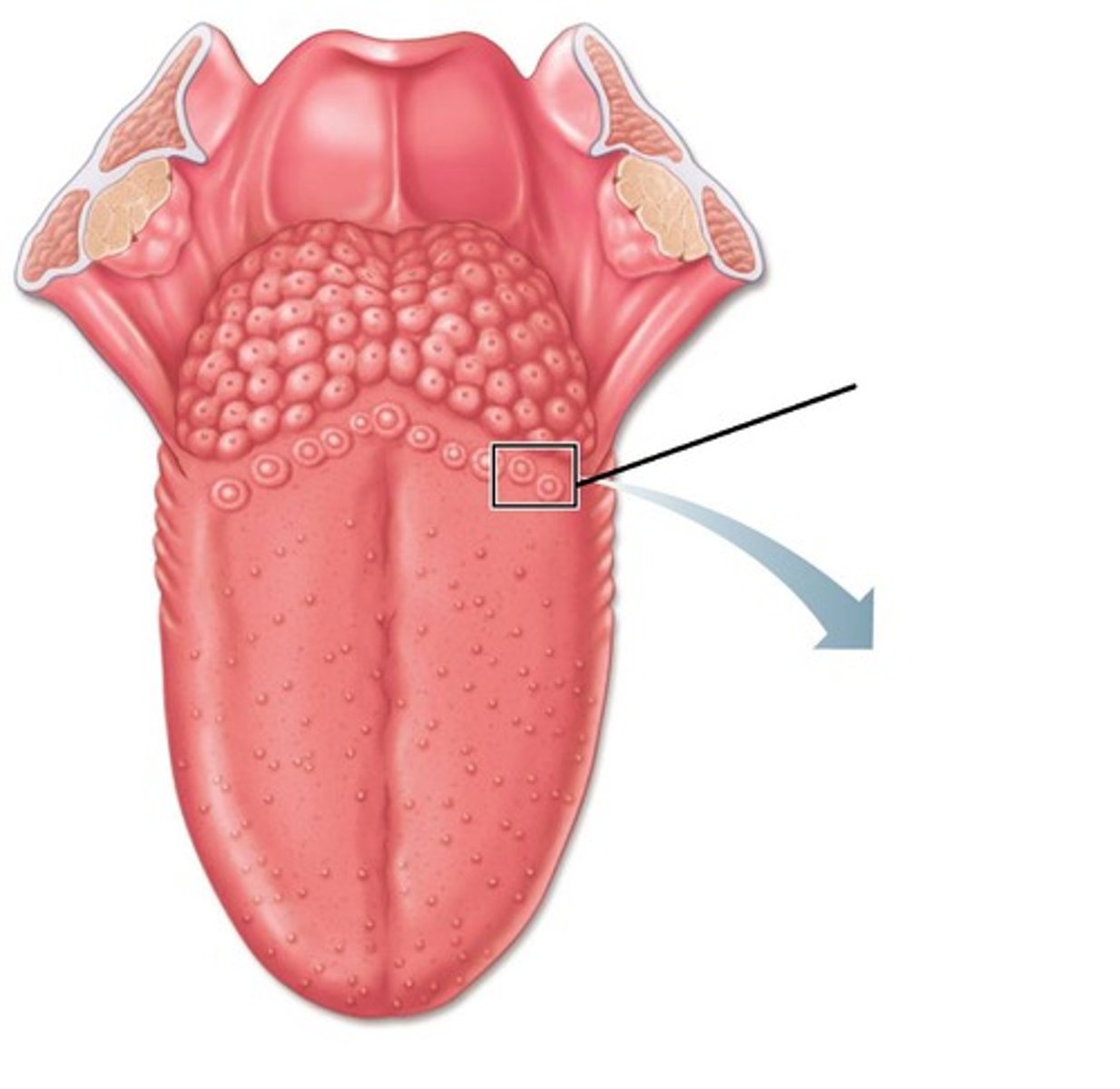
tongue
muscular projection anchored to the floor of the mouth
vallate (circumvallate) papilla
enlarged v shaped bumps on posterior surface of tongue
medulla oblongata
round most inferior portion of the brainstem located between the pons and spinal cord
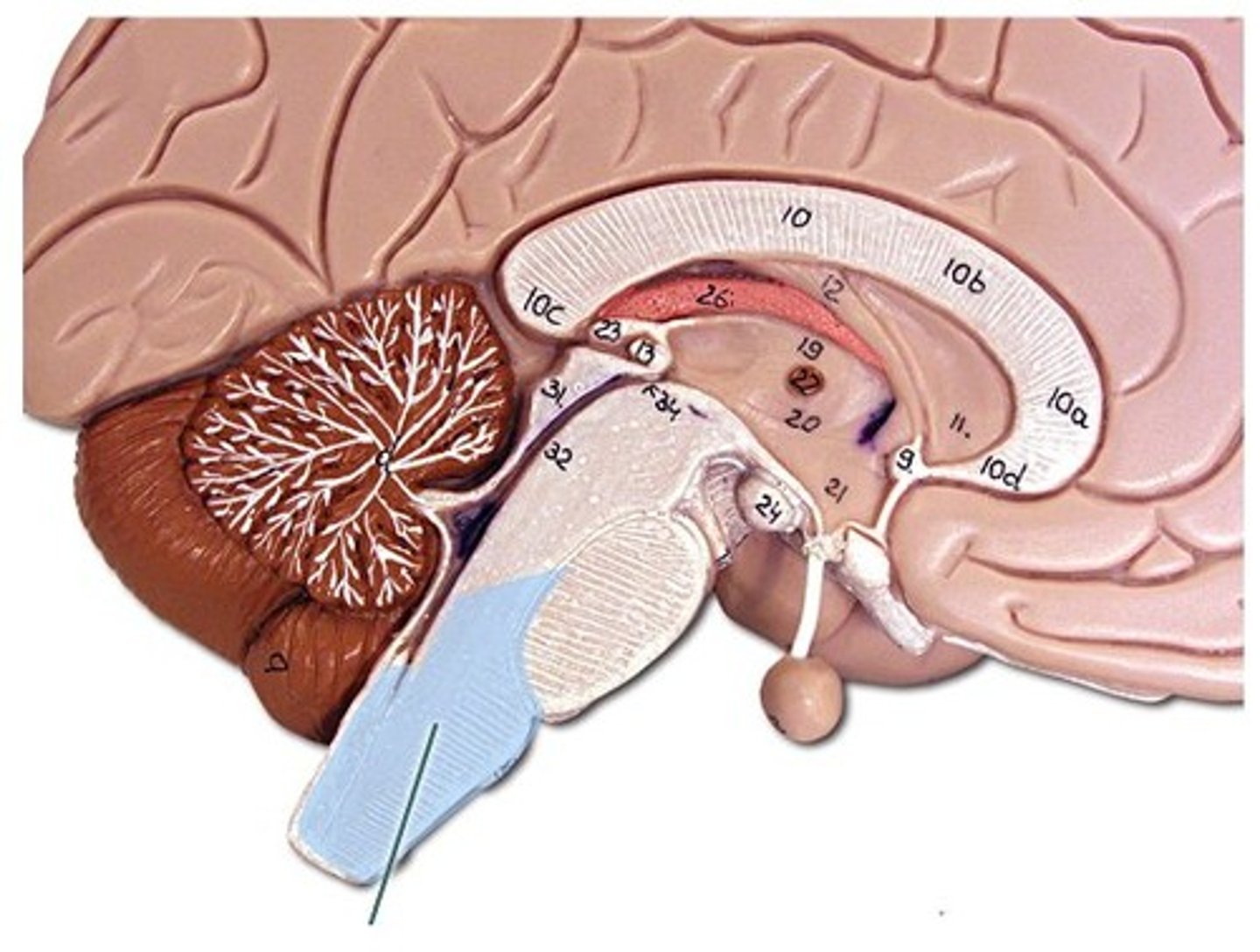
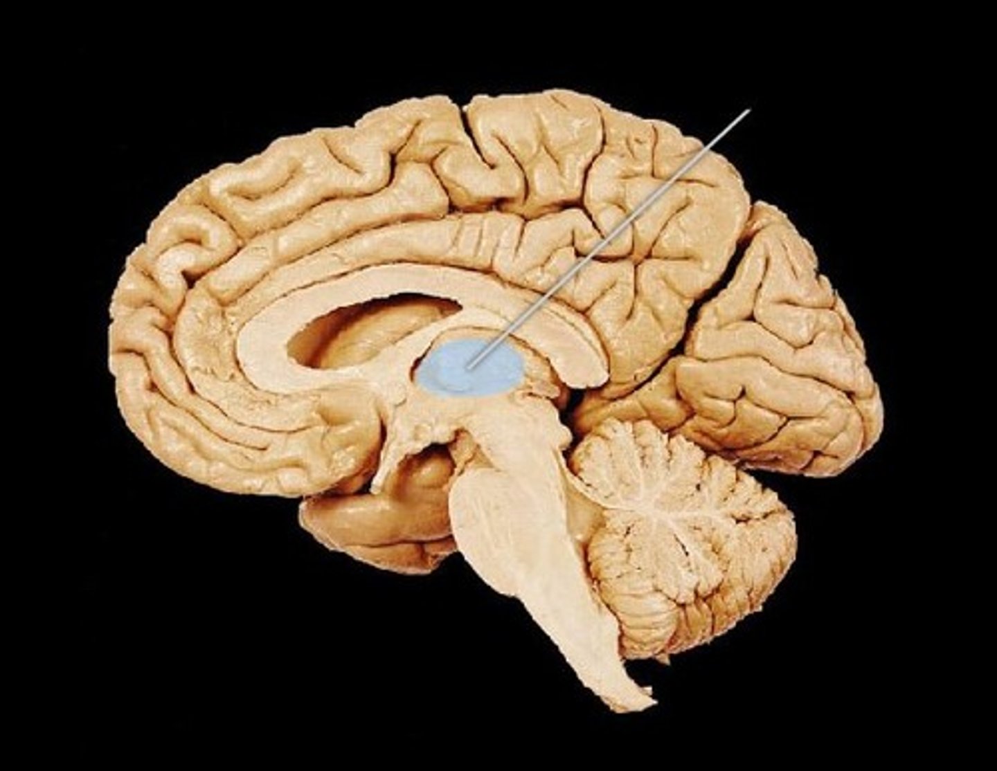
thalamus
in diencephalon region of the brain, separated by the third ventricle
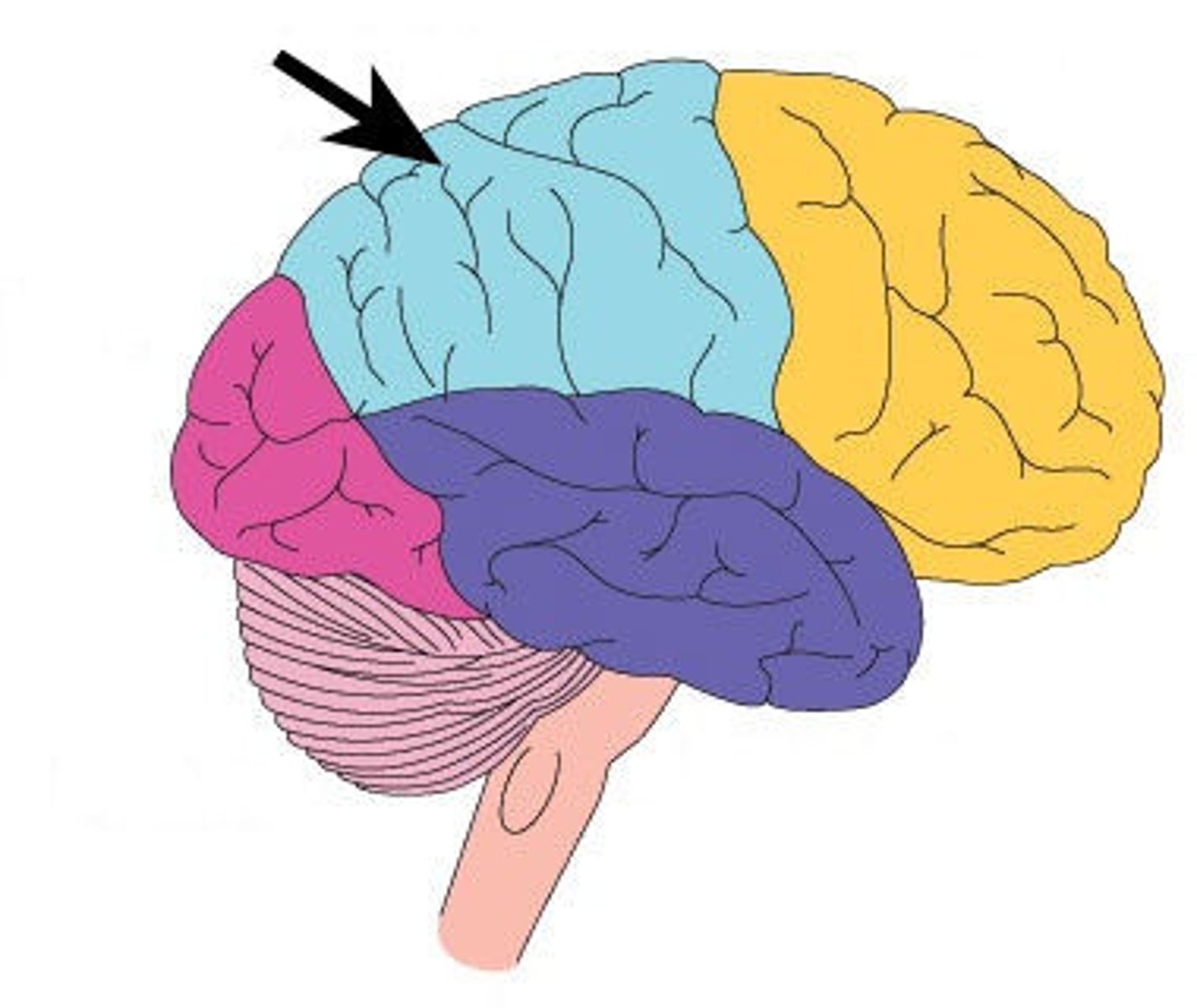
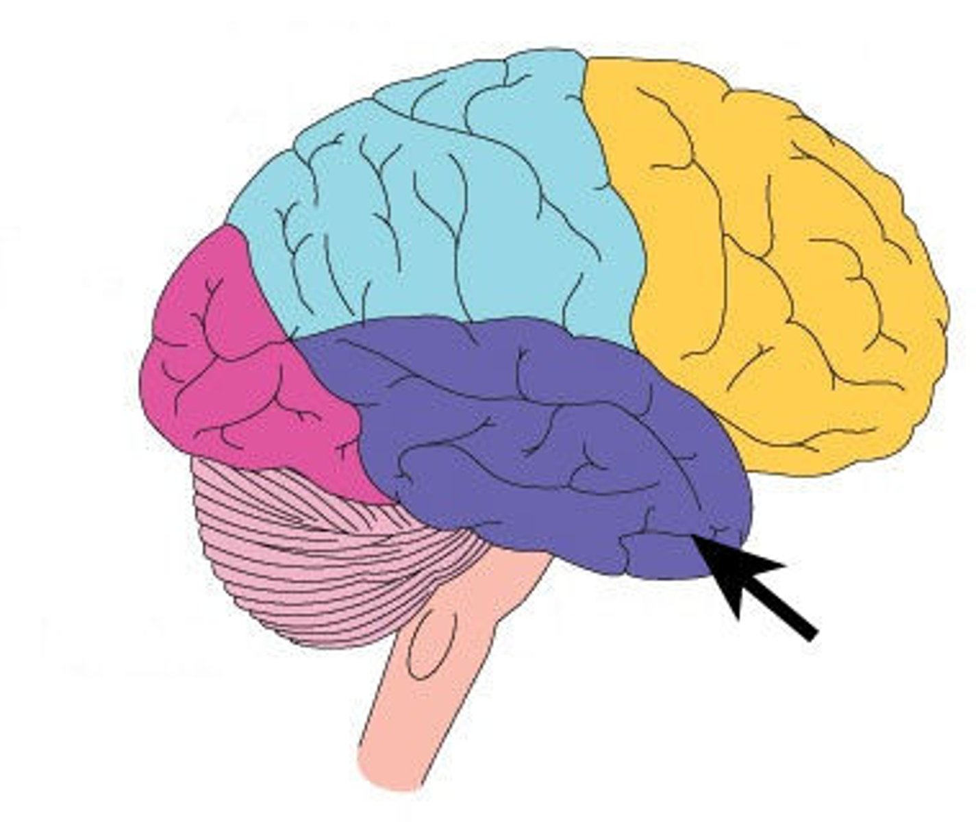
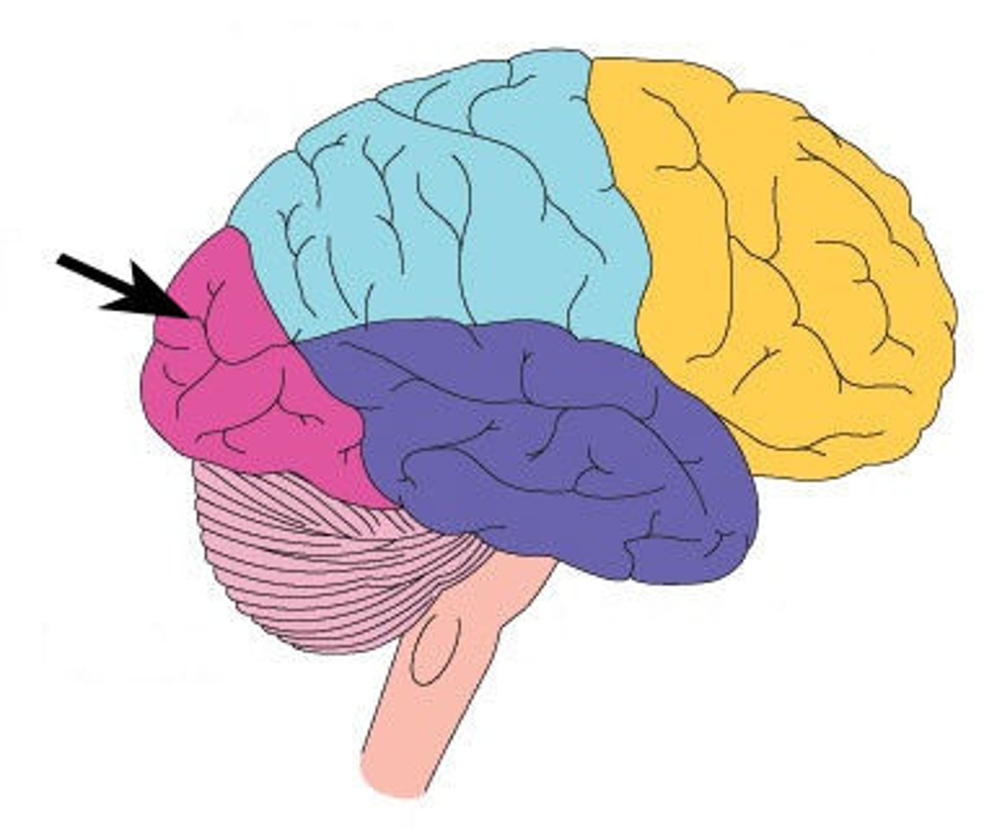
parietal lobe
lateral surfaces of cerebral hemispheres tat articulate with the parietal bones
olfactory foramina
microscopic spaces in the cribriform plate
temporal lobe
lateral and inferior surfaces of the cerebral hemispheres that articulate with temporal bone
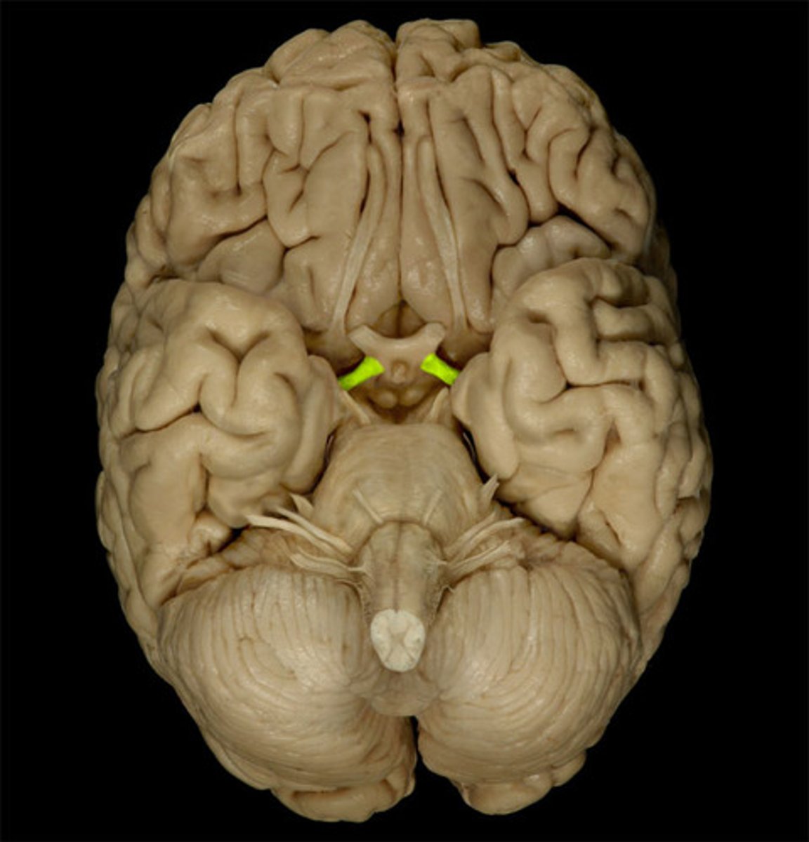
optic nerve (CNII)
cranial nerve from the optic chiasm
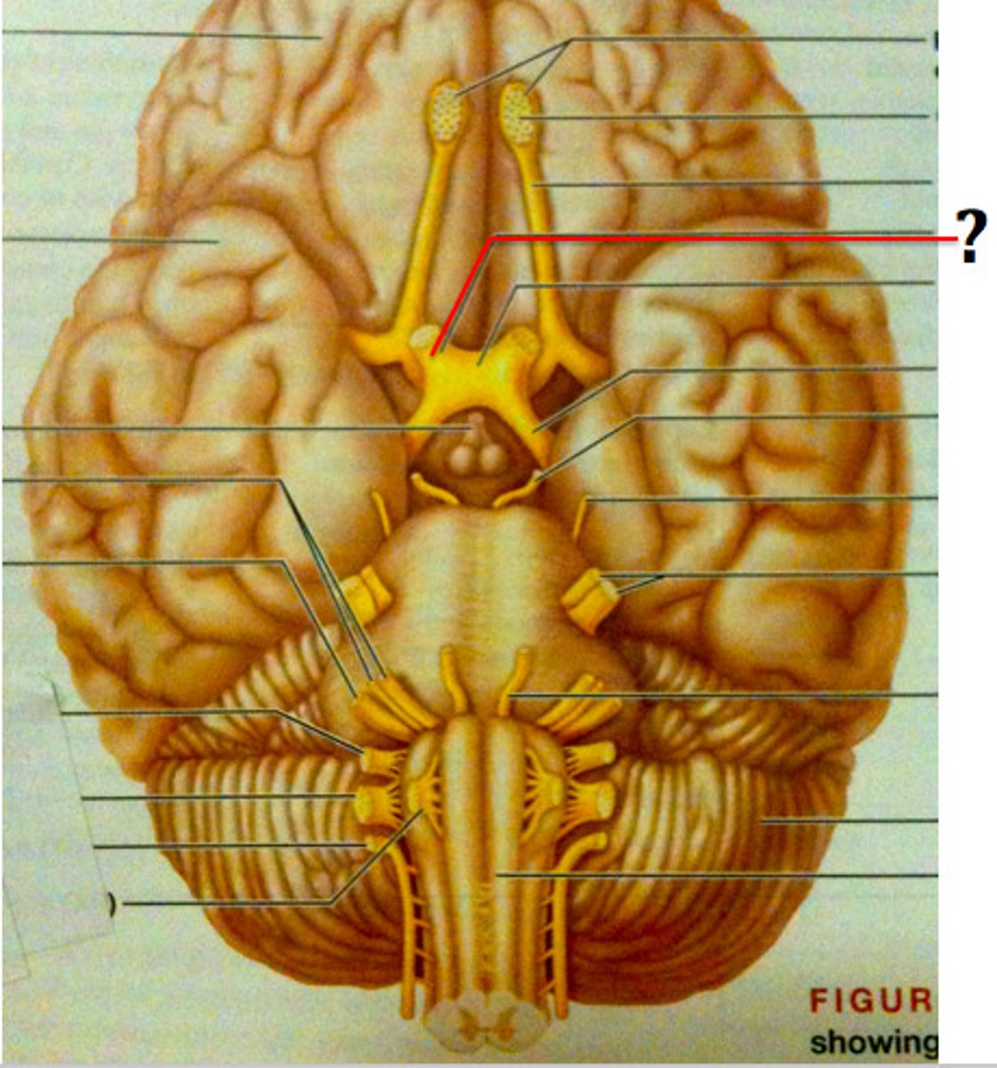
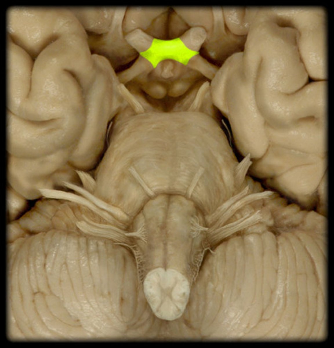
optic chiasm
expanded area between optic nerve and tracts that covers the sells turcica
optic tract
bundle of axons extending from the optic chasm to the occipital lobe, sensory impulses are interpreted
occipital lobe
posterior and inferior surface of the cerebral hemisphere that articulate with the occipital bone
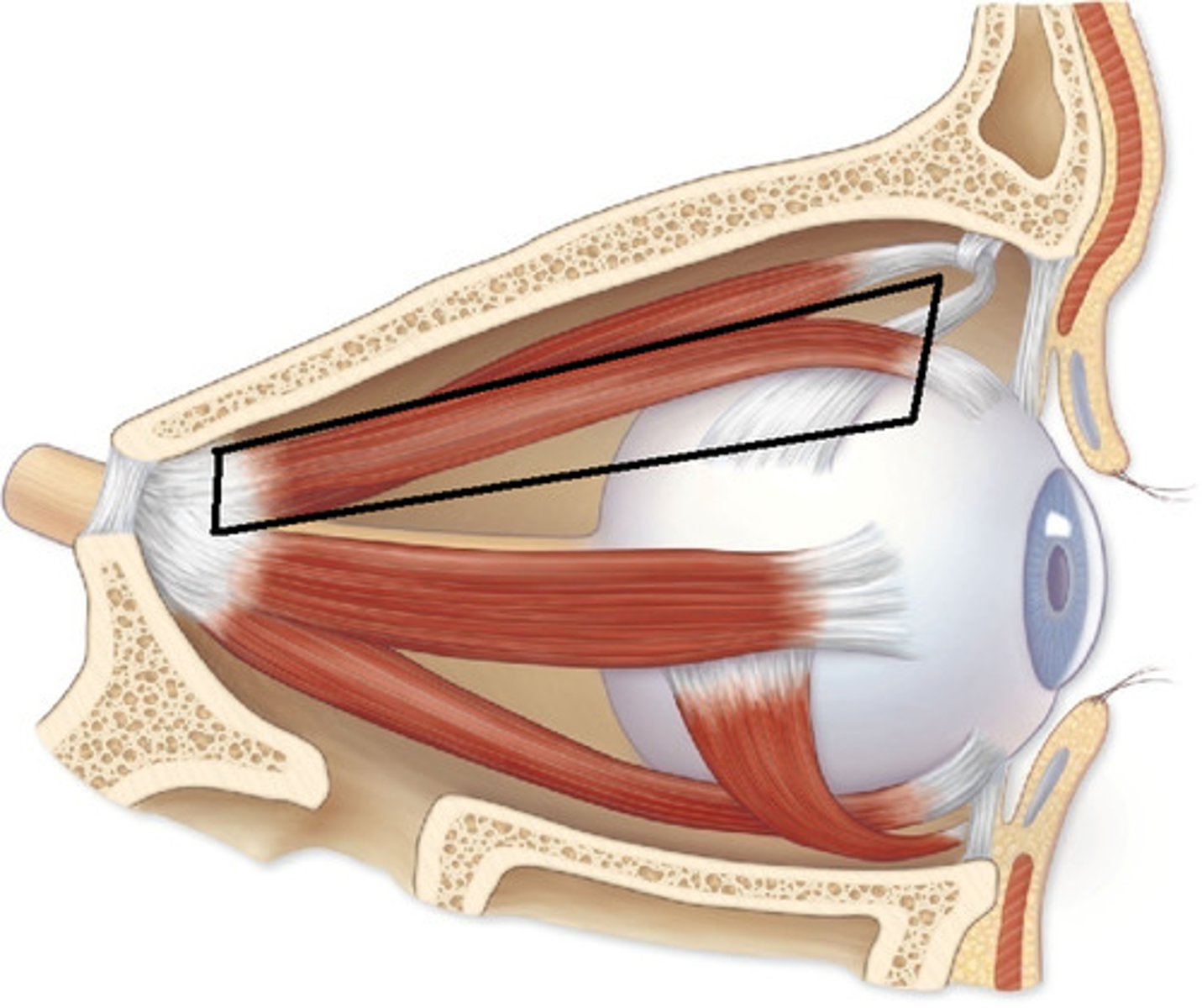
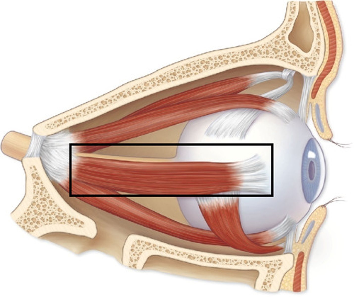
superior rectus
thin muscle attaching to the midline superior surface of the eyeball
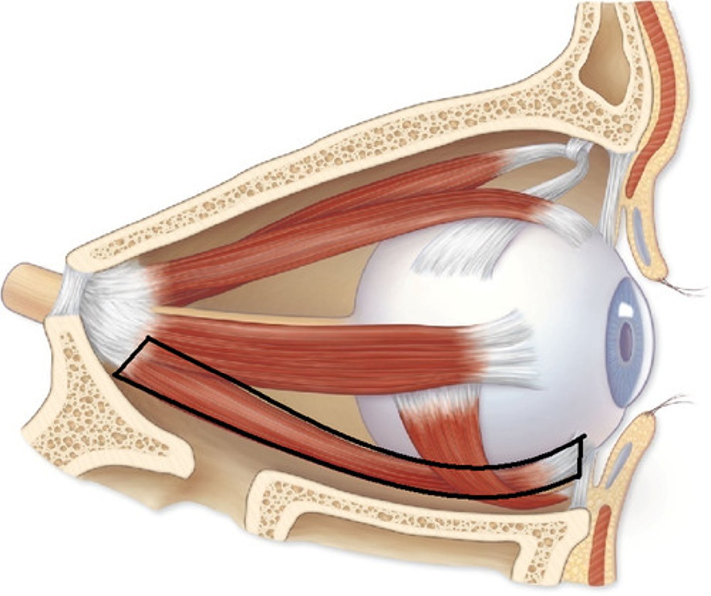
inferior rectus
thin muscle attaching to the midline inferior surface of the eyeball
medial rectus
thin muscle attaching to the medial surface of eyeball
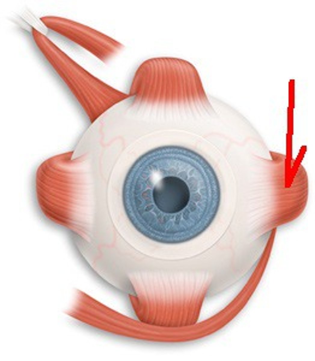
lateral rectus
thin muscle attaching to the lateral surface of the eyeball
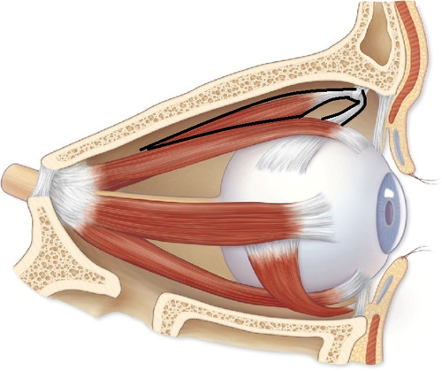
superior oblique
thin muscle attaching to the medial, superior surface of the eyeball
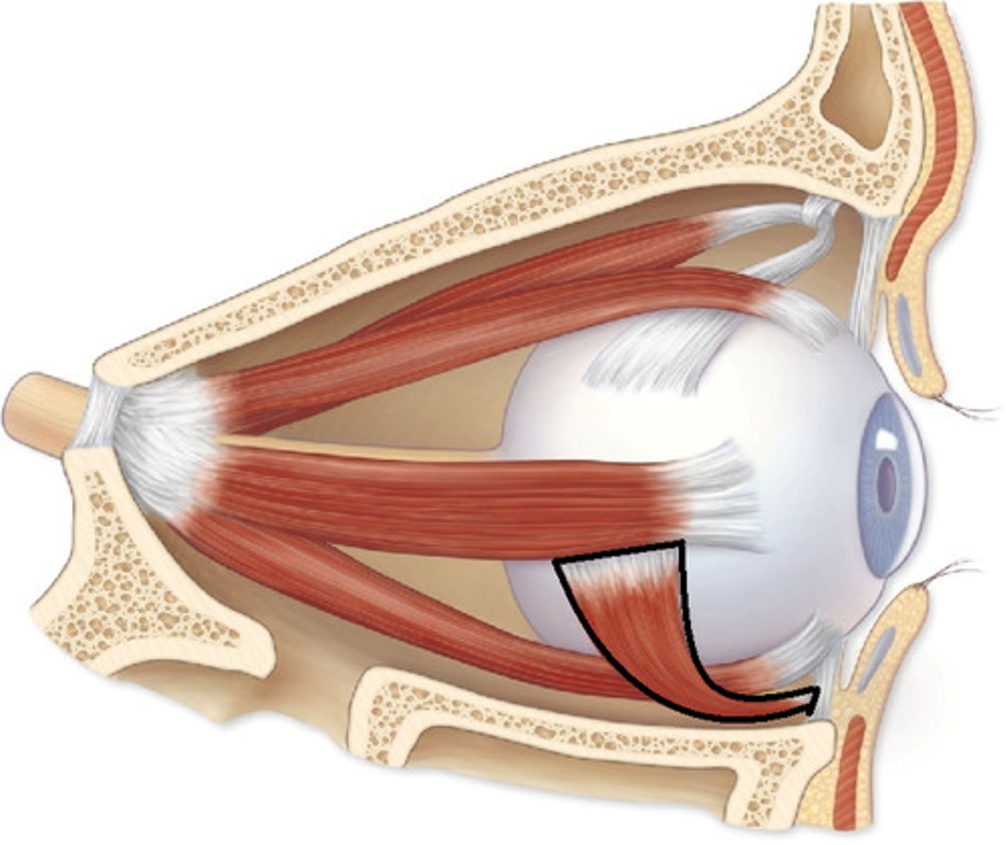
inferior oblique
thin muscle attaching to the lateral inferior surface of the eyeball
eyelids
palpebral conjunctiva
thin membrane coating the inside of the eyelid
ocular (bulbar) conjunctiva
thin membrane covering the sclera
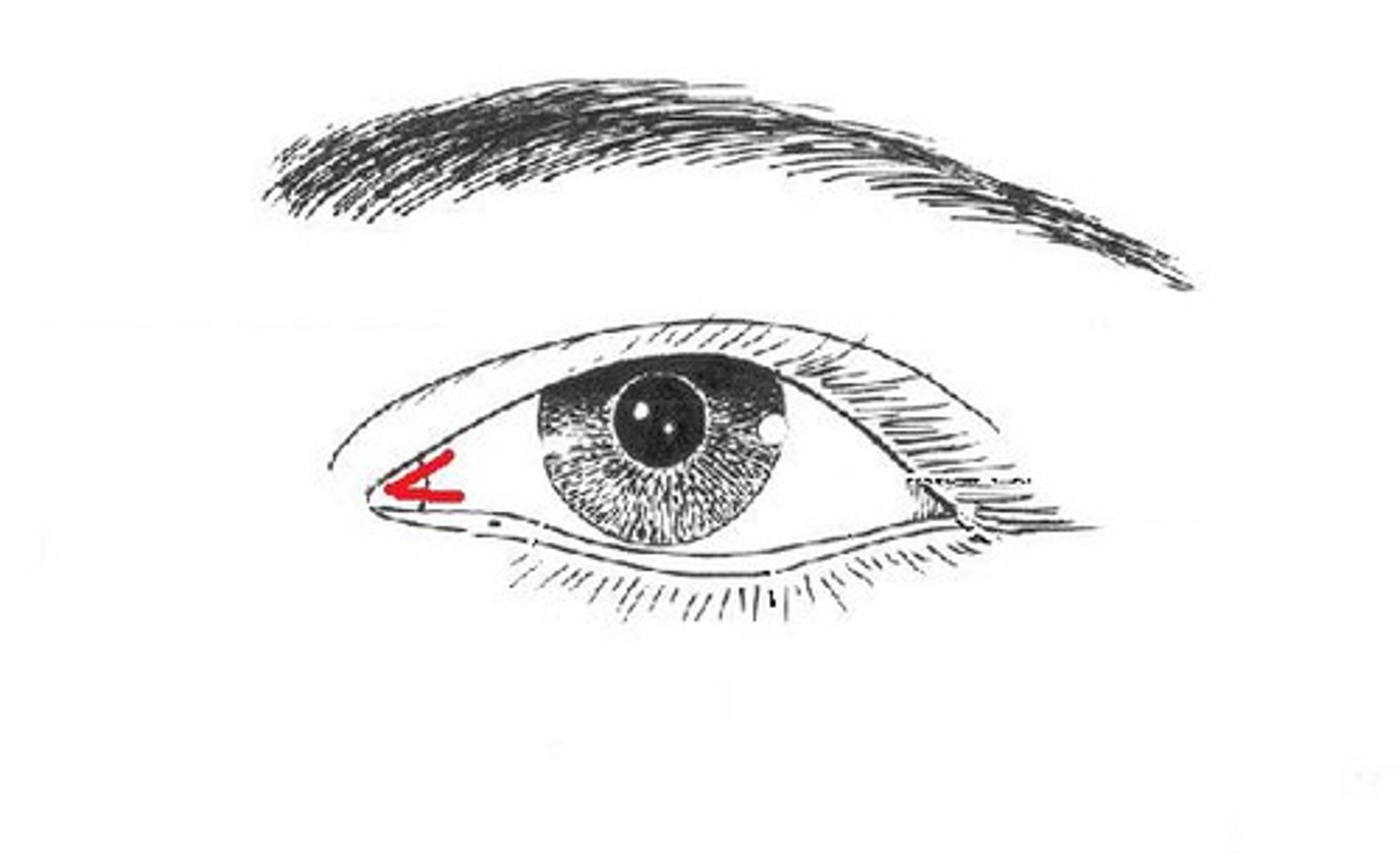
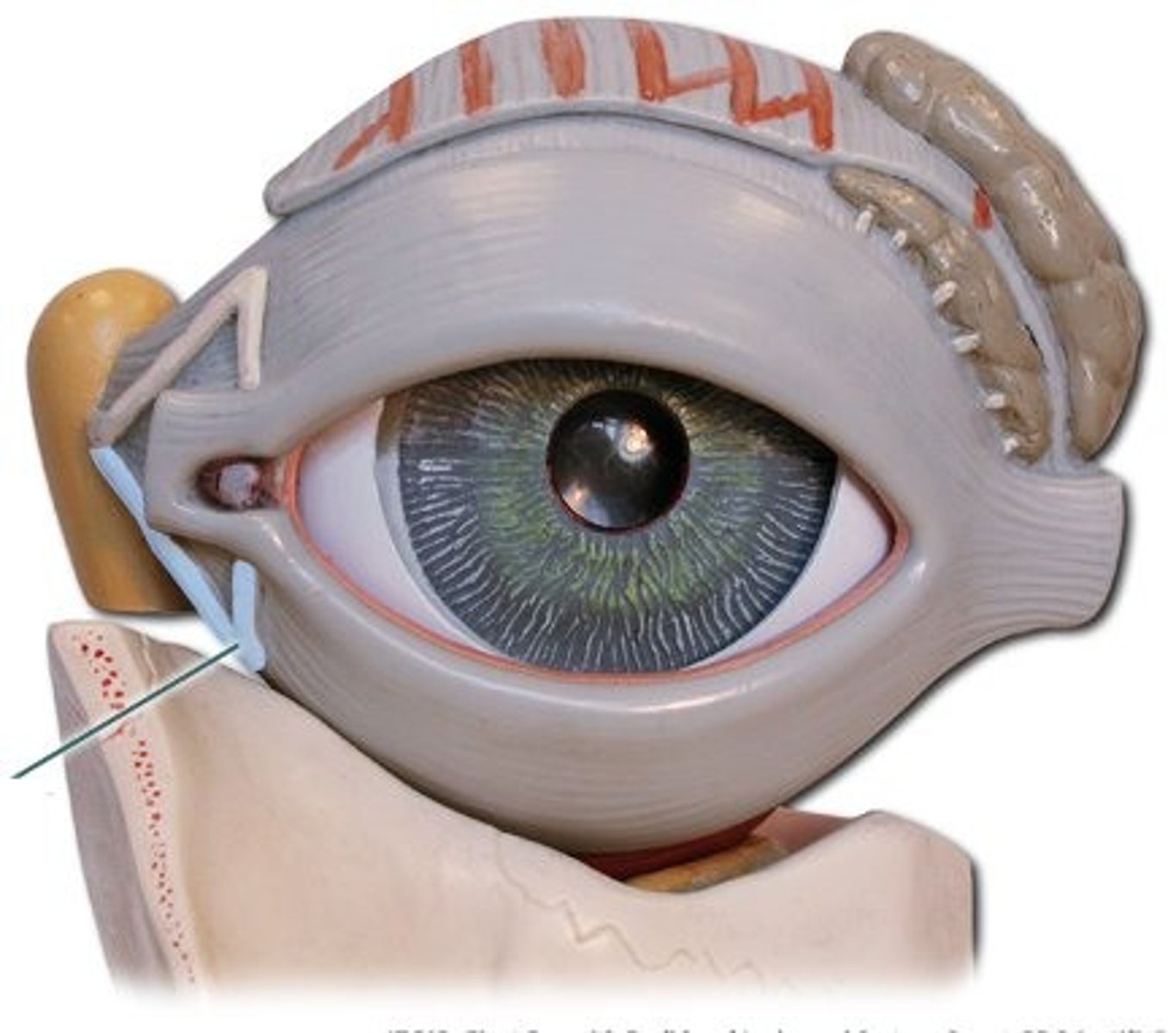
medial canthus
medial point (closest to the nose) where eyelids meet
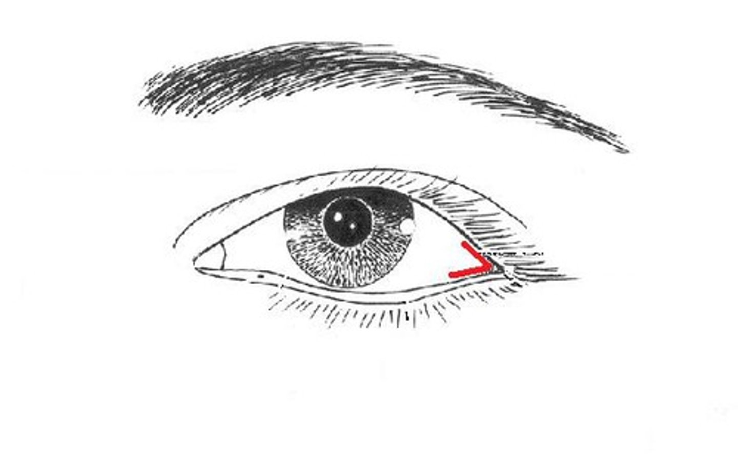
lateral canthus
lateral point where eyelids meet
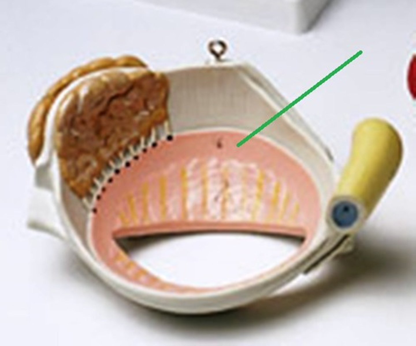
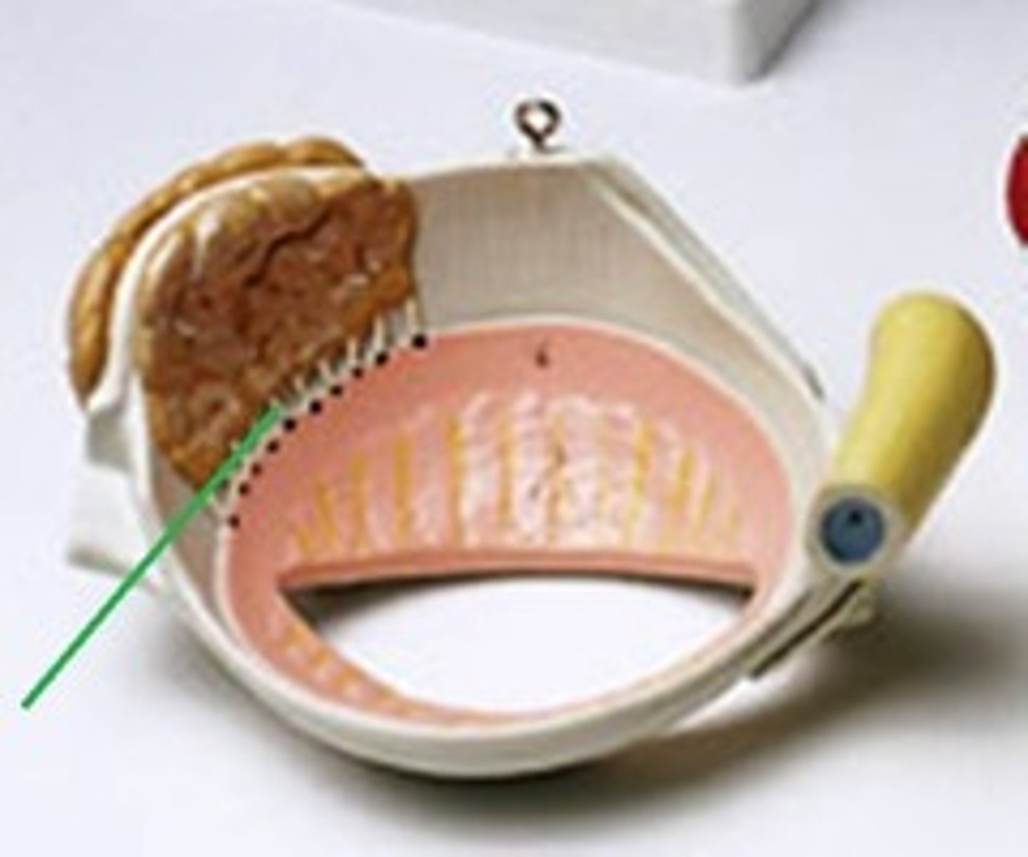
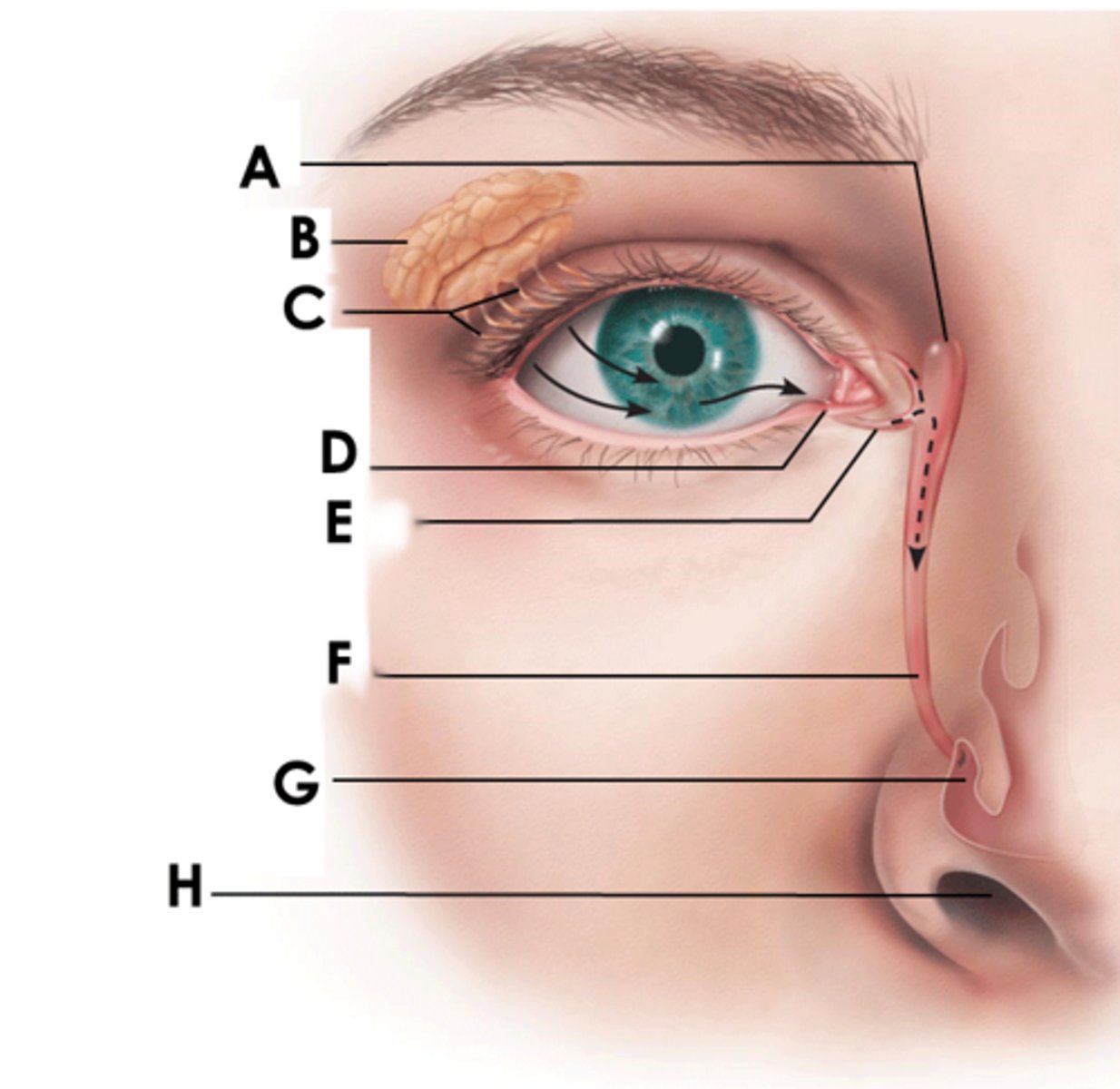
lacrimal gland
almond shaped gland in the superolateral orbit

lacrimal gland ducts
ducts connecting the gland to the surface of the eye
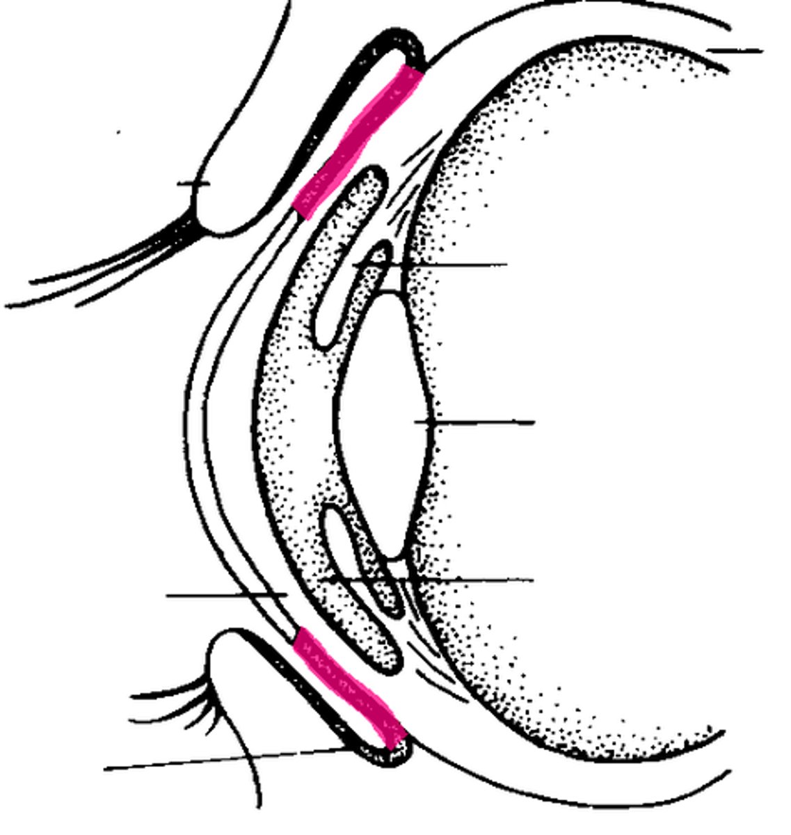
lacrimal punctum
two small holes in the medial canthus
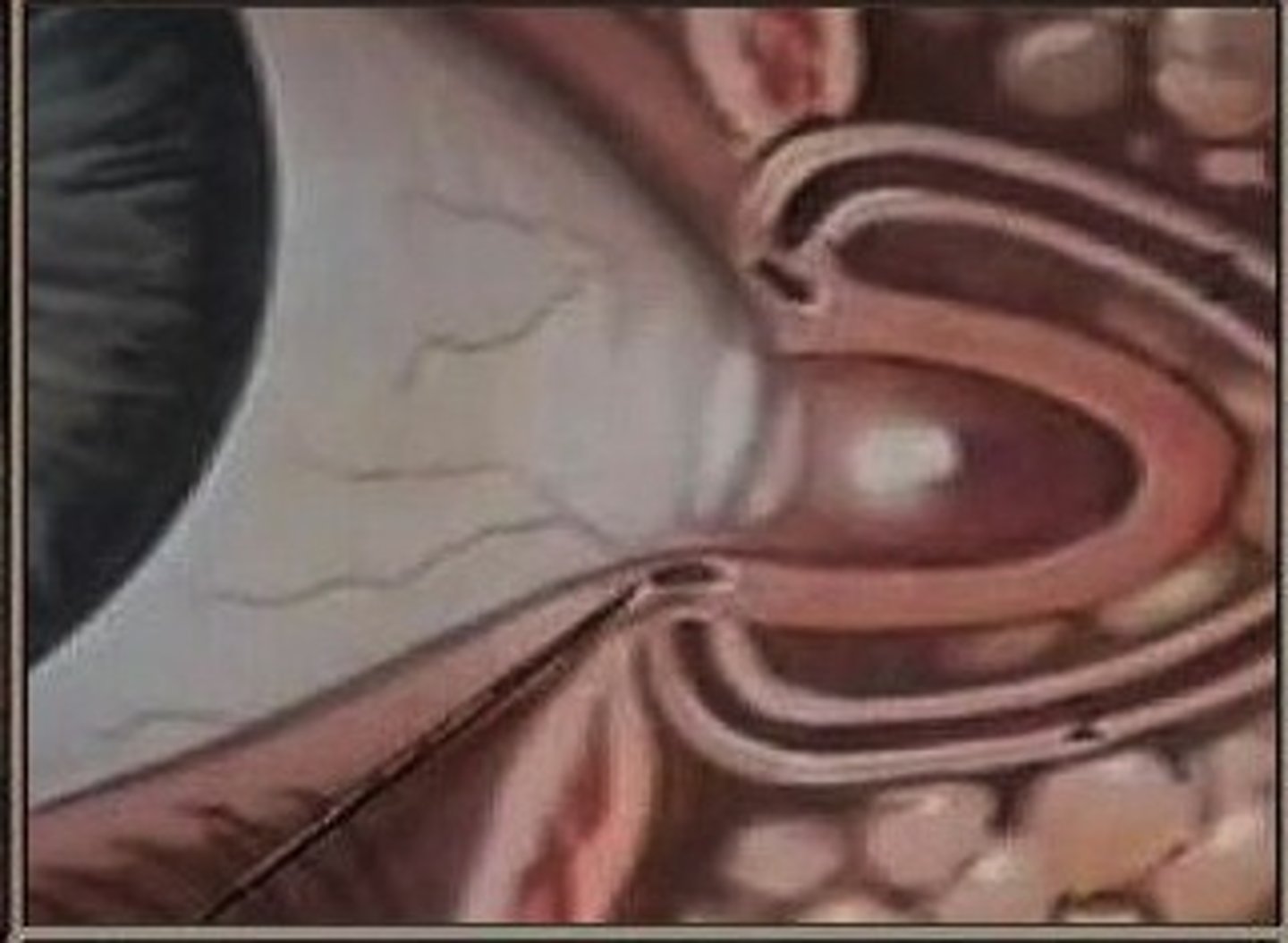
superior lacrimal canaliculus
thin superior duct draining the lacrimal punctum to the nasolacrimal duct

inferior lacrimal canaliculus
thin inferior duct draining the lacrimal punctum to the nasolacrimal duct
nasolacrimal duct
duct in medial nose where the canaliculus drain (F)
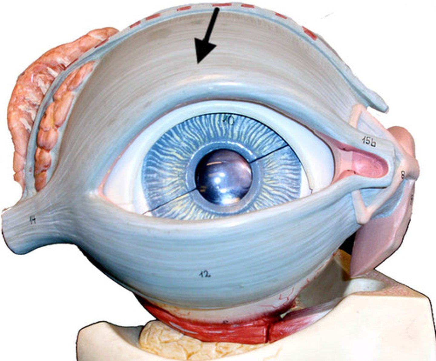
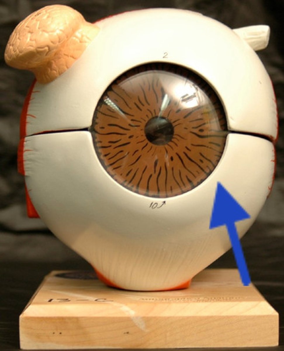
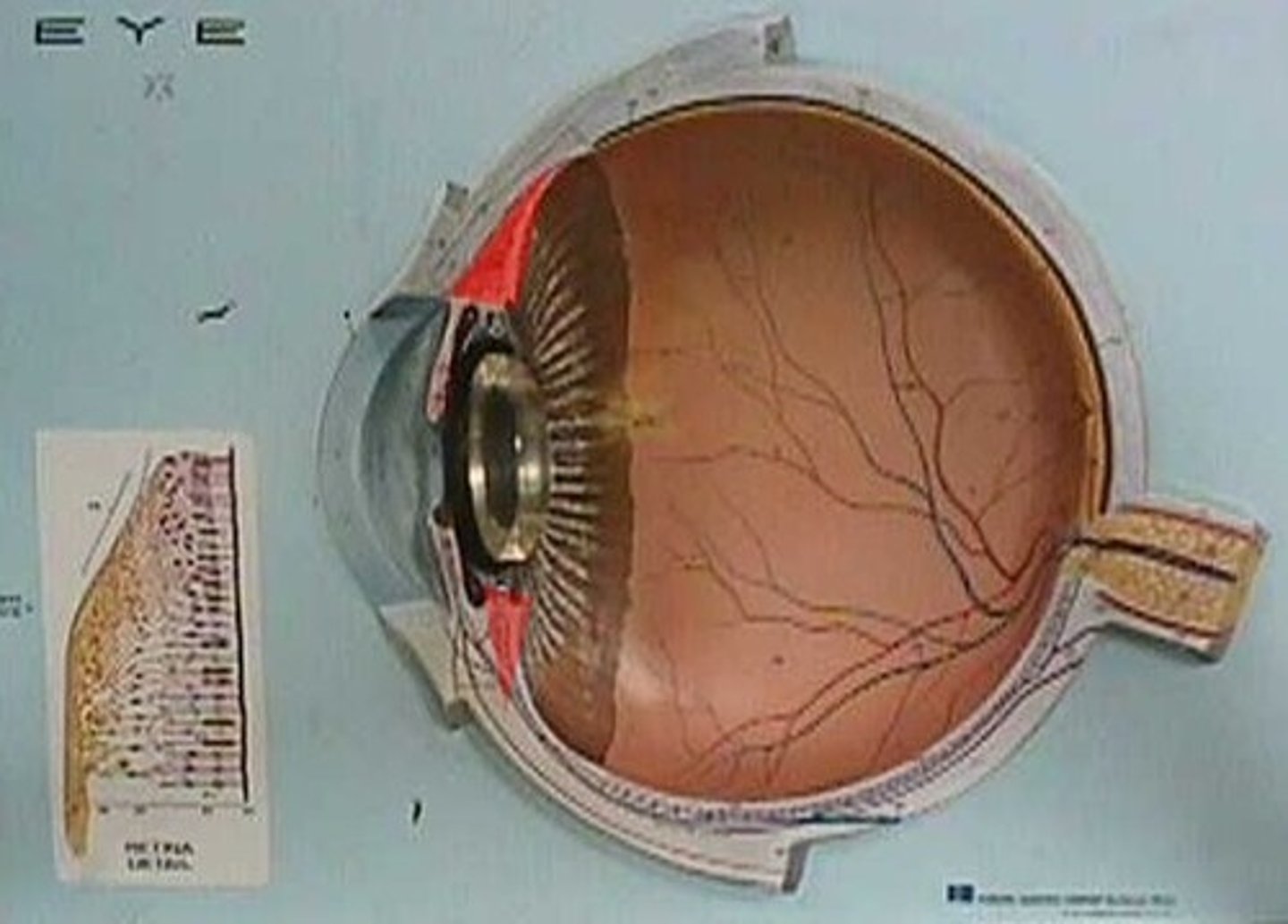
sclera
white fibrous outermost layer
choroid
middle vascular layer, contains blood supply to eye
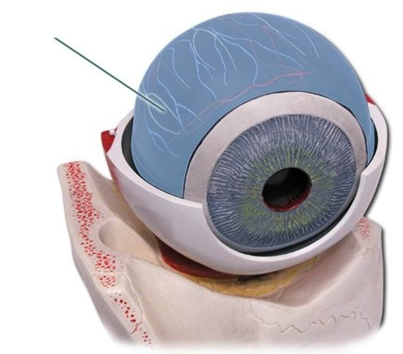
retina
brown neural innermost layer, contains rods and cones
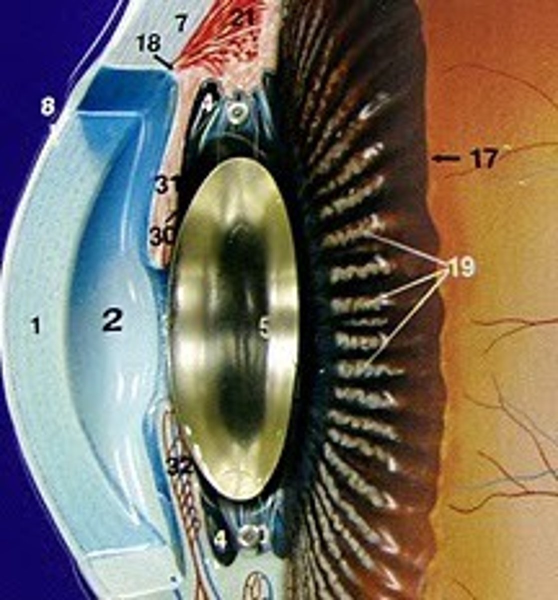
cornea
clear dome shaped covering of the eye, protects iris and pupil

anterior cavity
space between the cornea and lens, decided into two chambers
aqueous humor
fluid secreted by the posterior chamber that fills the anterior cavity
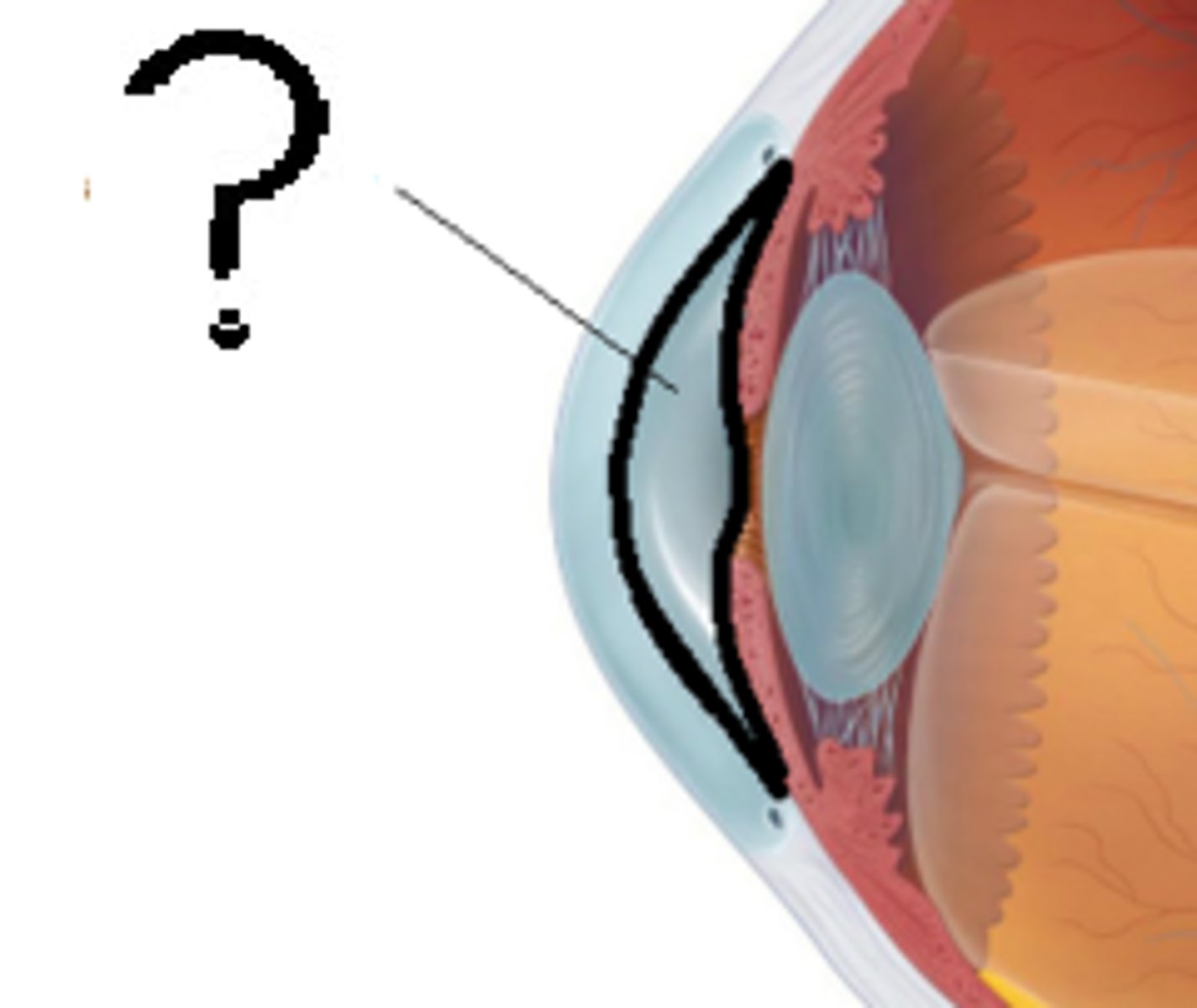
anterior chamber of the anterior cavity
the space between the cornea and iris (2)
posterior chamber of anterior cavity
the space bw the iris and lens (4)

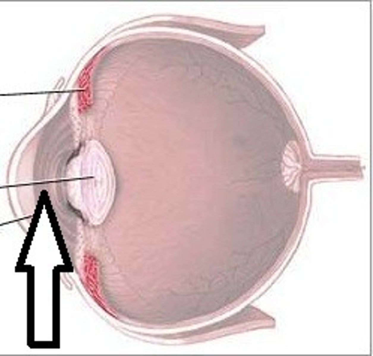
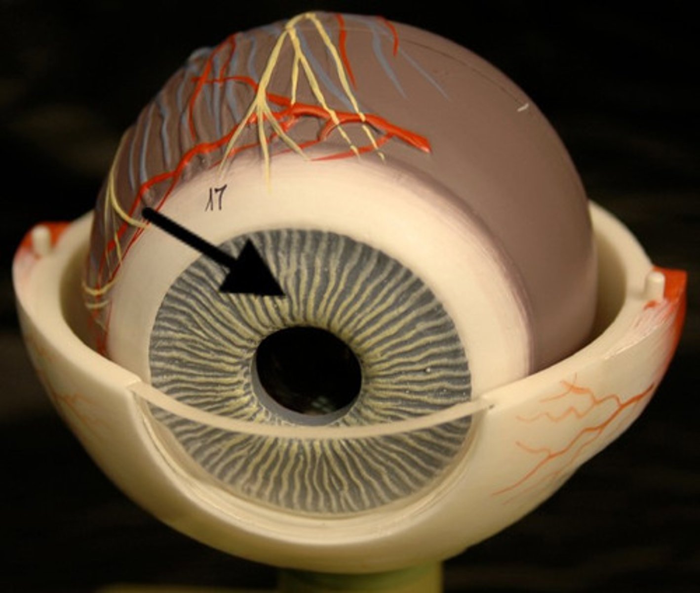
iris
ring of usually colored tissue behind the cornea
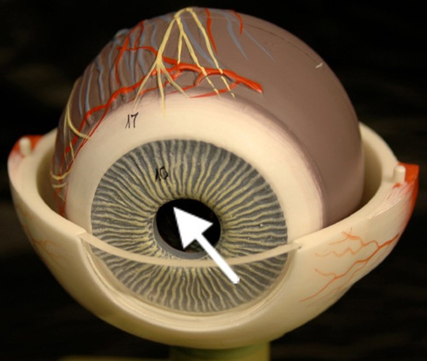
pupil
space in the center of iris, where light enters
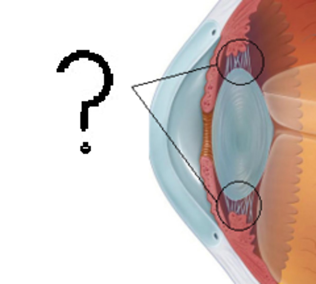
lens
transparent biconvex elastic disc separating the anterior and posterior cavities
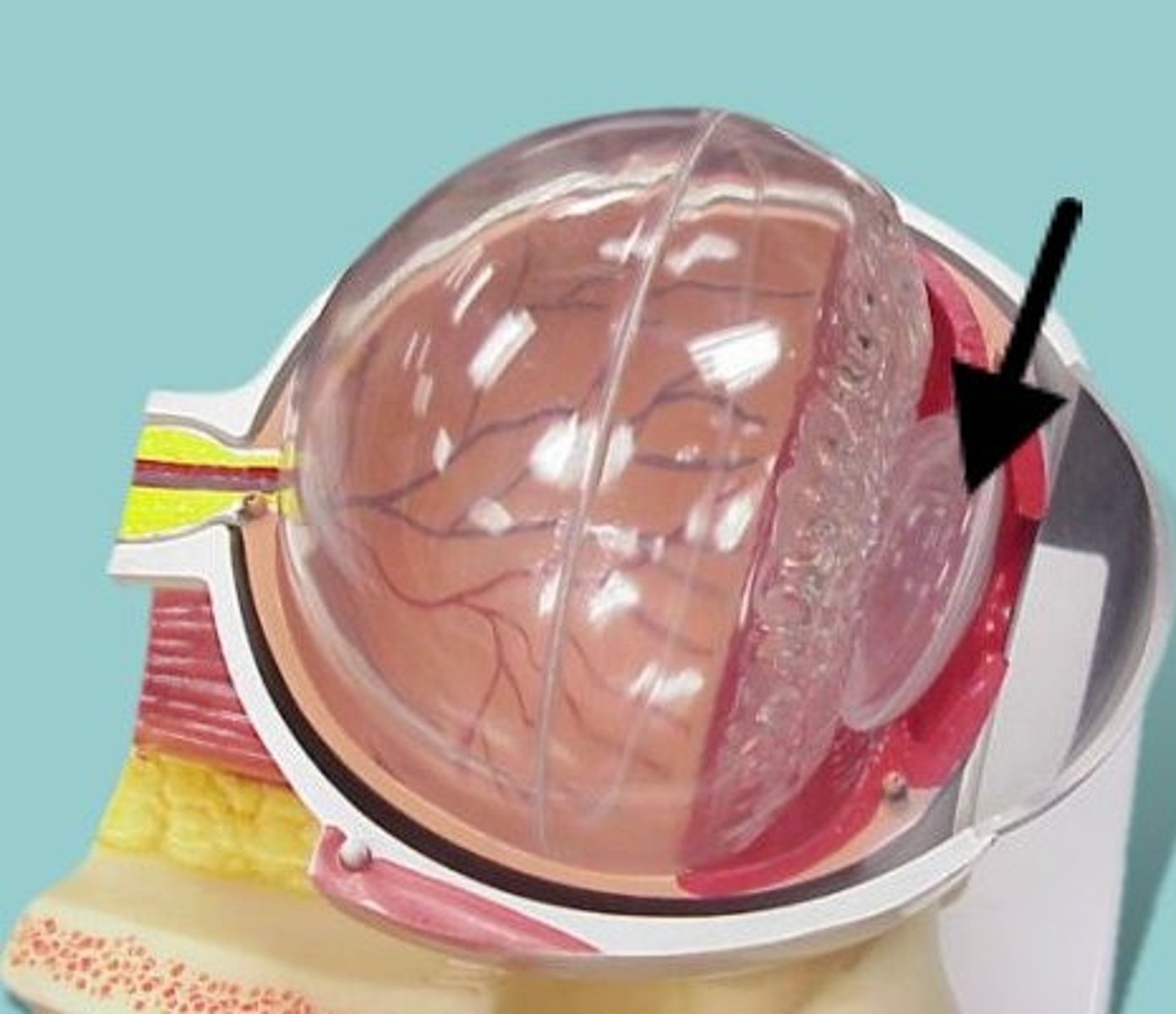
ciliary body
composed of the ciliary process and ciliary muscle which control the shape of the lens
suspensory ligament
clear elastic ligaments suspending the lens from the ciliary body
posterior cavity, eye
space posterior to the lens
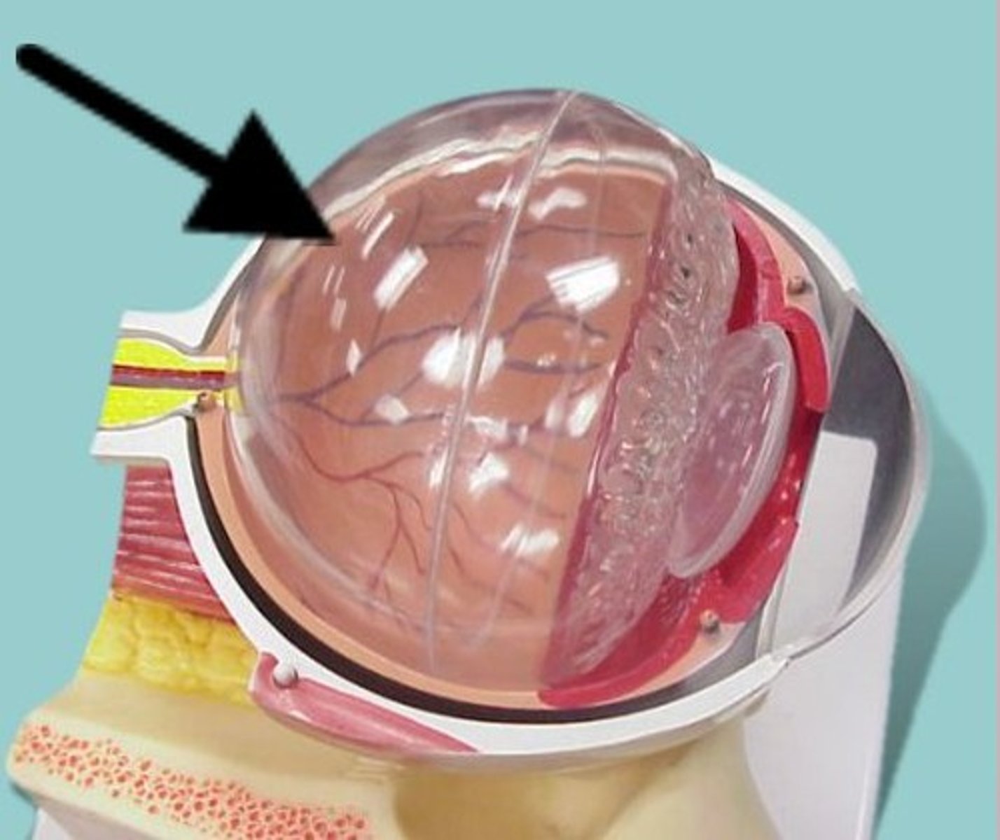
vitreous humor
clear gelatinous substance filling the posterior cavity

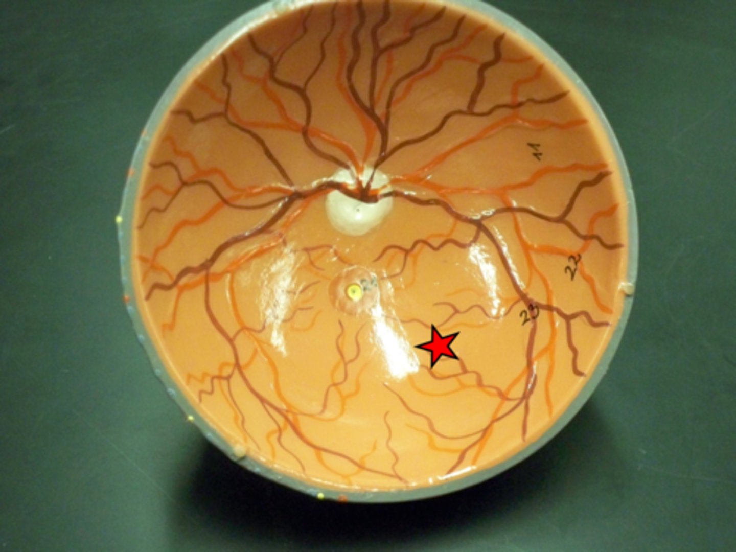
optic disc
area on retina where the optic nerve enters
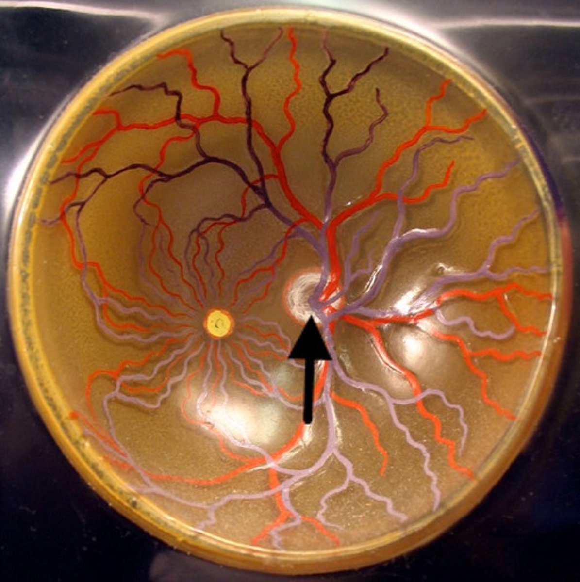
fovea
region on retina where the cones (color receptors) are concentrated

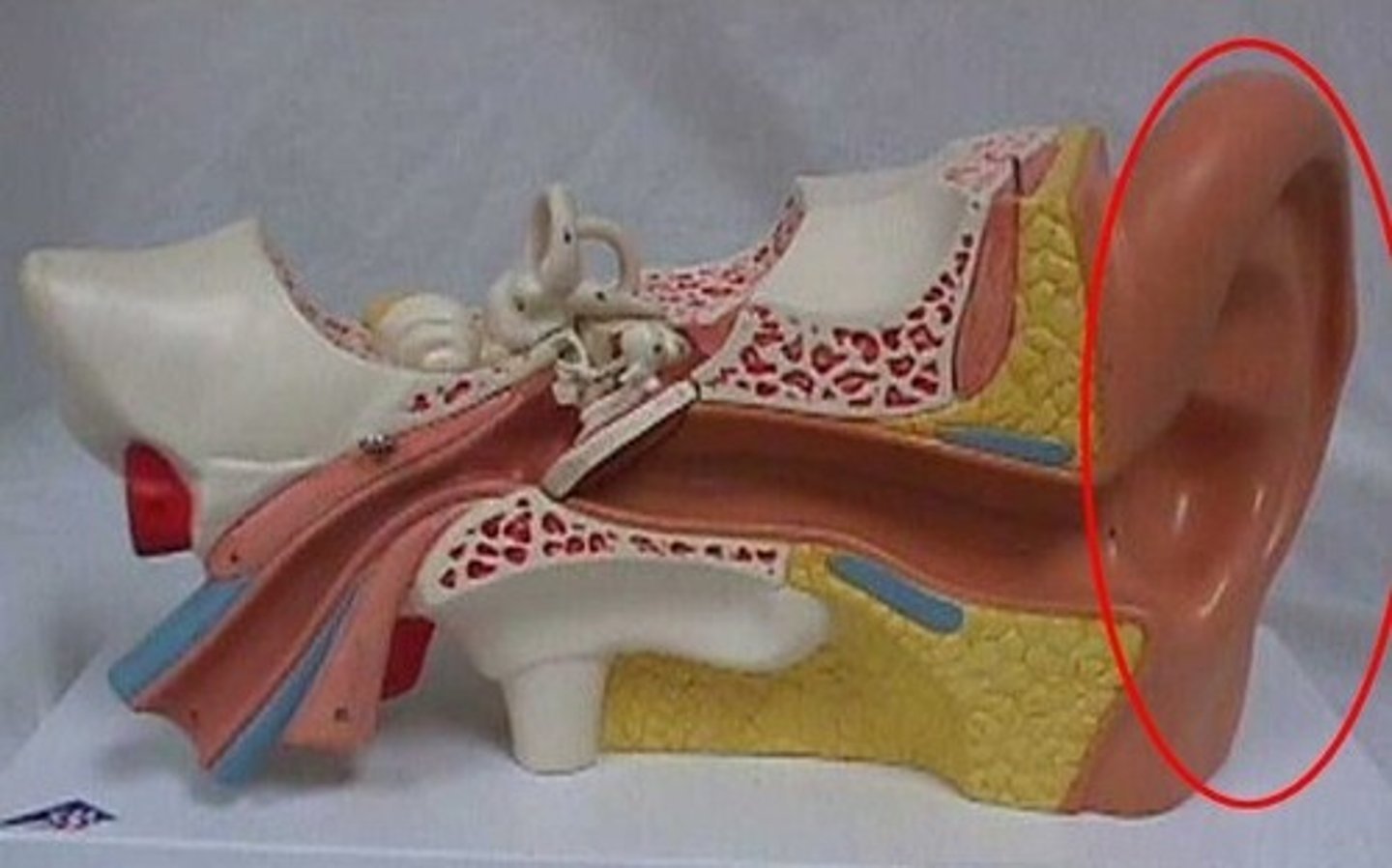
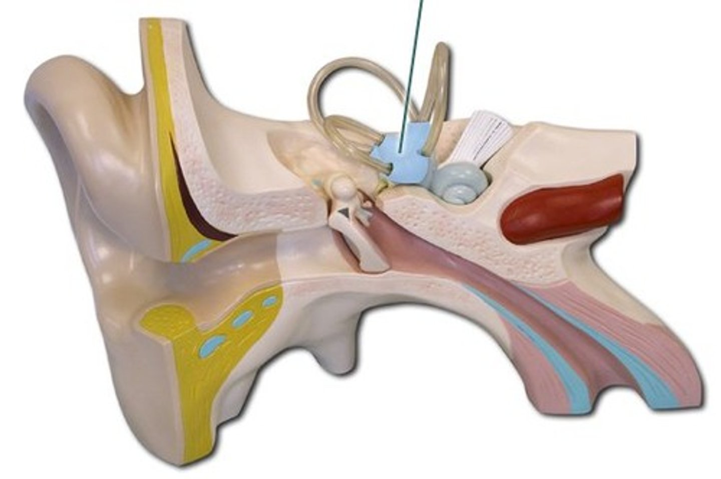
auricle (pinna)
c-shaped elastic cartilage covered in the skin
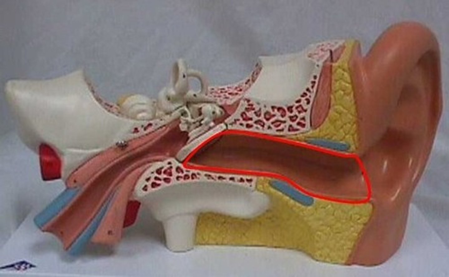
external acoustic meatus
canal leading inward from the auricle to middle ear
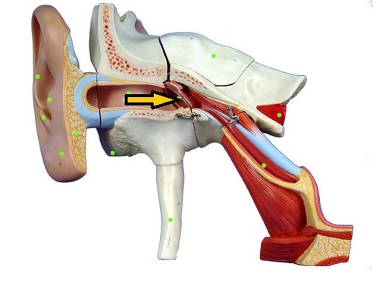
tympanic membrane
thin oval membrane separating the auditory canal from the middle ear, eardrum
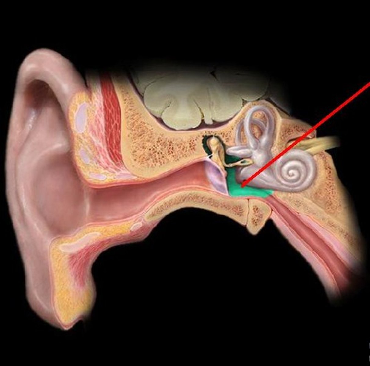
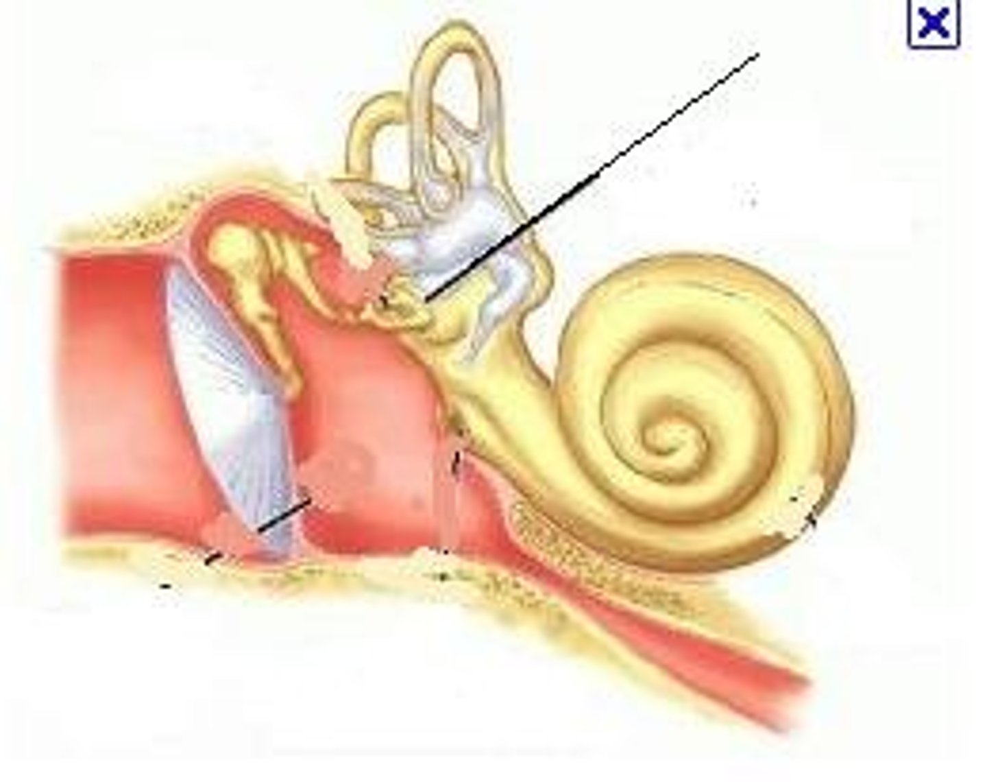
tympanic cavity (antrum)
air filled chamber between external and inner ear
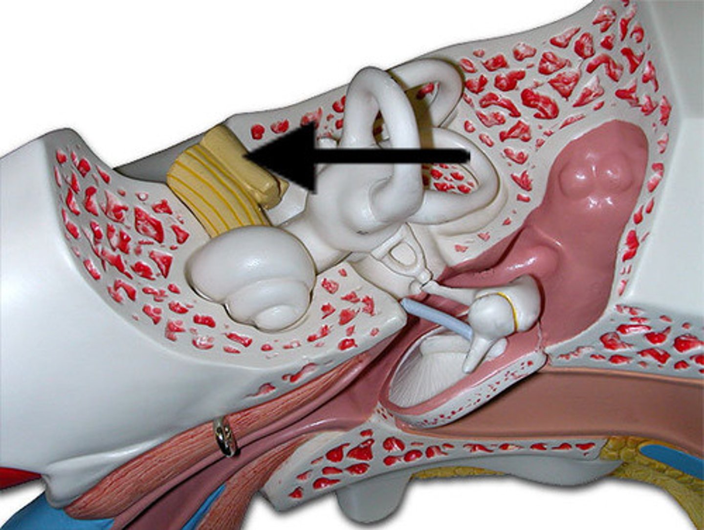
auditory (Eustachian) tube
passageway of the middle ear to the nasopharynx
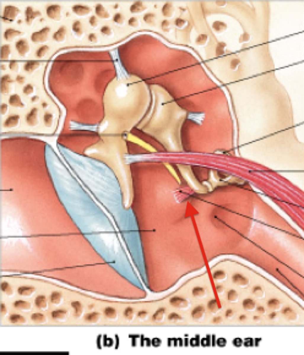
tensor tympani muscle
muscle along the eustachian tube attaching to malleus

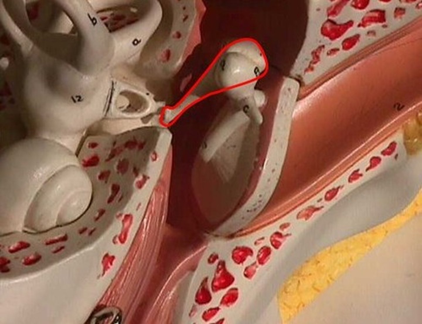
auditory ossicles
three bones of the ear
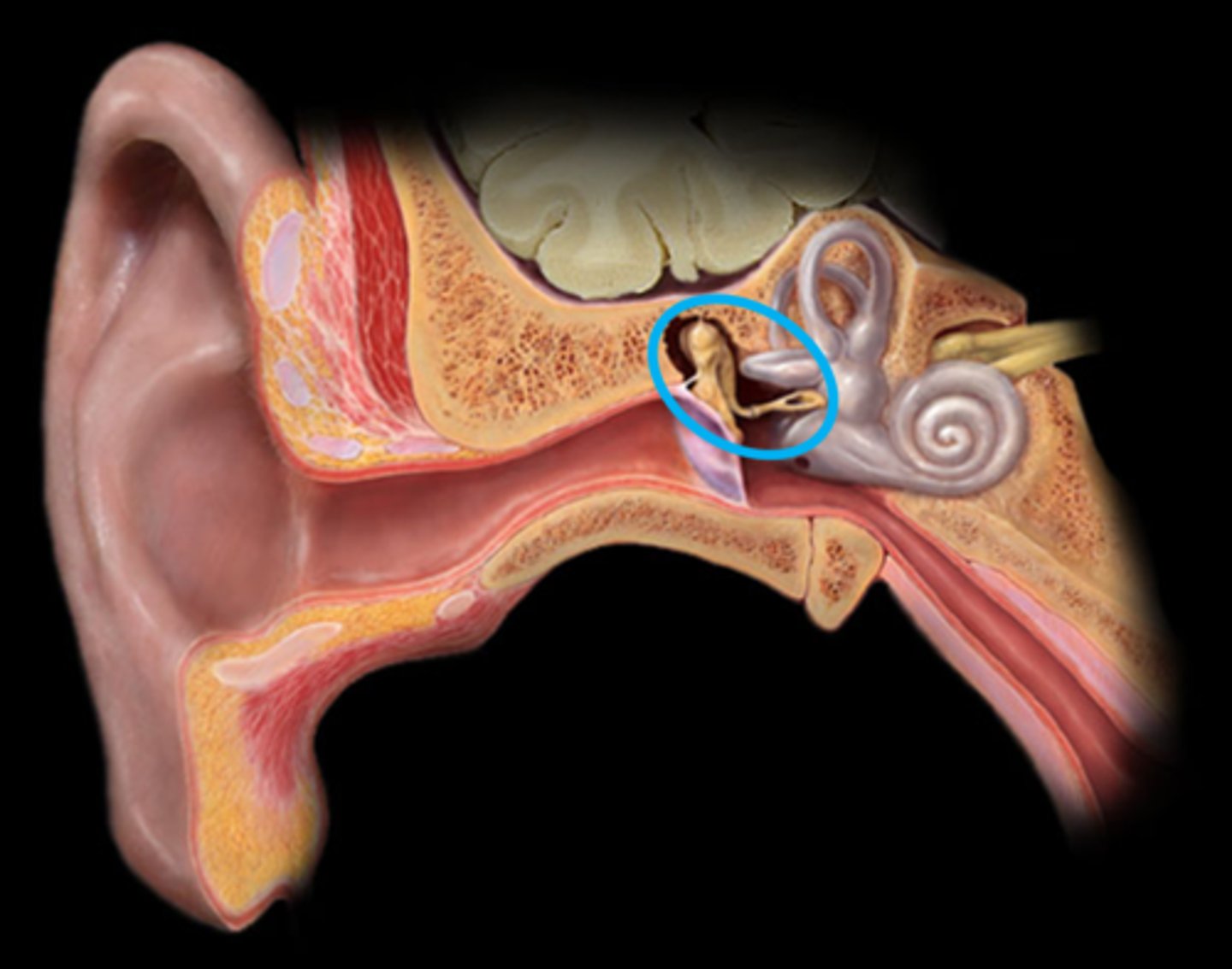
malleus
mallet shaped bone
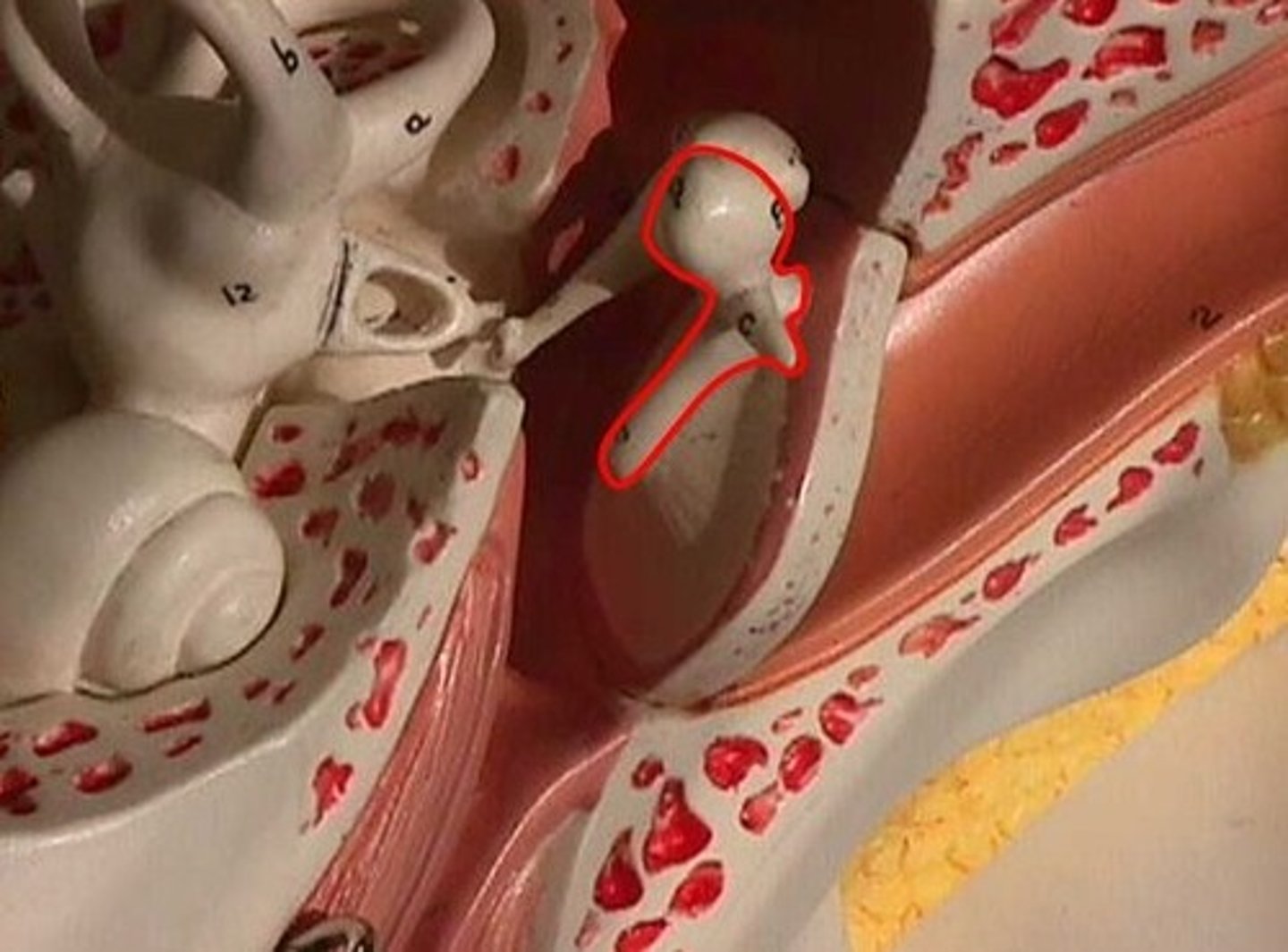
incus
anvil shaped bone between the malleus and stapes
stapes
stirrup shaped bone articulating with the oval window
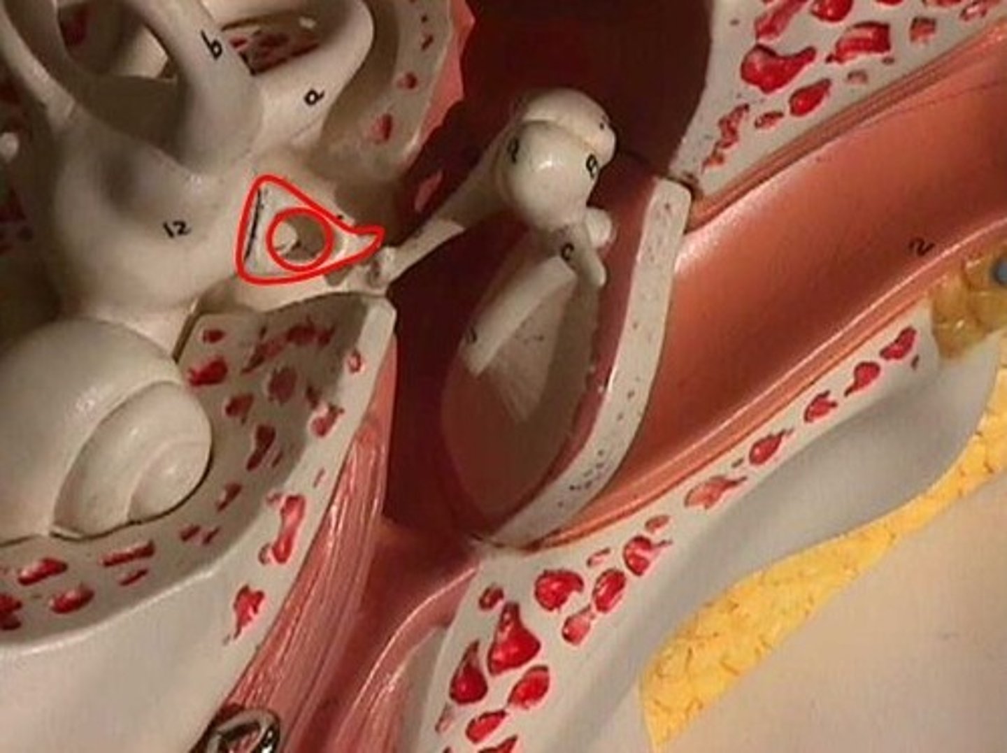
oval window
oval shaped membrane and opening into the inner ear at the base of stapes
round window
round shaped membrane and opening into the inner ear on the cochlea

stapedius muscle
smallest mice of the body attaching to the stapes
vestibule of inner ear
composed of the utricle, saccule and semicircular canals
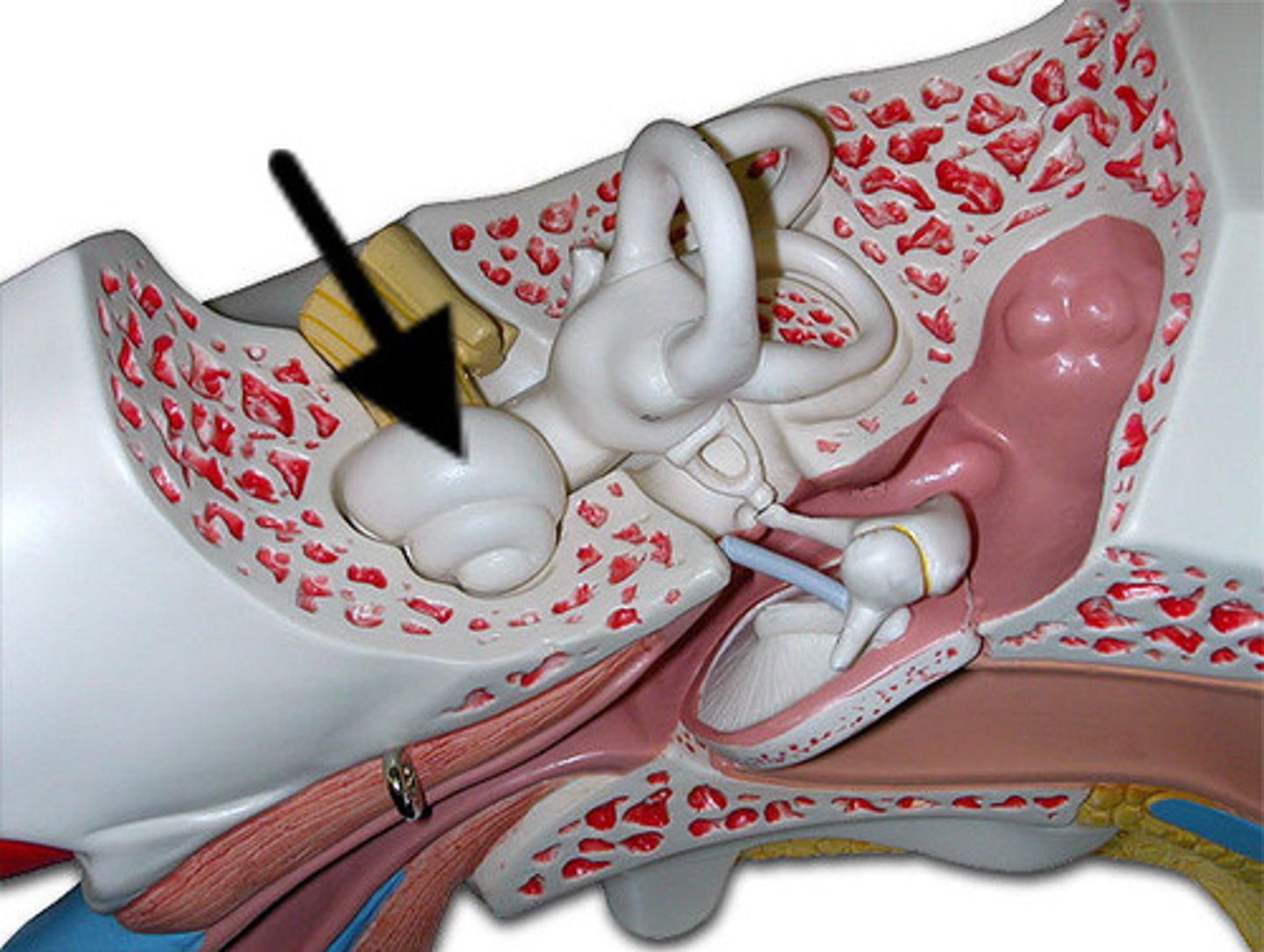
utricle
larger membraneous sac at base of semicircular canals
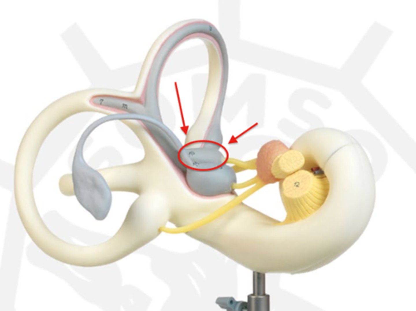
saccule
smaller membranous sac between the utricle and cochlea
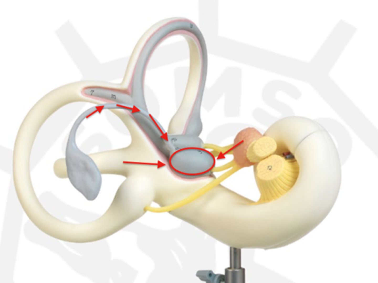
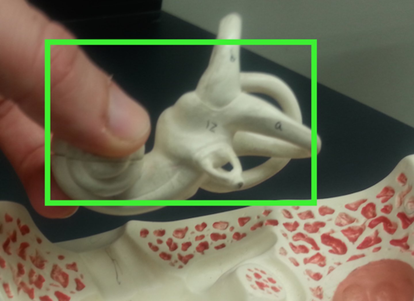
semicircular canals
anterior posterior and lateral membraneous loops surrounded by bony labyrinth
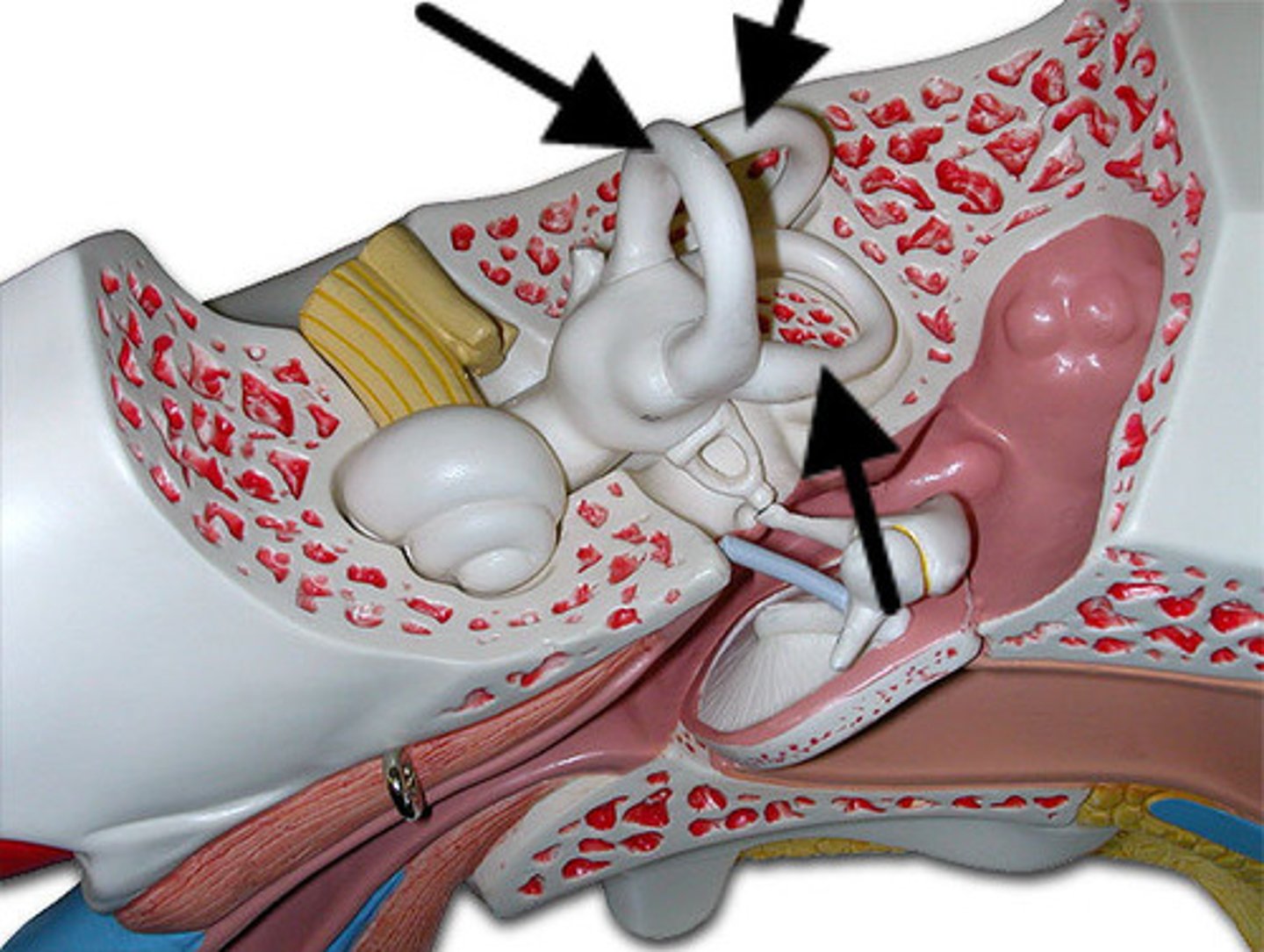
bony labyrinth
tubes and sacs of membrane containing the hearing and equilibrium apparatus
membraneous labyrinth
tubes and sacs of membrane containing the hearing and equilibrium apparatus

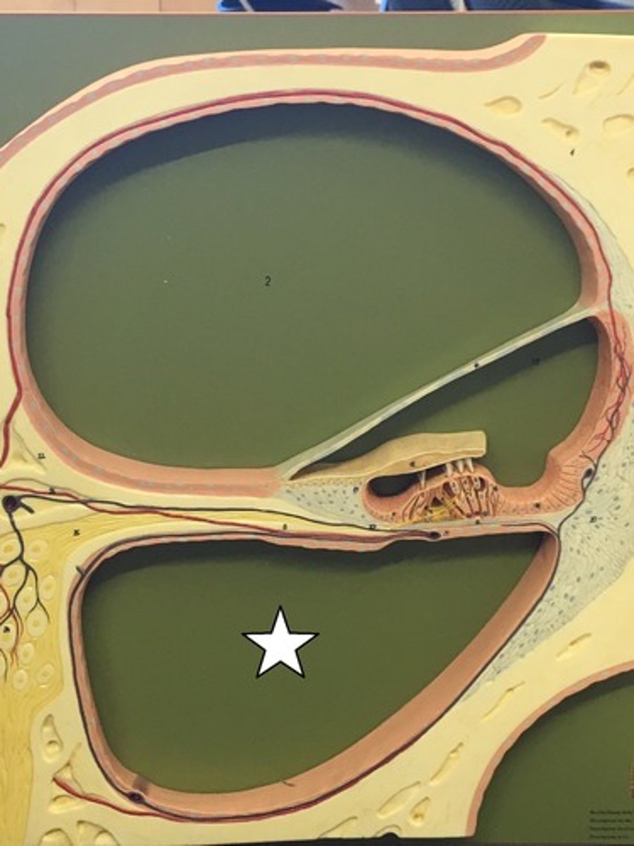
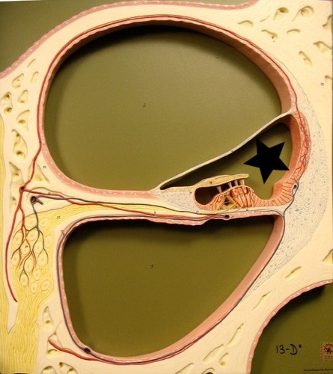
cochlea
snail shaped structure of coiled tubes
vestibular duct
perilymph filled tube
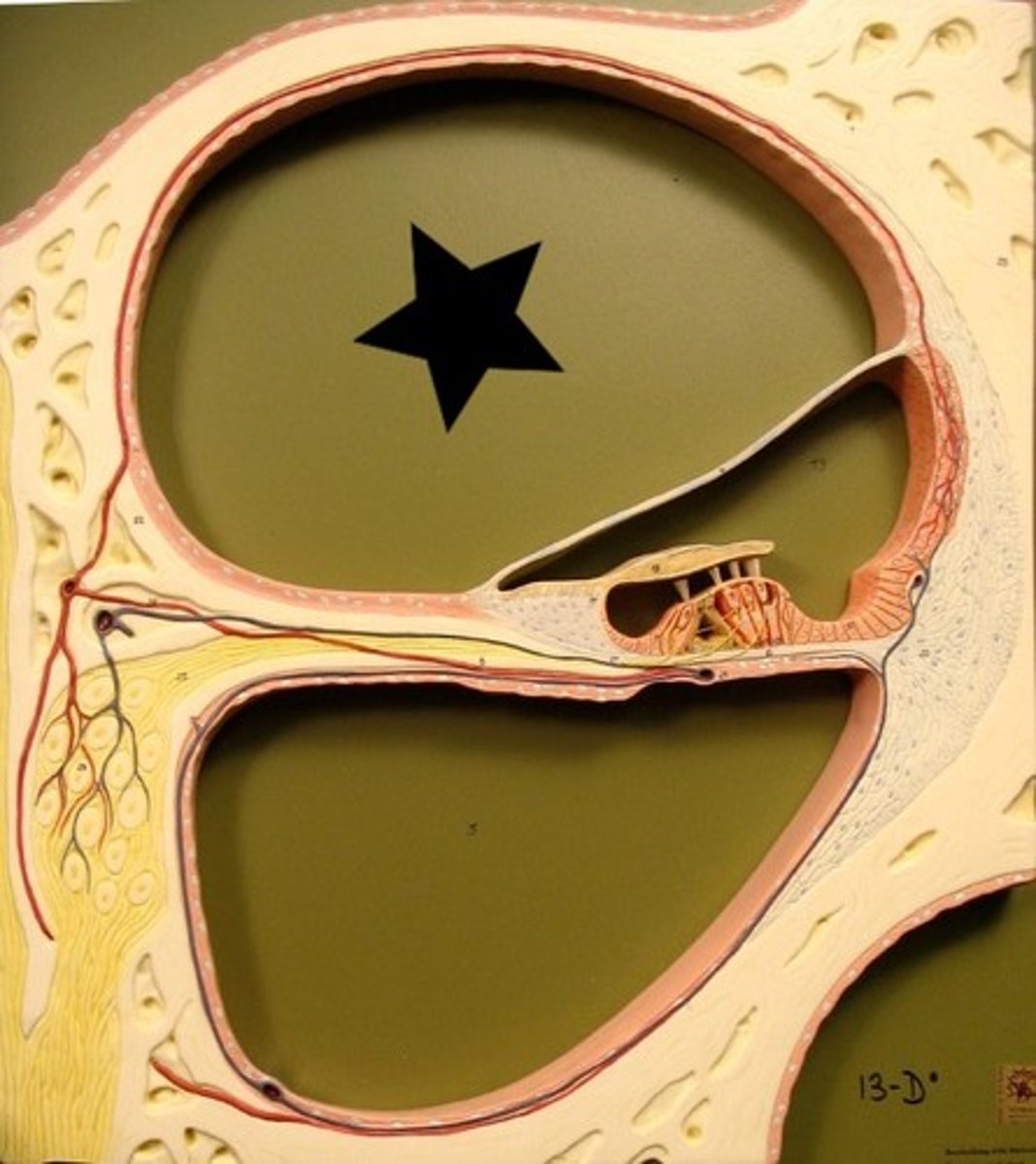
vestibular membrane
membrane between the vestibular and cochlear ducts
cochlear duct
smallest endolymph filled tube between the vestibular and tympanic ducts
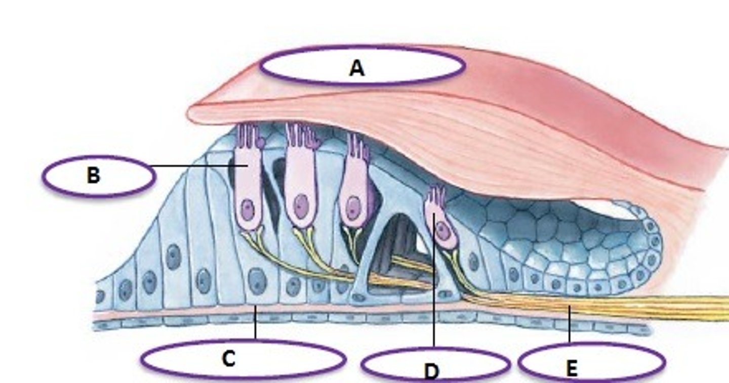
basilar membrane
membrane bw the cochlear and tympanic ducts
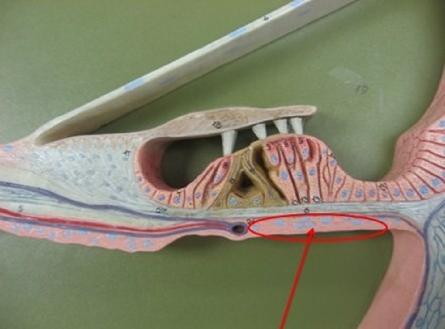
organ of corti
organ of hearing resting on the basilar membrane
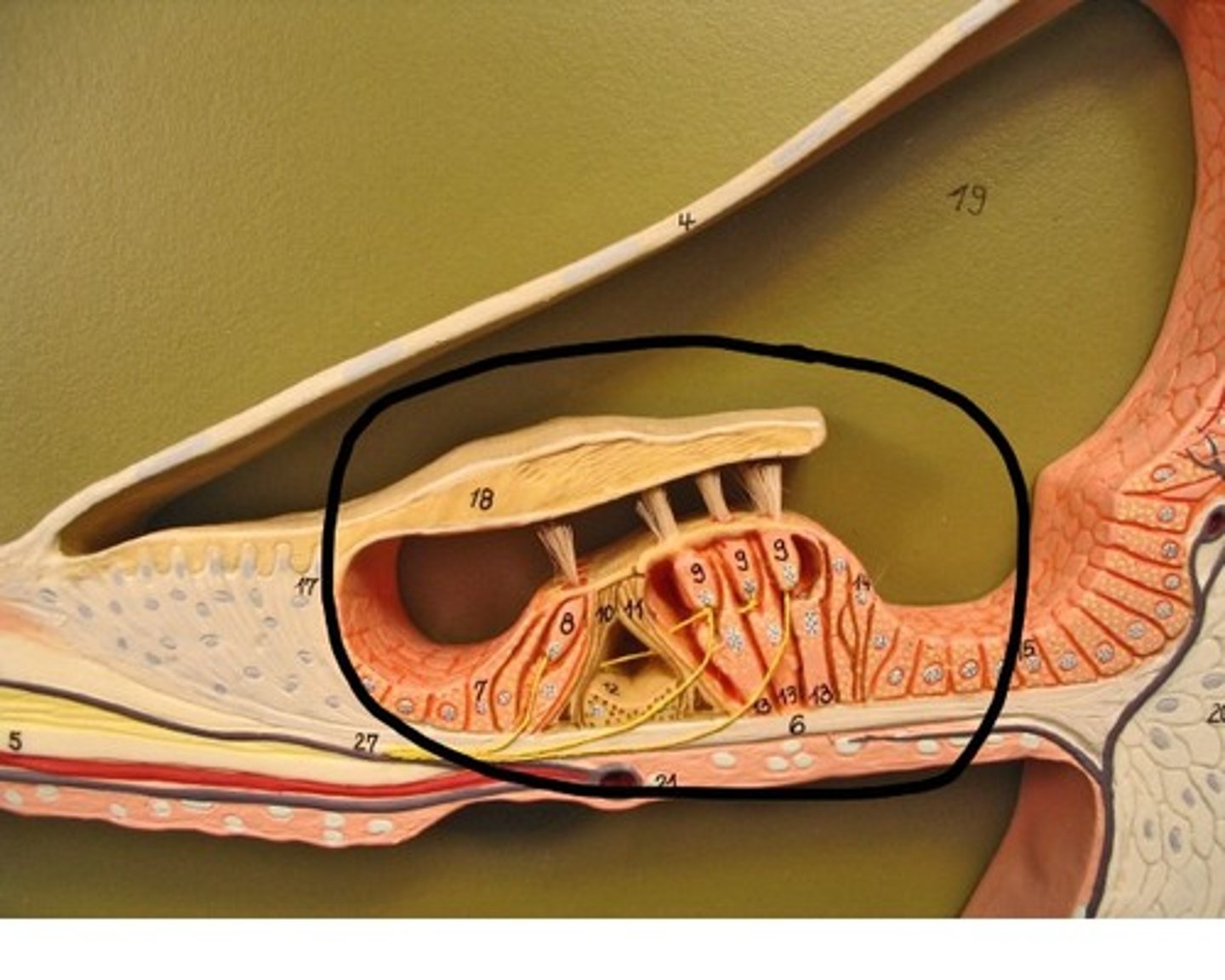
tectoral membrane
membrane covering the organ of corti (A)
tympanic duct
perilymph filled tube