intro to respiratory system
 Call Kai
 Learn
Learn Practice Test
 Spaced Repetition
 Match
Match Flashcards
 Knowt Play
1/36

There's no tags or description
Looks like no tags are added yet.
Name | Mastery | Learn | Test | Matching | Spaced | Call with Kai | Chat |
|---|
No analytics yet
Send a link to your students to track their progress
37 Terms
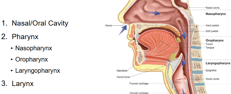
Upper respiratory tract parts
Nasal cavity Oral cavity Pharynx Larynx

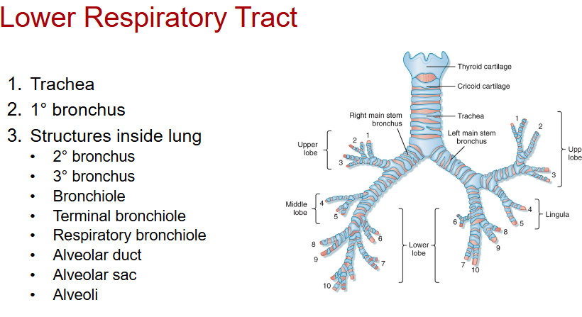
Lower respiratory tract
Trachea
1/2/3rd bronchus
bronchiole
terminal bronchiole
respiratory bronchiole
alveolar duct → sac → alveoli
Different parts of the Pharynx
Nasopharynx (behind nose)
Oropharynx (behind mouth)
Laryngopharynx
Which structure in Lower Respiratory Tract are inside lung?
2nd bronchus and below
Functions of Respiratory Tract
Ventilation
Waste elimination
filtration of foreign particles from air
humidity control
temp control
Upper respiratory system function
Olfaction
Draining sinuses
Producing and resonance of speech
Larynx
Voice box
Pharynx function
dual passage way for esophagus and trachea
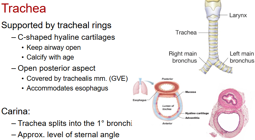
Trachea
Supported by C shaped hyaline cartilages rings
prevents collapse
posterior/back side open for esophagus
trachealis muscle closes posterior gap
Whats the trachealis muscle innervated by in trachea?
GVE (visceral efferent, autonomic)
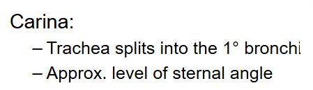
Carina
The split where trachea divides
(left and right rbonchus)
Around sternal angle landmark
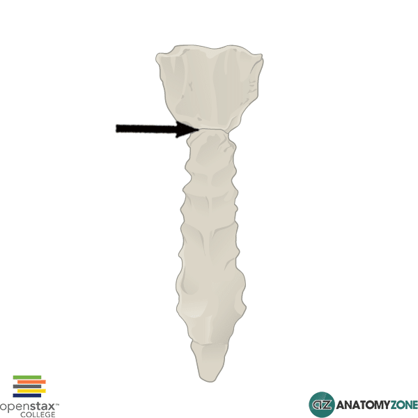
Sternal Angle / Angle of Louis
place where top of sternum meets body
Right vs Left Primary Bronchus
Right
Wider
Shorter
More vertical
Left
Narrower
Longer
More horizontal
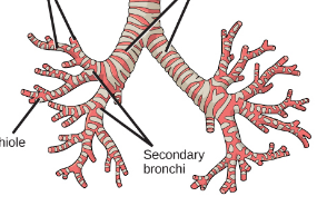
Clinical Correlate for left / right bronchus
a foreign object would enter the right bronchus due to it being WIDER and VERTICAL
Right vs Left lung lobes
Left
2 lobes (Superior, Inferior)
has 2 due to space of heart
Right
3 lobes (Superior Middle Inferior)
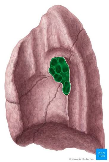
Hilum
doorway of the lung
depression where blood vessels, nerves, and ducts leave an organ
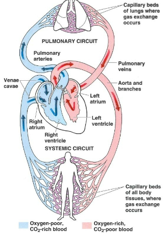
Pulmonary circulation
Right ventricle → pulmonary arteries (deoxy blood) → lungs → pulmonary veins (oxy blood) → left atrium
Which structures enter/leave lymph node?
Bronchi
Pulmonary arteries
Pulmonary veins
Lymphatics
Nerves
Pulmonary Pleurals
Parietal Pleura (covers chest wall)
Visceral Plerua (covers lung surface)
Whats between pulmonary pleuras?
Pleural fluid to reduce friction
Pleural cavity
Very thin
nearly No Space
has some serous fluid
Clinical Correlate: Pleurisy/Pleuritis
Inflammation of pleura
Pain with breathing
Increased friction
Clinical Correlate: Pleural Plaques
thickening of pleura that attaches to ribs
happens due to asbestos exposure
Clinical Correlate: Mesothelioma
cancer of the serous tissues that lines body cavities
abestos exposure
Systemic circulation
arteries(oxy blood)→system organs →Veins (deoxy blood)
Inspiration AND its main muscle
Diaphragm
Diaphragm contracts → Moves downward → Thoracic volume increases → Air enters lungs
Expiration and muscle
Diaphragm relaxes -> Thoracic volume decreases -> Air exits lungs
Quiet breathing main muscle
Diaphragm AND intercostal muscles for inspiration
elastic recoil of alveoli for expiration
Forced breathing main muscle
Neck muscles
SCM
Scalenes
Intercostals
Serratus muscles
Pectoralis muscles
Abdominal muscles
Diaphragm innervation
C3, C4, C5
"C3, 4, 5 keep the diaphragm alive."
intercostal innervation
T1-T11d
Diaphragm muscle openings
Caval opening
Esophageal hiatus
Aortic hiatus
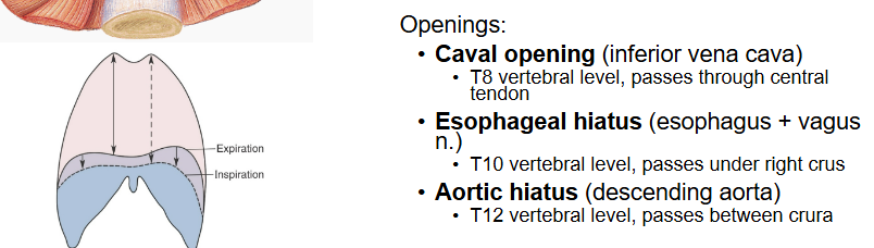
Caval opening part and innervation
Inferior vena cava
T8 vertebral
Esophageal hiatus part and innervation
esophagus + vagus nerve (T10)
Aortic hiatus part and innervation
descending aorta (T12)
3 layers of intercostal muscles from external to internal
external intercostal
internal intercostal
innermost intercostal
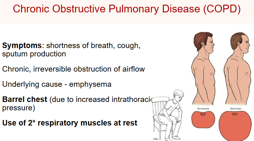
COPD
caused by smoking usually
Damaged alveoli
Difficult expiration
Air trapping
Barrel chest
Accessory muscles used even at rest