Neuroanatomy week 1- Overview of the Nervous System
 Call Kai
 Learn
Learn Practice Test
 Spaced Repetition
 Match
Match Flashcards
 Knowt Play
1/115

Earn XP
Description and Tags
https://www.youtube.com/watch?v=Tp25wrm-AoA
Name | Mastery | Learn | Test | Matching | Spaced | Call with Kai | Chat |
|---|
No analytics yet
Send a link to your students to track their progress
116 Terms
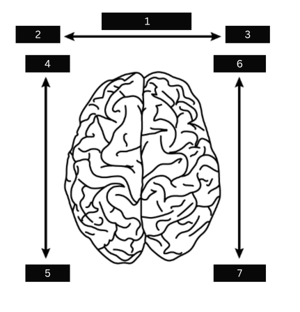
what is figure 1?
Medial

what is figure 2?
lateral
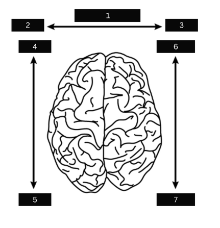
what is figure 3?
Lateral
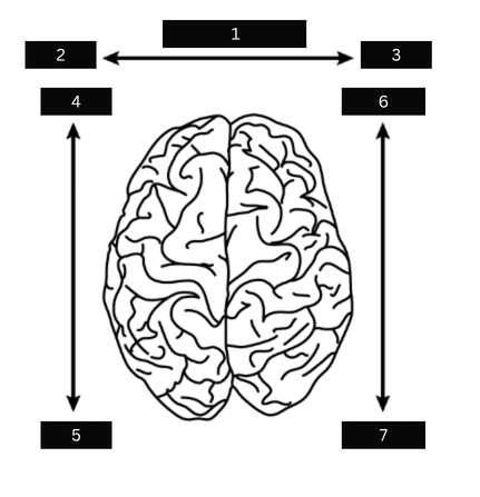
what is figure 4
Rostral
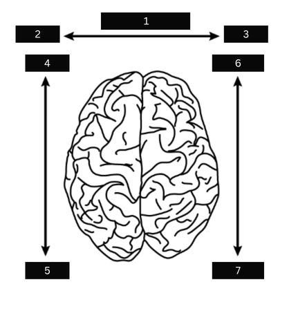
what is figure 5
Caudal
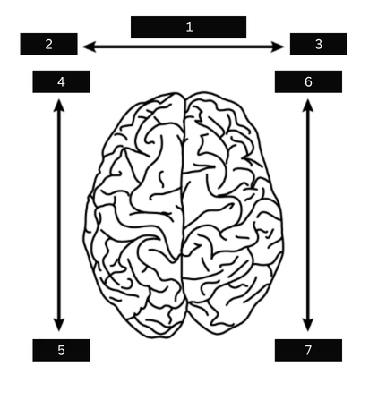
what is figure 6
Anterior
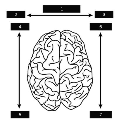
what is figure 7
Posterior
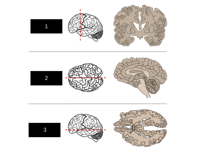
what is figure 1
frontal or coronal plane
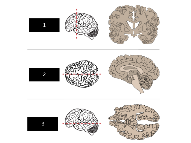
what is figure 2
sagittal plane
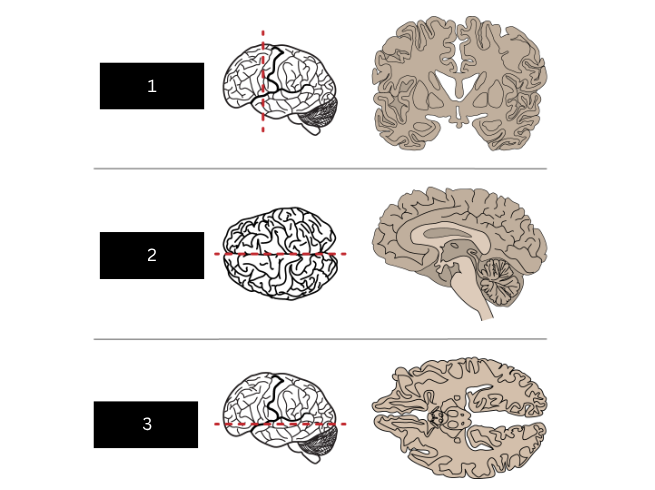
what is figure 3
Horizontal plane
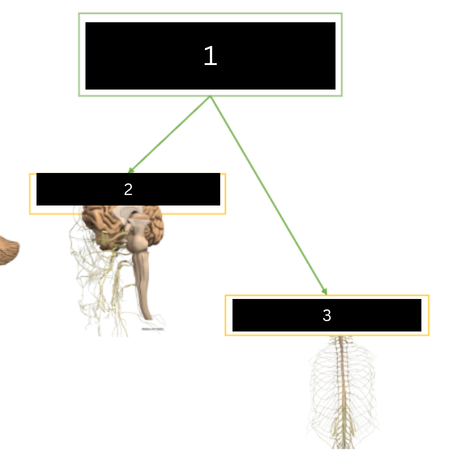
what is figure 1
Peripheral nervous system PNS
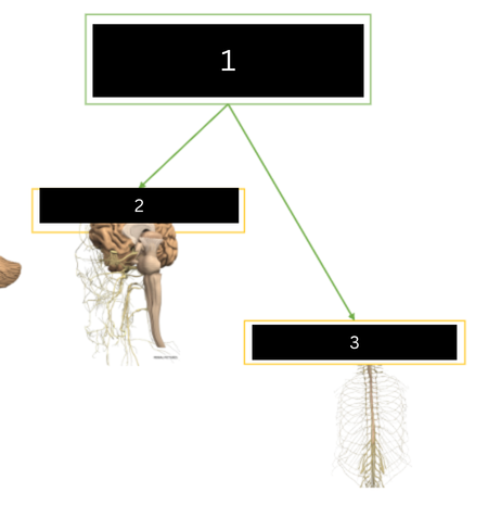
what is figure 2
cranial nerves

what is figure 3
spinal nerves
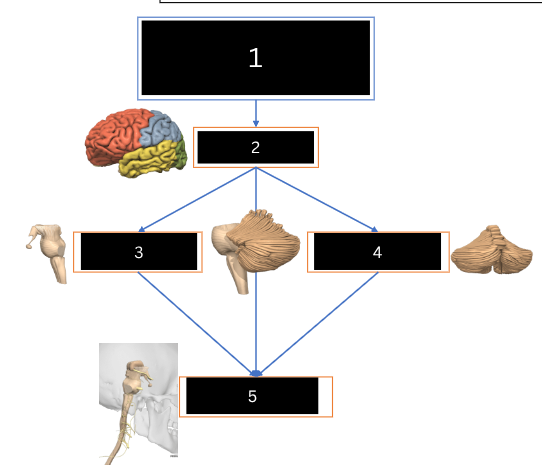
what is figure 1
Central nervous system CNS
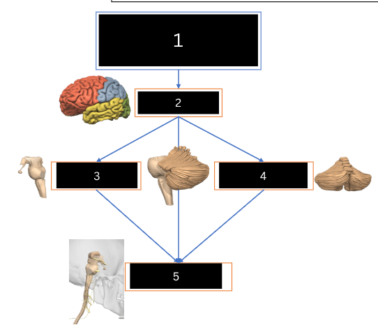
what is figure 2
cerebrum
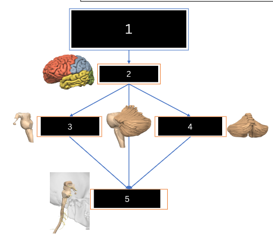
what is figure 3
brainstem
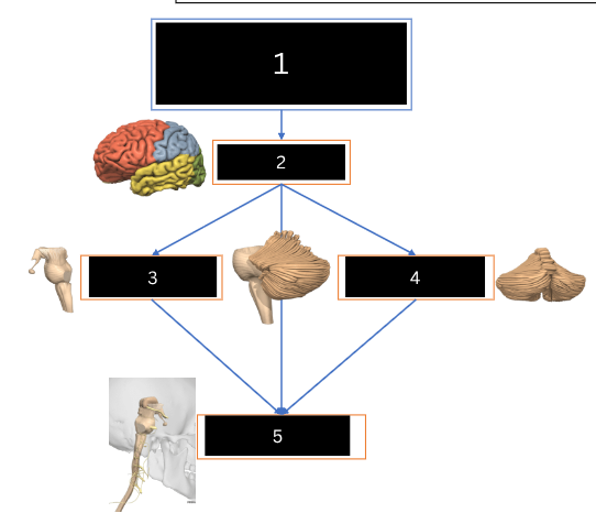
what is figure 4
cerebellum
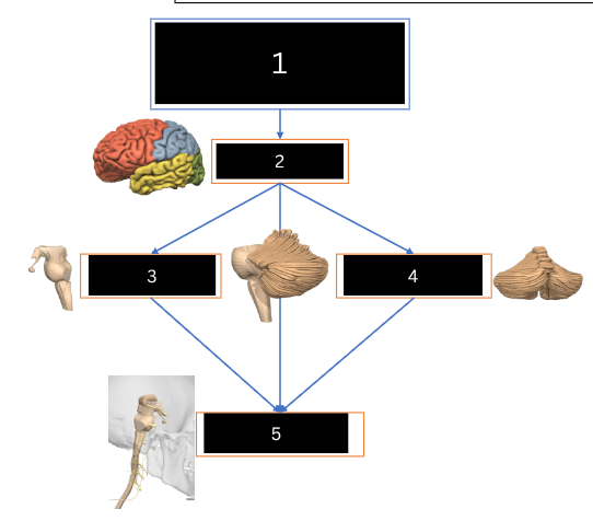
what is figure 5
spinal cord
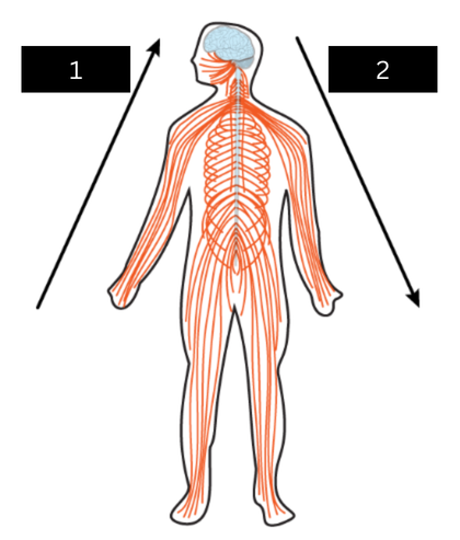
what is figure 1
Afferent
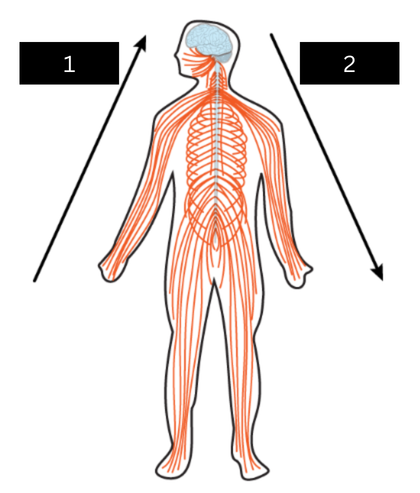
what is figure 2
Efferent
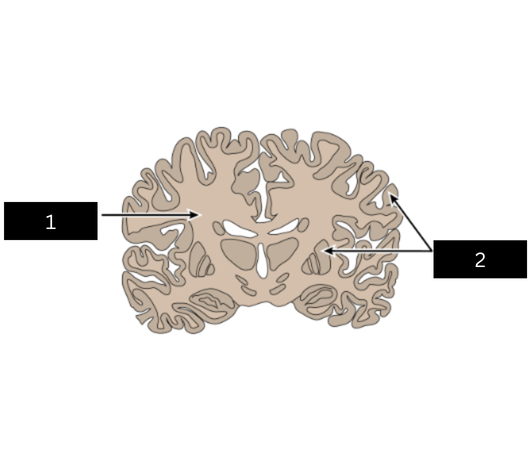
what is figure 1
White matter (axons)
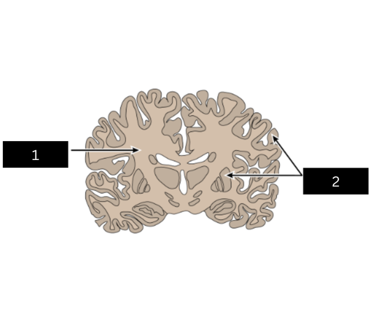
what is figure 2
Grey matter (cell bodies)
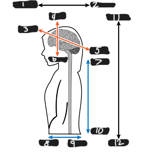
what is figure 1
anterior
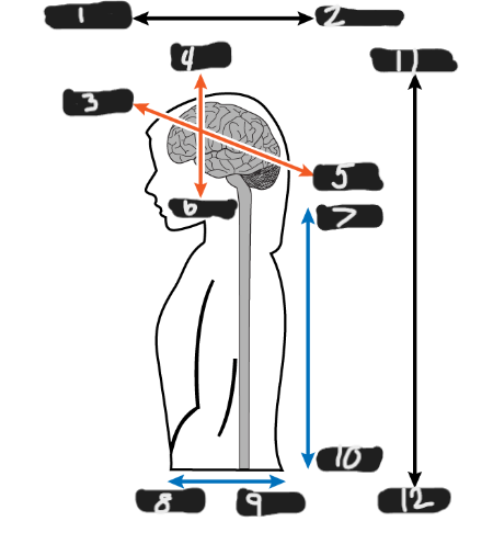
what is figure 2
posterior

what is figure 3
rostral
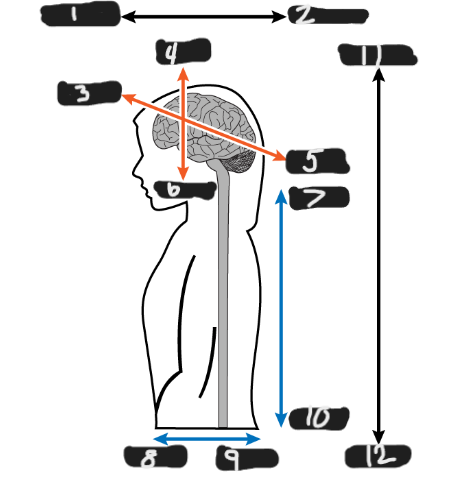
what is figure 4
dorsal
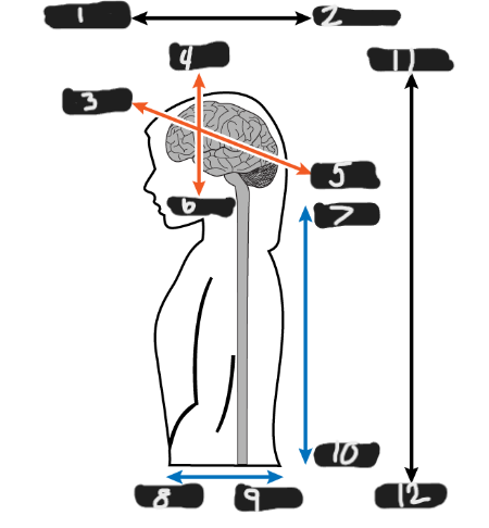
what is figure 5
caudal
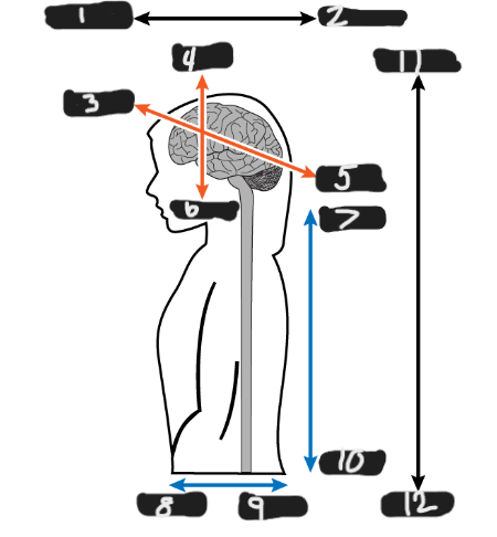
what is figure 6
ventral
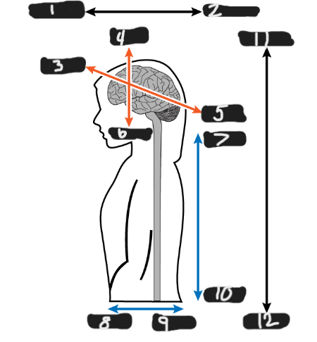
what is figure 7
rostral
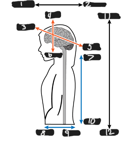
what is figure 8
ventral
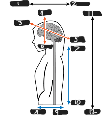
what is figure 9
dorsal
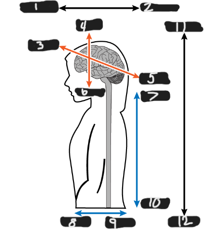
what is figure 10
caudal
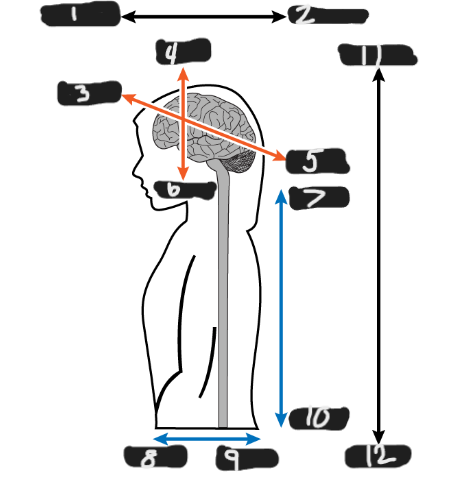
what is figure 11
superior
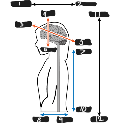
what is figure 12
inferior
Autonomic nervous system consists of the
sympathetic system and parasympathetic system
The Autonomic nervous system is located in the
prefrontal cerebral cortex and subcortical regions
Sympathetic nervous system
Fight, Flight, and Freeze
Parasympathetic nervous system
slows heart rate, constricts pupils, and lowers blood pressure
The Somatic Nervous system is made up of
skeletal muscles, pyramidal or direct system, extrapyramidal or indirect system, and conscious or controlled movements.
Pyramidal or Direct system
control of conscious controlled skilled movement
Extrapyramidal or Indirect system
control of background, supportive movements
Embryonic period →
Weeks 1–8 of development
Fetal period →
Weeks 9–37 of development
What embryological structure does the nervous system develop from?
The neural plate
Neural tube
A structure that begins forming by about 4 weeks gestational age and gives rise to structures of the nervous system
When does the neural tube begin forming?
Around 4 weeks gestational age (GA)
What develops from the neural tube?
Structures of the nervous system
Neural crest cells
Cells that move to the periphery of the neural tube and form structures including the cranial and spinal nerves
What happens around 4 weeks to the neural tube?
It “pinches” to form three bulges
Three primary brain vesicles- P
Prosencephalon
Three primary brain vesicles- M
Mesencephalon
Three primary brain vesicles- R
Rhombencephalon
Prosencephalon
The forebrain; develops into the telencephalon and diencephalon

Prosencephalon- develops into the (1) T
telencephalon
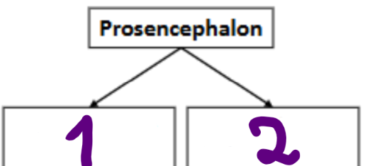
Prosencephalon- develops into the (2) D
diencephalon
Prosencephalon develops into: →
Telencephalon + Diencephalon
Telencephalon →
Develops into the cerebral hemispheres and subcortical structures
Cerebral hemispheres
The two major halves of the cerebrum
Subcortical structures
Structures located below the cerebral cortex.
Diencephalon
Develops into structures including the thalamus, hypothalamus, and pituitary gland
Thalamus →
A major structure that develops from the diencephalon
Hypothalamus
A structure that develops from the diencephalon
Pituitary gland
A structure associated with the diencephalon in the slide's developmental organization.
Mesencephalon develops into:
midbrain
Midbrain →
Part of the brainstem
Cerebral peduncles →
Structures associated with the midbrain that connect to the cerebellum according to the slides
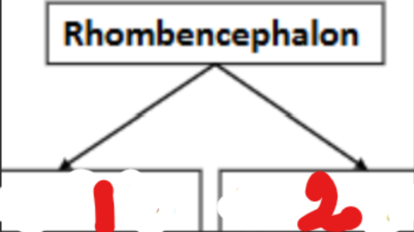
Rhombencephalon develops into: → (1) Me
Metencephalon

Rhombencephalon develops into: → (1) My
Myelencephalon
Metencephalon →
Develops into the pons and cerebellum.
Pons →
A structure that develops from the metencephalon.
Cerebellum →
A structure that develops from the metencephalon
Myelencephalon →
Develops into the medulla oblongata
Medulla oblongata →
A structure that develops from the myelencephalon
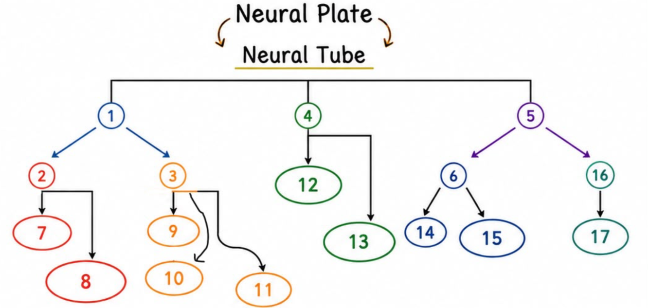
1
Prosencephalo

2
telencephalon
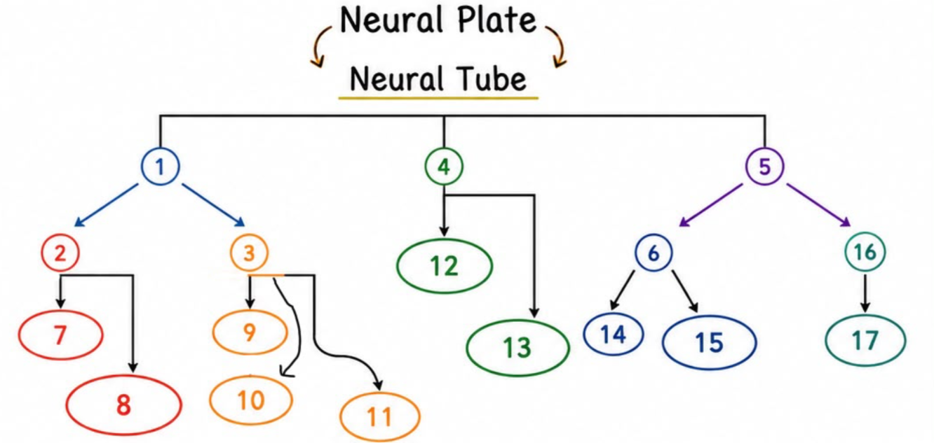
7
cerebran hemispheres
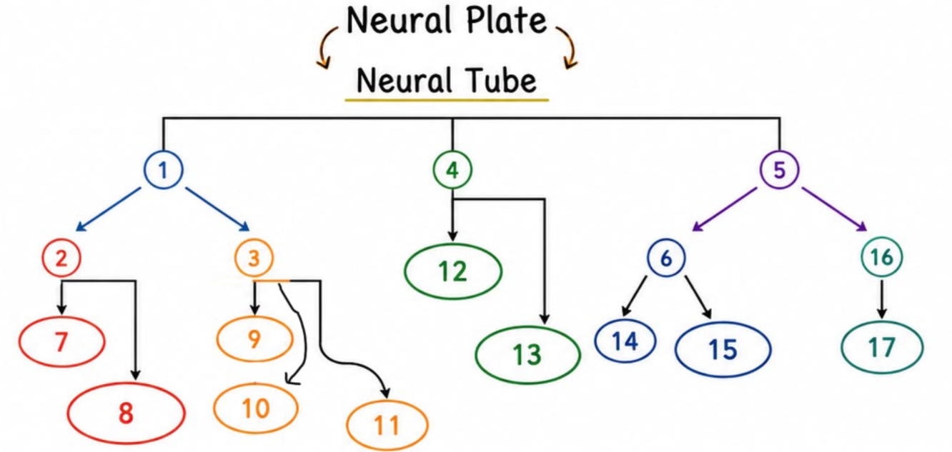
8
subcortical structures

3
Diencephalon

9
thalamus
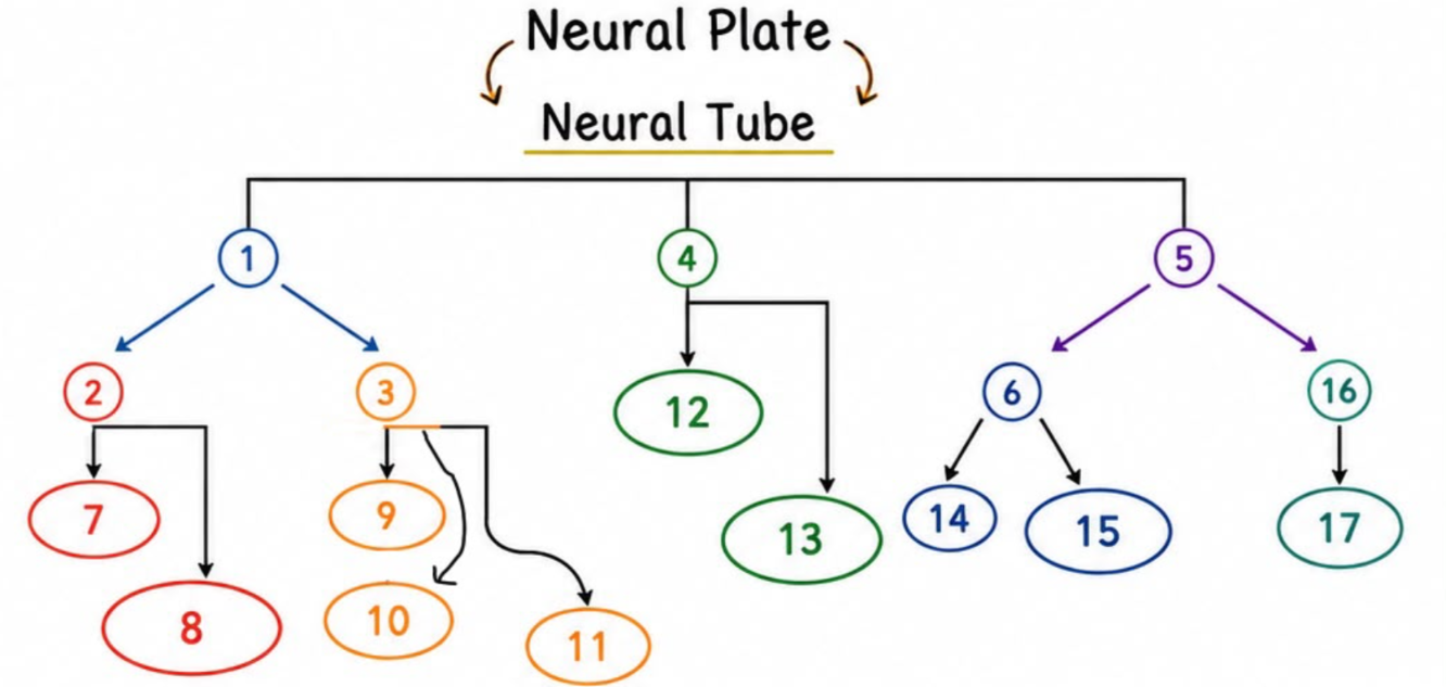
10
hypothalamus
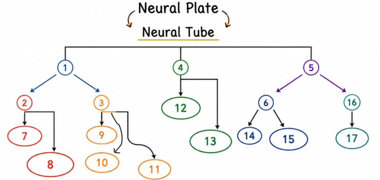
11
pituitary grand

4
Mesencephalon

12
Midbrain (brainstem)
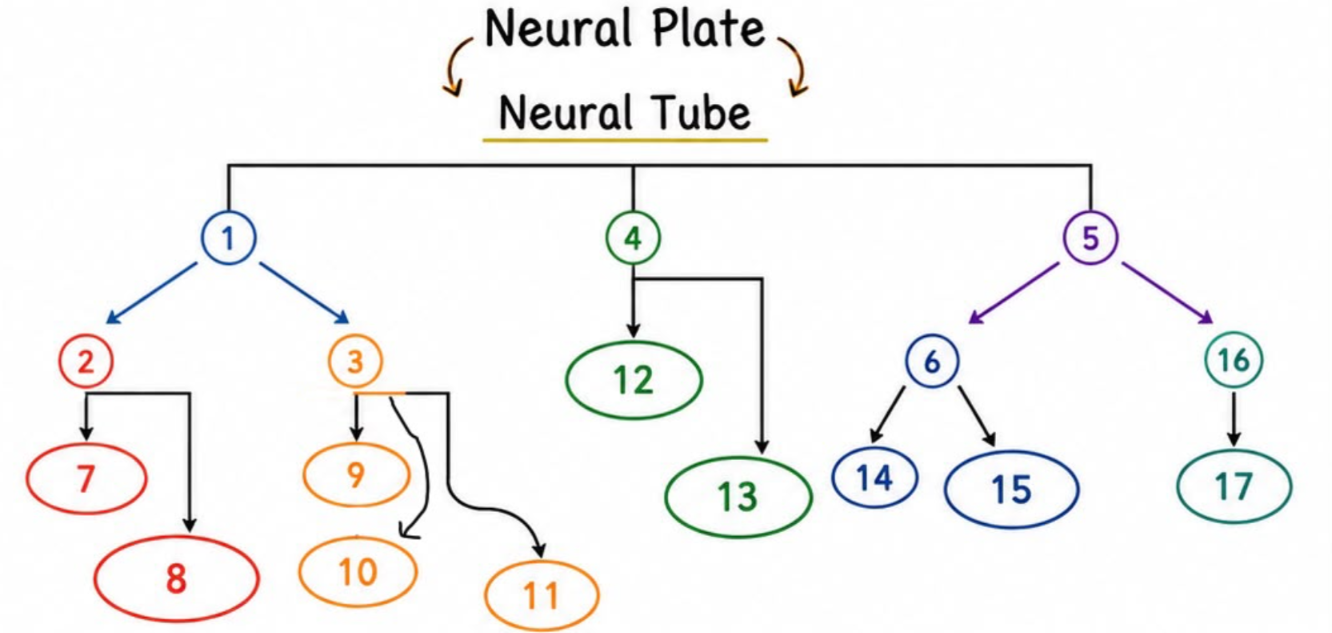
13
Cerebral peduncles (connected to cerebellum)

5
Rhomencephalon

6
metencephalon
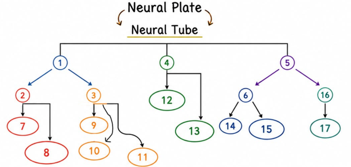
14
pons

15
cerebellum
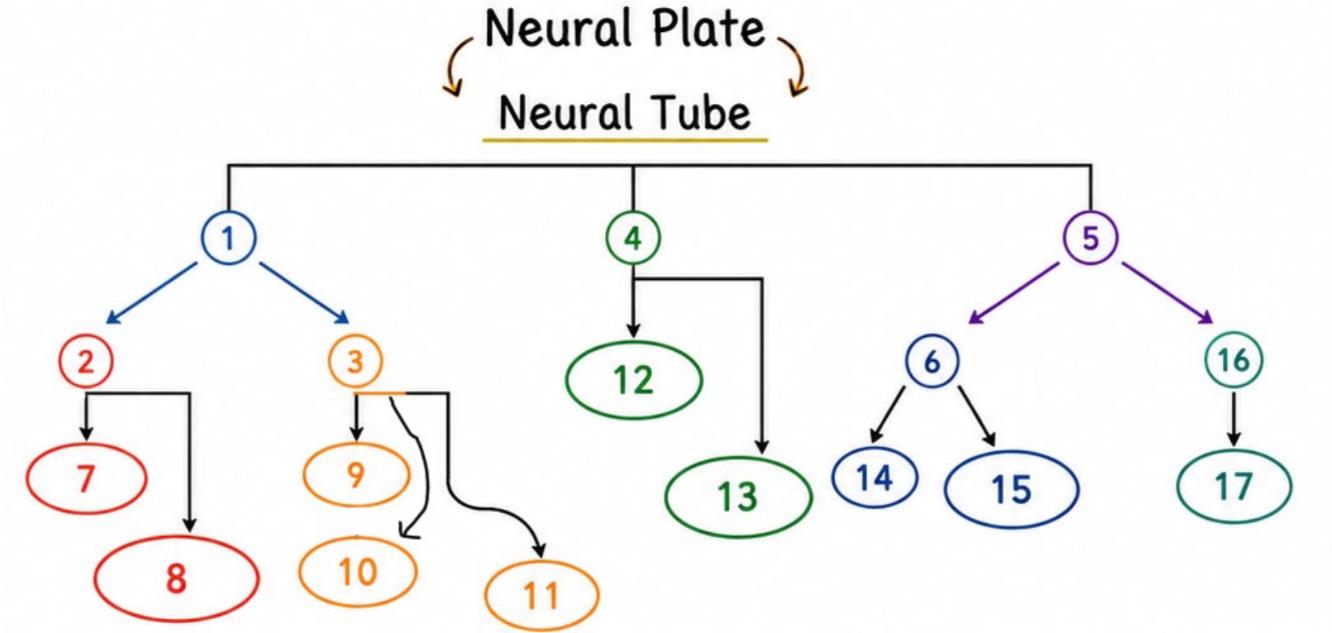
16
myelencephalon
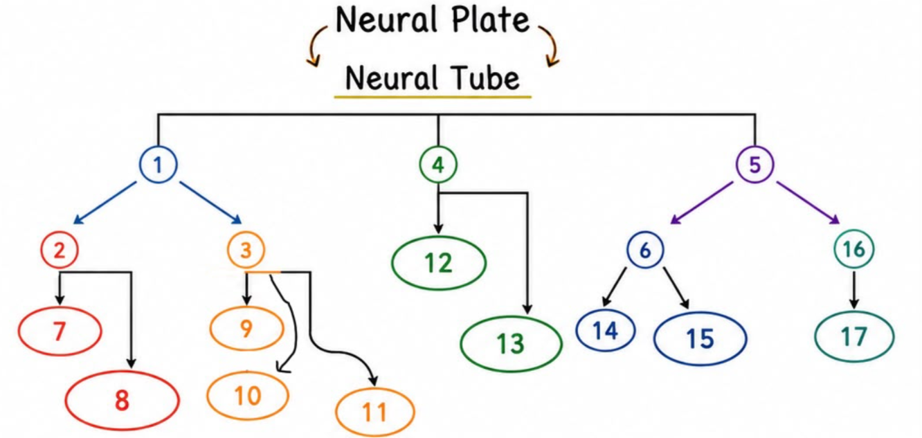
17
medulla oblongata
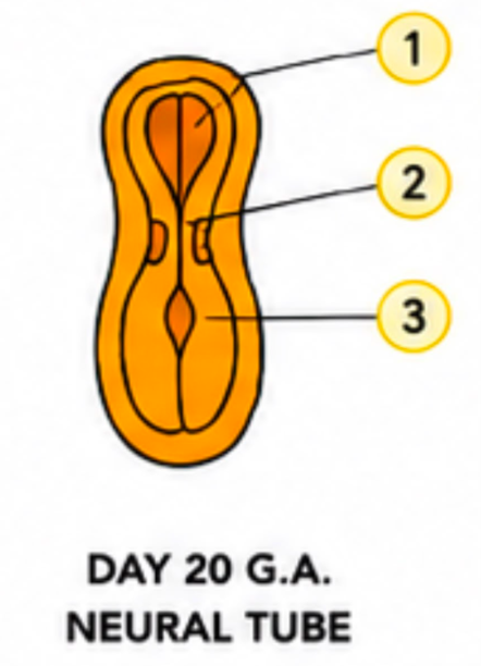
1
neural fold of forebrain

2
neural plate
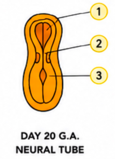
3
neural groove
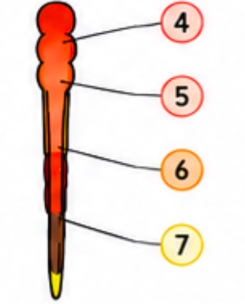
4
Prosencephalon (forebrain)
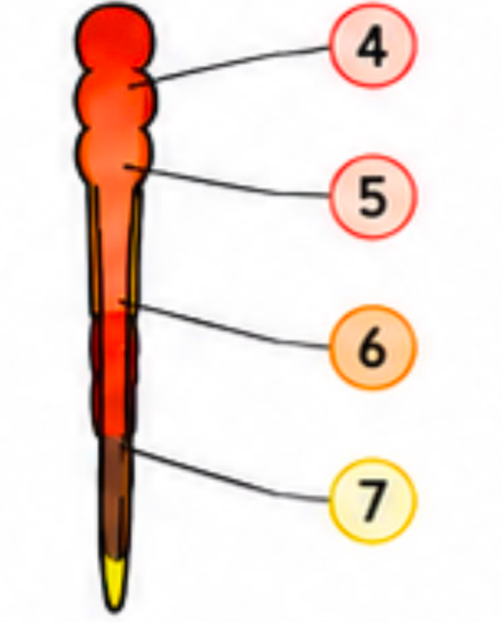
5
Mesencephalon (midbrain)
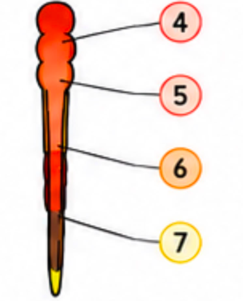
6
rhombencephalon
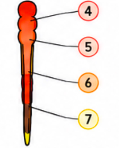
7
spinal cord
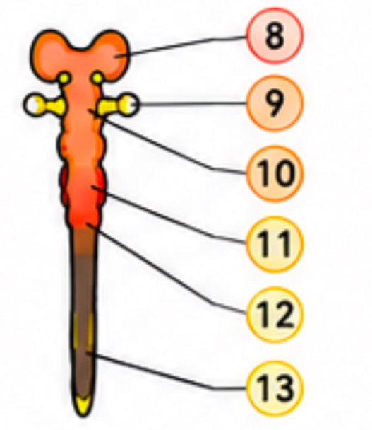
8
telencephalon

9
optic vesicle
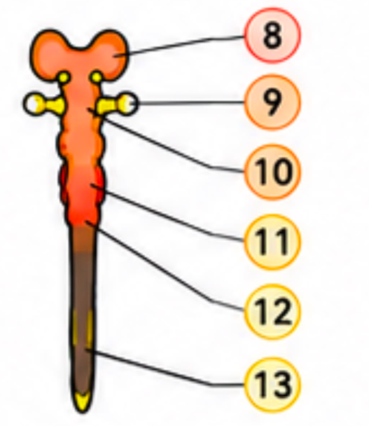
10
diencephalon