Anatomy exam 1 (Chapter 4)
 Call Kai
 Learn
Learn Practice Test
 Spaced Repetition
 Match
Match Flashcards
 Knowt Play
1/63

There's no tags or description
Looks like no tags are added yet.
Name | Mastery | Learn | Test | Matching | Spaced | Call with Kai | Chat |
|---|
No analytics yet
Send a link to your students to track their progress
64 Terms
What is the biological definition of a tissue?
Any grouping of similar living cells that work together to perform a common or specific physiological function.
What are the 4 primary tissue types in the human body, and what is the one-word primary function of each?
1) Nervous tissue (Control).
2) Muscle tissue (Movement).
3) Epithelial tissue (Covering).
4) Connective tissue (Support).
How do neurons and supporting cells (glial cells) differ in function within nervous tissue?
Neurons generate and transmit electrical impulses throughout the body in response to stimuli. Supporting cells protect, insulate, and nourish neurons, but cannot generate electrical impulses.
How do skeletal muscle, cardiac muscle, and smooth muscle differ in voluntary control and striations?
Skeletal muscle is voluntary and striated. Cardiac muscle is involuntary and striated. Smooth muscle is involuntary and non-striated.
Describe the cell shape and nucleus count of skeletal muscle tissue
Long, thread-like cells with multiple nuclei (multinucleate).

Describe the cell shape and nucleus count of smooth muscle tissue
Spindle-shaped cells with a single nucleus (uninucleate).

Describe the cell shape and nucleus count of cardiac muscle tissue
Branched cells with a single nucleus (uninucleate).

Where in the body are cardiac muscle located
walls of the heart

where in the body is skeletal muscle tissue found
in skeletal muscles attached to bones
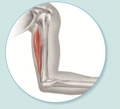
where in the body is smooth muscle tissue found
mostly in the walls of hallow organs
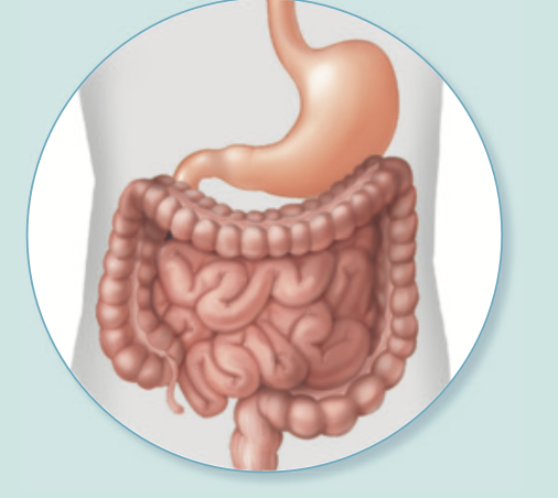
What are the two major functional forms of epithelial tissue in the human body?
1) Covering and lining epithelium (covers body surfaces, lines cavities and hollow organs).
2) Glandular epithelium (forms exocrine and endocrine glands).
What is apical-basal polarity in epithelial tissue?
Epithelial cells have two distinct sides: an apical surface that faces an open space (lumen or outside environment) and a basal surface attached to underlying tissue.
describe the structure and function of microvilli.
finger-like plasma membrane extensions that increase surface area for absorption/secretion.
describe the structure and function of cilia
hair-like projections that beat rhythmically to propel substances across the cell surface.
What are the 3 major functions of the epithelial basement membrane?
1) Anchors/holds epithelial cells in place.
2) Reinforces the epithelial sheet to resist tearing/stretch
3) Defines the physical boundary between epithelia and underlying connective tissue.
in which major functional type of epithelium are tight junctions and desmosomes exclusively found?
Covering and lining epithelia ONLY (they are absent in glandular epithelia).
What does it mean that epithelial tissue is "avascular but innervated"?
Avascular means it lacks its own blood vessels. Innervated means it is supplied with nerve fibers for sensation.
How do epithelial cells receive oxygen and nutrients and dispose of metabolic waste if they lack blood vessels?
Oxygen and nutrients diffuse upward from blood vessels in the underlying connective tissue through the basement membrane into epithelial cells; wastes diffuse in the opposite direction.
Why does epithelial tissue possess a high capacity for cellular regeneration?
because its exposed to abrasion frequently and chemical conditions, it requires rapid mitotic division to replace lost cells and prevent tissue loss
How is an epithelial tissue named based on cell layers and apical cell shape?
The first word describes layer thickness (Simple = 1 layer; Stratified = 2+ layers). The second word describes apical cell shape (Squamous = flat; Cuboidal = cube/box-like; Columnar = tall).
Why are simple epithelia specialized for absorption, filtration, and secretion rather than protection from abrasion?
because it’s a single layered tissue, absorption, filtration and secretion can be done easily with minimal distance (for fast passage of gases, fluids, and nutrients across tissue)
what is mesothelium?
simple squamous epithelium lining serous membranes (pleura, pericardium, peritoneum)
What is endothelium?
slick simple squamous lining inside blood vessels and heart chambers.
what are the 2 types of simple squamous epithelium?
endothelium and mesothelium
what specific epithelial tissue lines lung alveoli, why is its structure vital for gas exchange, and what would happen if it became multi-layered?
simple squamous epithelium (mesothelium). Its single layer of flat cells provides an ultra-thin barrier for rapid, efficient gas exchange. If it became multi-layered (thickened), gas diffusion speed would drop significantly, creating a severe functional disadvantage (hypoxia).
Where is simple columnar epithelium typically found in the digestive system, and what apical modification enhances its absorptive function?
Found lining the stomach and intestines (e.g., small intestine). Its apical surface contains microvilli, which increase surface area to maximize nutrient absorption.
Why is pseudostratified columnar epithelium termed "false," and how can it be distinguished from true stratified columnar tissue?
It appears multi-layered because cell nuclei sit at varying heights, but it is "false" because every single cell rests directly on the basement membrane (making it a single simple layer).
Why are stratified epithelia specialized for protection from abrasion rather than absorption or secretion?
Multiple cell layers form a thick, sacrificial barrier where superficial apical layers can rub off during friction without damaging underlying tissues.
What structural change causes apical cells in transitional epithelium (urothelium) to alter their shape, and where is this tissue found?
Stretching of the organ wall as urine accumulates causes apical cells to flatten from dome/cuboidal shapes into squamous-like shapes. It is found exclusively in the urinary system (bladder, ureters).
What do exocrine glands release/secrete and where?
they release non-hormone secretions onto body surfaces or into cavities via ducts
What do endocrine glands release/secrete and where?
they release hormones directly into extracellular fluid and blood.
How does a merocrine gland release its secretion, and what happens to the cell during this process?
Secretion is released via exocytosis. The cell remains completely intact and undamaged during secretion (e.g., eccrine sweat glands, salivary glands).
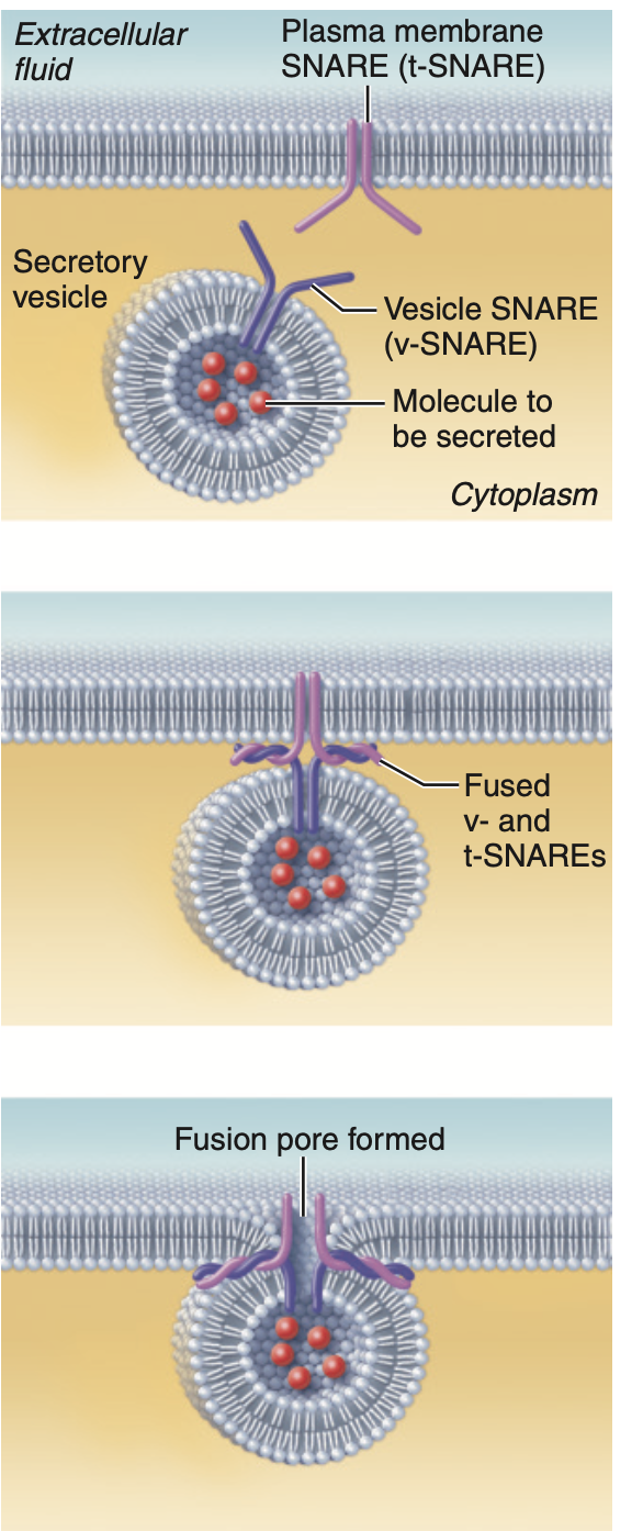
what is this picture showing
exocytosis
How does a holocrine gland release its secretion, and what is the only body example of this gland type?
Secretory products accumulate inside the cell until the entire cell ruptures and dies to release the product. Sebaceous (oil) glands are the only example.
How does an apocrine gland release its secretion, and what is a primary body example?
Secretory products accumulate at the apical cell tip, and the apical portion pinches off (partial cellular rupture), after which the plasma membrane quickly repairs itself. Mammary glands are a primary example.
What structural feature distinguishes connective tissue from all other primary tissue types, and what two main components make up its nonliving matrix?
they are widely separated by an abundant nonliving extracellular matrix (ECM). The ECM is composed of ground substance and fibers.
From what embryonic tissue do all classes of connective tissue originate?
Mesenchyme
What are the 3 chemical components of connective tissue ground substance?
1) Interstitial fluid (ISF).
2) Cell adhesion proteins (glue holding cells to ECM).
3) Proteoglycans (protein-carbohydrate complexes that trap water).
connective tissue is composed of….
1) fibers 2) ground substance 3) cells
How does adding proteoglycans to ground substance affect its physical viscosity (resistance to flow)?
binds more water, making the ground substance thicker, firmer, and more gel-like (e.g., cartilage matrix).
What structural characteristics give collagen fibers their high tensile strength?
They are composed of thick strands of collagen protein that assemble and cross-link with neighboring collagen fibers, resisting pulling and tearing forces.
What physical capability do elastic fibers provide to tissues, and why is this capability important in organs like lungs and skin?
allow tissues to stretch under tension and recoil back to their original shape without tearing, enabling repeated expansion and contraction.
What is the structural arrangement and function of reticular fibers, and where are they located?
Short, fine, extensively branched collagenous fibers that form a net-like internal framework (stroma) supporting free blood cells in lymph nodes, the spleen, and red bone marrow.
How do "-blast" cells differ functionally from "-cyte" cells in connective tissue?
-blast = immature cell , mitotically active
-cyte = mature , mitotically inactive to monitor blast cells
What are the chief cell type and main functions of areolar loose connective tissue?
Fibroblasts are the chief cell type. Functions include supporting, wrapping, and cushioning tissues, nourishing neighboring cells, and storing fluid (found almost everywhere in the body).
What is the chief cell type of adipose tissue
the adipocyte, filled with triglycerides.
what lipid fills adipose tissue, and where is subcutaneous fat located?
triglycerides; located deep to the dermis, providing thermal insulation, shock absorption, and fast energy storage.
How does the fiber arrangement in dense regular connective tissue compare to dense irregular connective tissue, and how does this affect tension resistance?
Dense regular has collagen fibers running parallel in one direction. high resistance to tension in 1 direction
Dense irregular has collagen fibers running in multiple directions. high resistance to tension in all directions
What are the chief cell types of cartilage?
chondroblasts (immature) and chondrocytes (mature)
why does cartilage take so long to heal?
because it is completely avascular and uninnervated, causing slow and difficult tissue healing.
What chemical composition makes osseous (bone) tissue rigid, and what are its chief cell types?
Inorganic calcium salts deposited on collagen fibers make bone matrix rigid. Chief cell types are osteoblasts and osteocytes.
Why is a rich blood supply to bone tissue physiologically essential?
Bone is highly metabolically active; blood delivers calcium/nutrients for constant remodeling, rapid fracture repair, and red bone marrow nourishment.
Why are fibers normally absent in circulating blood, and under what specific condition do blood fibers form?
Fibers are absent during normal circulation to keep blood fluid. Fibers (fibrin mesh) form only during blood clotting to stop blood loss following vessel injury.
unlike epithelial tissue, connective tissue…
has nonliving ECM (ground substance + fibers), and vascularized
unlike connective tissue, epithelial tissue…
has specialized cell junctions (desmosomes, tight junctions), and avascularized with no ECM
What events occur during Step 1 of tissue repair
Severed vessels bleed, a blood clot forms to seal the wound, and the clot surface dries into a scab
in tissue repair, why do surrounding undamaged blood vessels become more permeable?
so white blood cells, fluid, clotting factors, and repair nutrients can easily enter the damaged area.
What happens during Step 2 of tissue repair to restore blood supply and initiate scar formation?
Blood clot is replaced by vascular granulation tissue. Fibroblasts lay down collagen fibers to form scar tissue, while macrophages phagocytize dead cells and debris.
what is final wound healing (Step 3) of tissue repair
Fibrosis replaces damaged tissue with dense collagen scar tissue, but tissue loses specialized function
How does tissue regeneration differ from fibrosis during final wound healing (Step 3)
Regeneration replaces damaged tissue with identical, fully functional parenchymal cells. Fibrosis cannot and replaces it with scar tissue (with no specialized function)
What is a neoplasm, and how do benign tumors differ from malignant tumors?
A neoplasm is an abnormal mass of tissue cells resulting from unregulated cell division. Benign tumors are slow-growing, localized, and encapsulated. Malignant tumors are unencapsulated, fast-growing, and invasive.
What does it mean if a malignant tumor becomes metastatic?
tumors spread out to establish secondary tumor sites
What is a mutated proto-oncogene called, and what role do tumor suppressor genes normally play in cell division?
oncogenes; tumor suppressor genes normally produce proteins that inhibit unregulated cell division and prevent tumor growth.
how does a mutation disrupting fibrillin (Marfan syndrome) affect elastic fiber function in the lungs and skin?
Elastic fibers lose recoil capability. Lungs remain permanently stretched out during breathing, impairing gas exchange. Skin loses elasticity and stretches permanently, forming stretch marks.