Neuro Quiz 5 whoop whoop! You got this!
1/61
There's no tags or description
Looks like no tags are added yet.
Name | Mastery | Learn | Test | Matching | Spaced |
|---|
No study sessions yet.
62 Terms
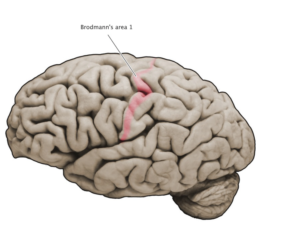
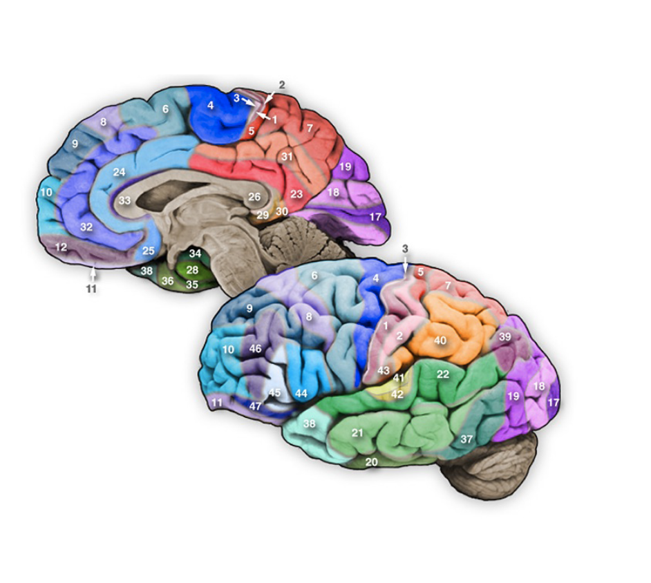
Brodmann's Area 1
Sensory cortical area in the crest of the postcentral gyrus; this area is a component of the primary somatosensory cortex.
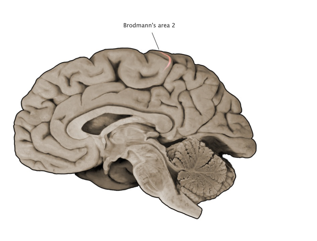
Brodmann's Area 2
Sensory cortical area in the crest and posterior bank of the postcentral gyrus; this area is a component of the primary somatosensory cortex.
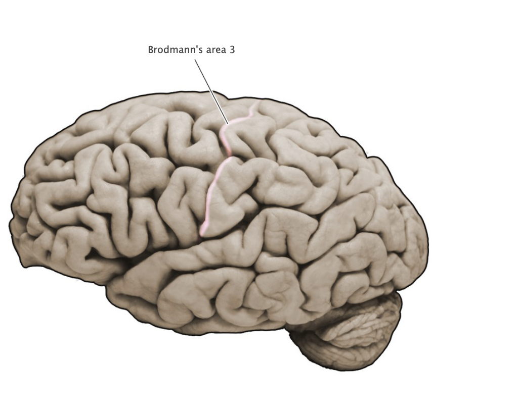
Brodmann's Area 3
Sensory cortical area in the posterior bank of the central sulcus (postcentral gyrus); this area is a principal component of the primary somatosensory cortex. Area 3 is further subdivided into area 3a, which receives proprioceptive signals that originate in deep receptors, and area 3b, which receives discriminitive mechanosensory signals that arise from cutaneous receptors.
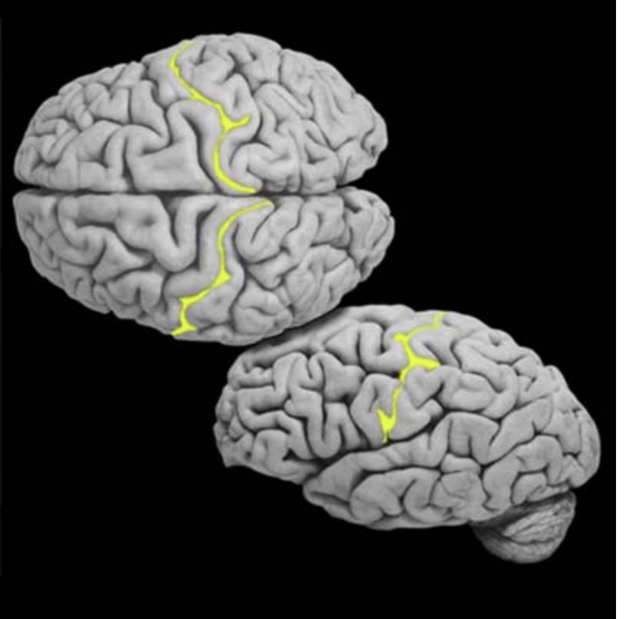
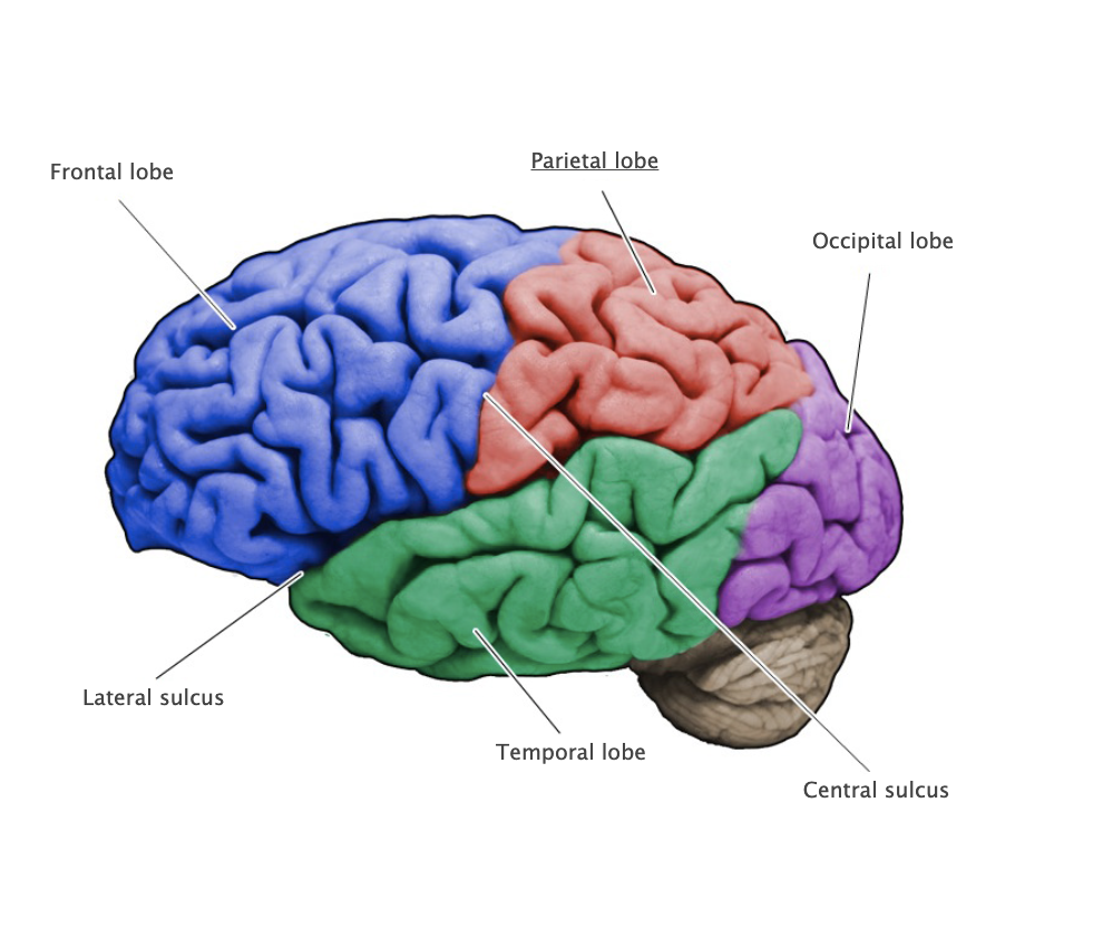
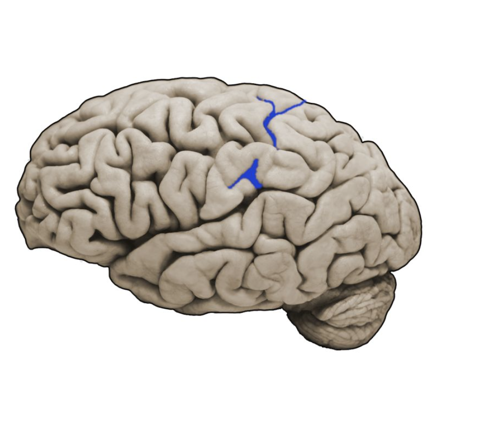
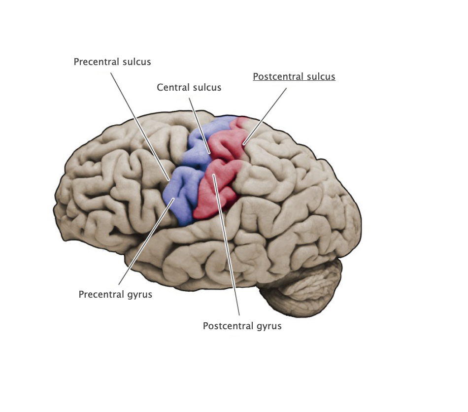
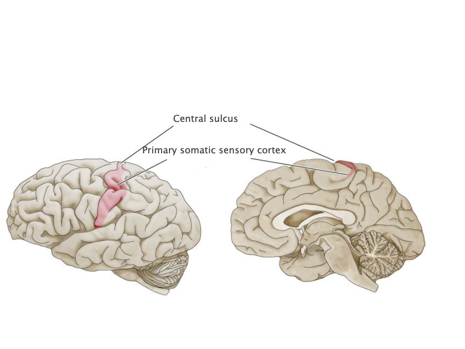
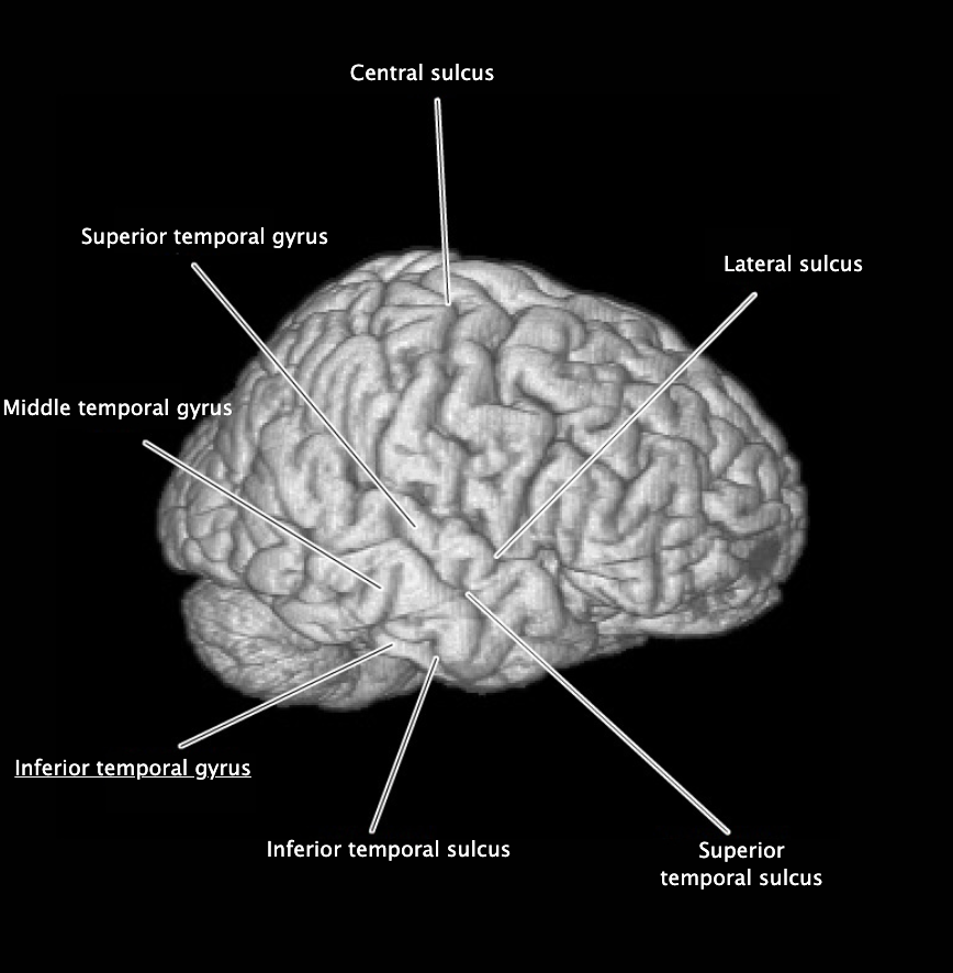
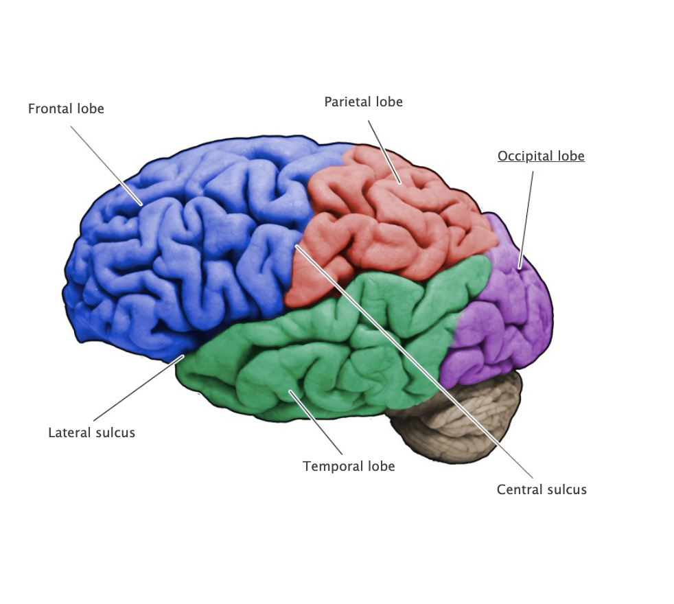
Central Sulcus
A prominent sulcus on the dorsal-lateral aspect of the cerebral hemispheres formed by that precentral and postcentral gyri, this sulcus defines the boundary between the frontal and parietal lobes.
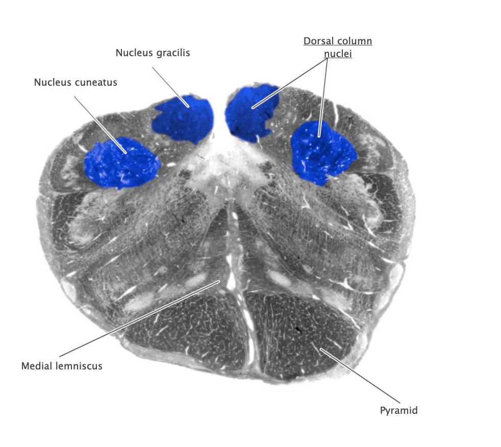
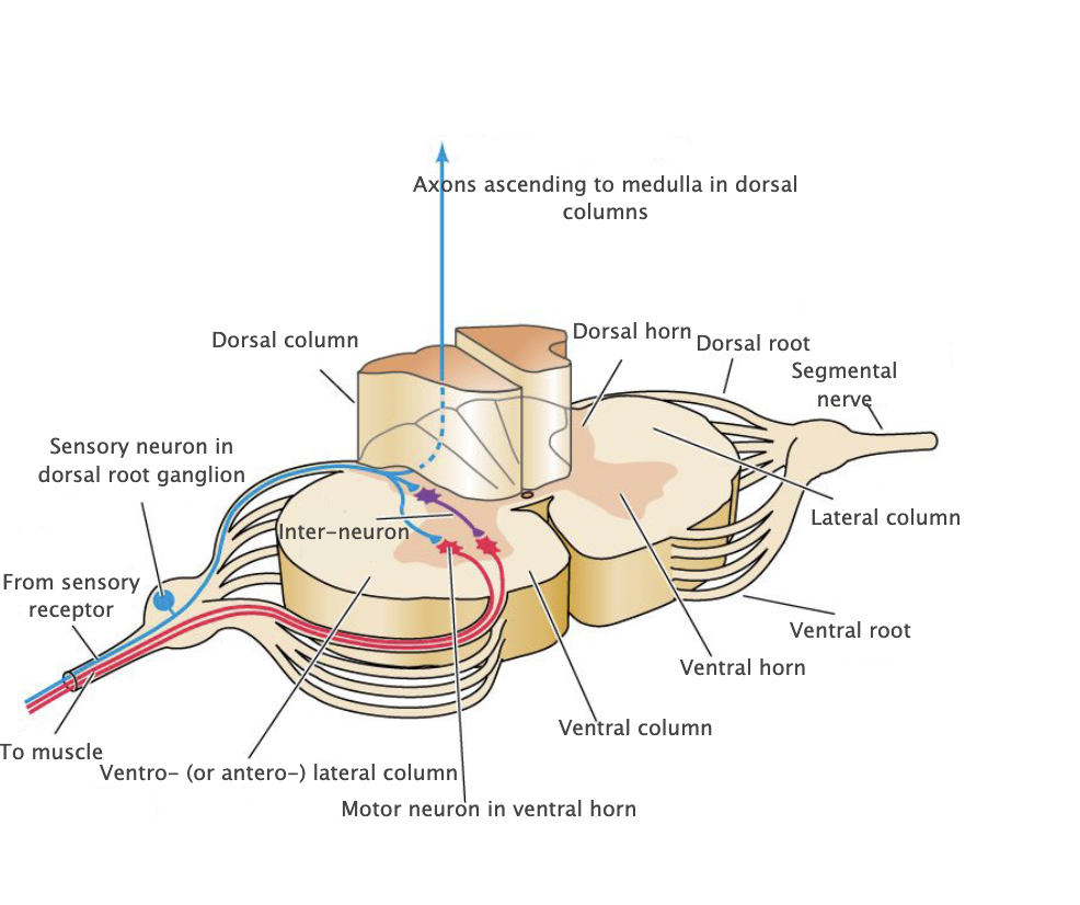
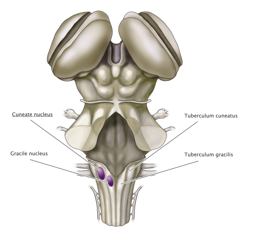
Dorsal column nuclei
Gracile nucleus and cuneate nucleus; contain the second-order sensory neurons that relay mechanosensory information from peripheral receptors in the body (excluding the face) to the thalamus via the medial lemniscus. The dorsal column nuclei are located in the lower medulla.

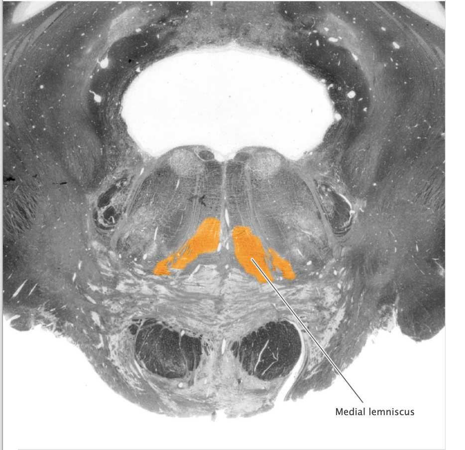
Dorsal column-medial lemniscal system
Ascending somatic sensory pathway that conveys tactile and proprioceptive information for the body and posterior third of the head.
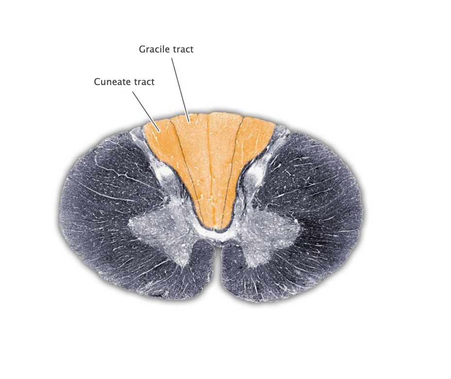
Dorsal columns
White matter region between the dorsal horns of the spinal cord containing the gracile and cuneate tracts.
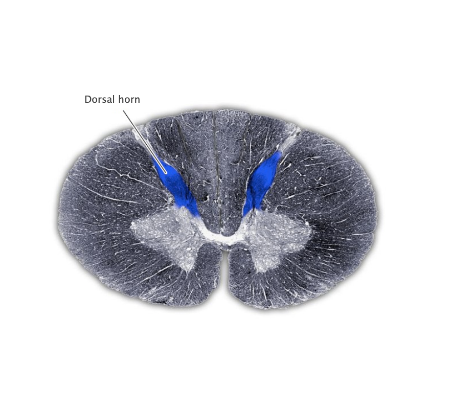
Dorsal horn
The dorsal (sensory) portion of the spinal cord gray matter.
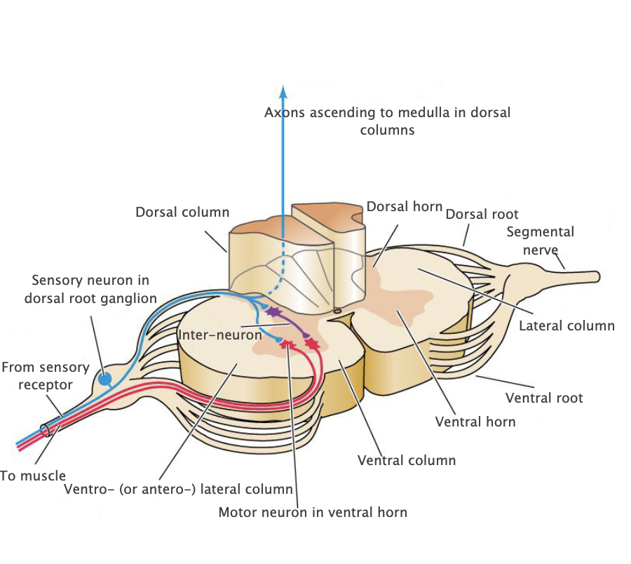
Dorsal root ganglia
Segmental sensory ganglia of the spinal cord.

Dorsal roots
Bundle of axons that runs from the dorsal root ganglia to the dorsal horn of the spinal cord carrying sensory information from the periphery.
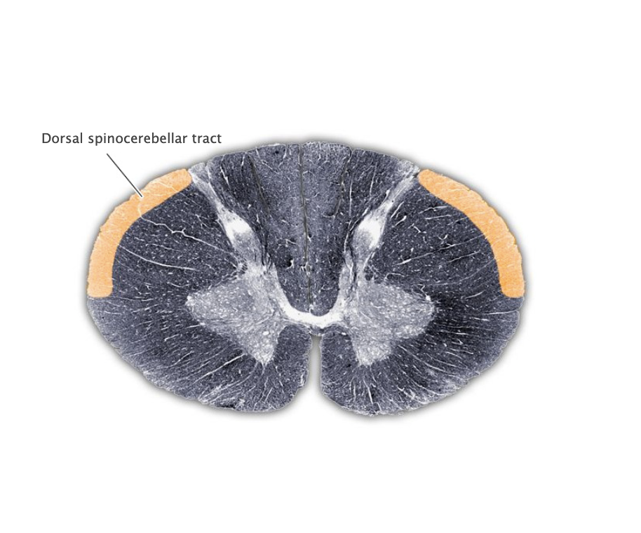
Dorsal spinocerebellar tract
Ascending sensory tract of the spinal cord originating in the dorsal nucleus of Clarke and terminating in the cerebellum (via the inferior cerebellar peduncle); this tract conveys proprioceptive information from the lower body.
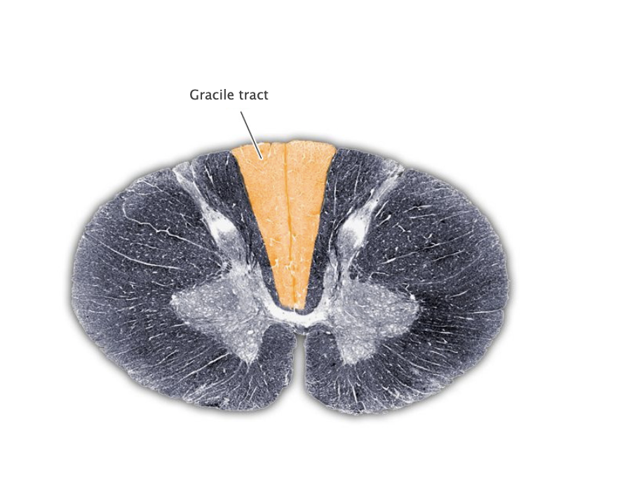
Gracile tract
Medial portion of the dorsal columns comprising the central processes of dorsal root ganglion axons that convey mechanosensory information from the lower body. Also referred to as the "fasciculus gracilis."
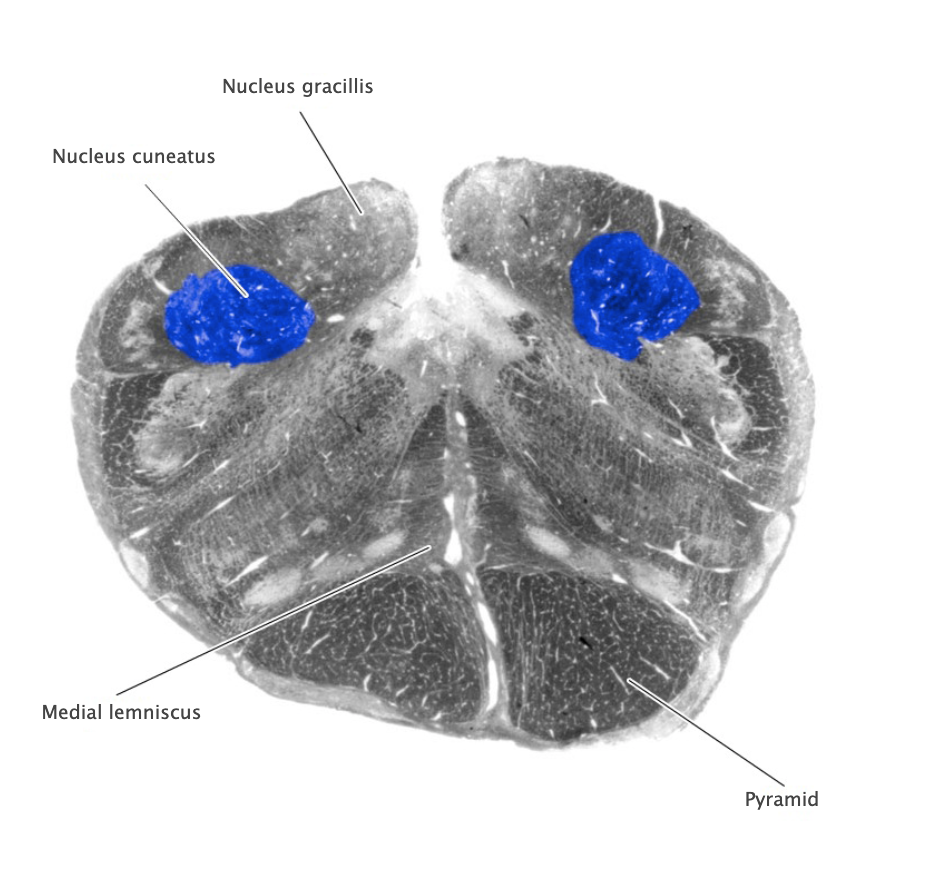
Nucleus cuneatus
Dorsal column nucleus in the lower medulla; contains second-order sensory neurons that relay mechanosensory information from peripheral receptors in the upper extremities to the thalamus via the medial lemniscus.

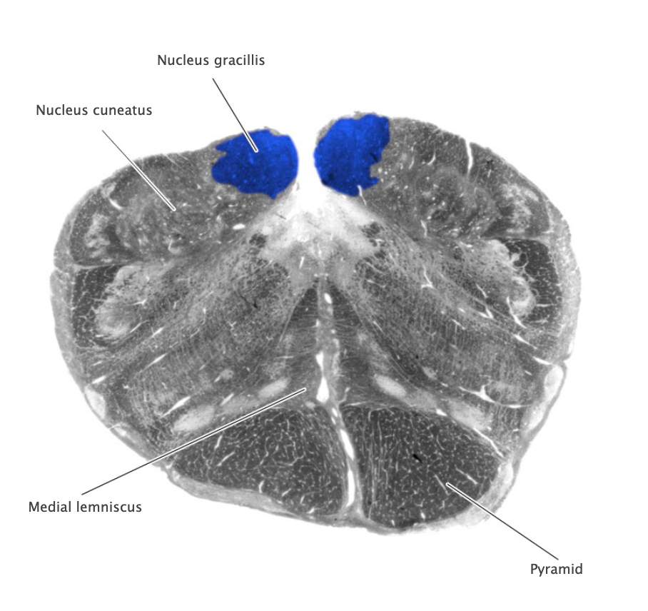
Nucleus gracilis
Nucleus containing the second-order sensory neurons that relay mechanosensory information from peripheral receptors in the lower body to the thalamus via the medial lemniscus, located in the lower medulla (also called the "gracile nucleus"). The nucleus gracilis also relays visceral pain information from second-order neurons in the central spinal cord to the thalamus.
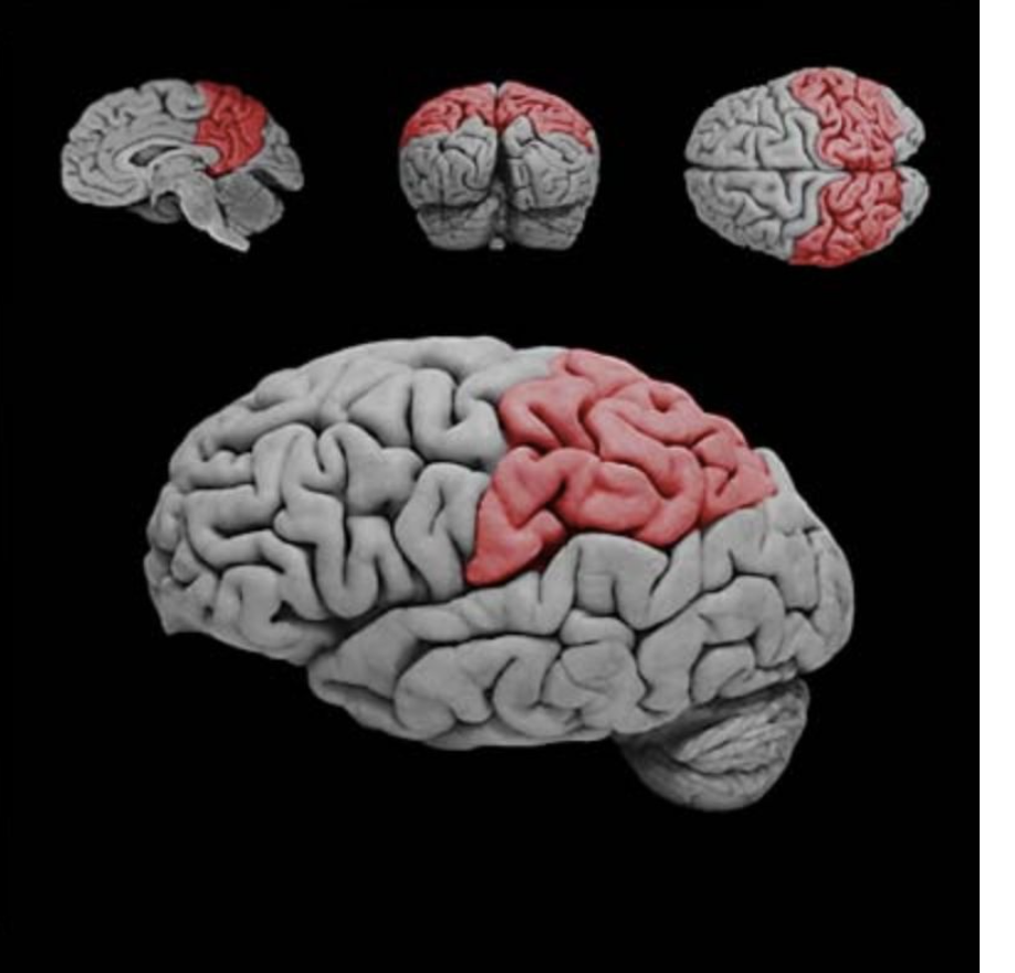
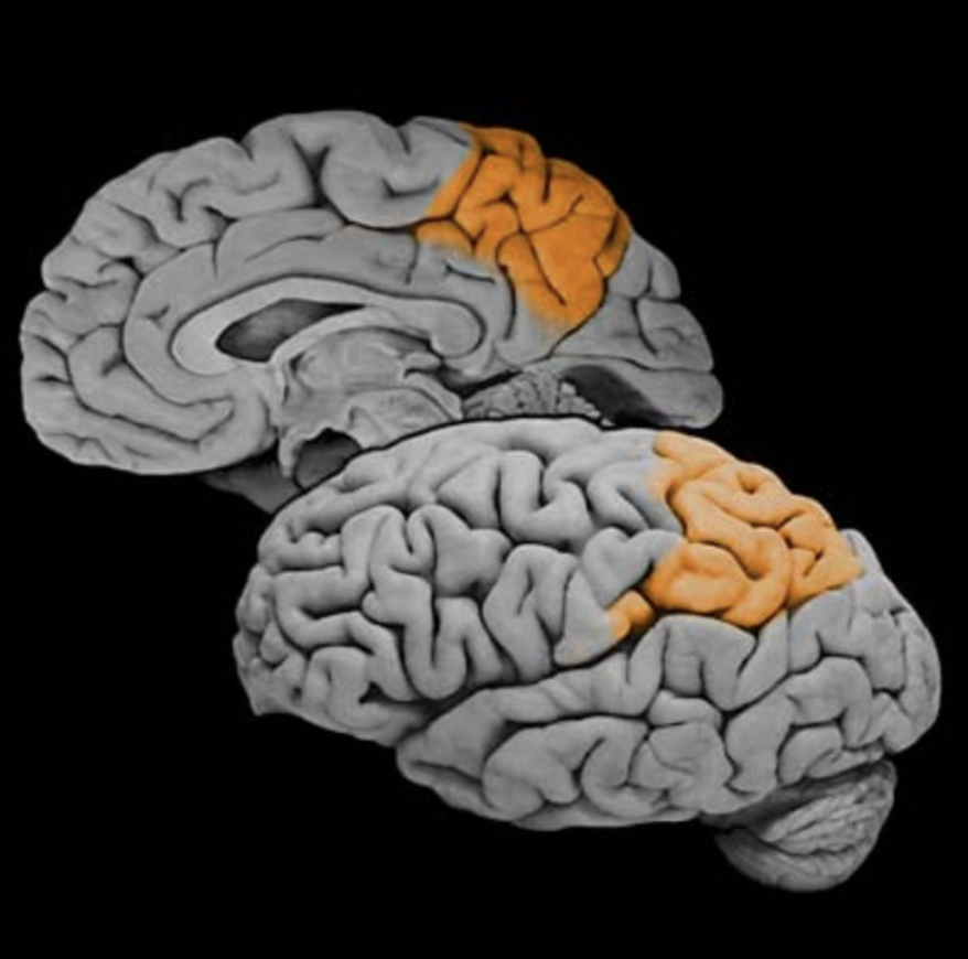
Parietal lobe
Region of the cerebral cortex posterior to the central sulcus, anterior to the occipital lobe and superior to the lateral fissure; involved in somatic sensation and numerous complex functions, including multimodal proprioception, language comprehension, attention, and spatial awareness.
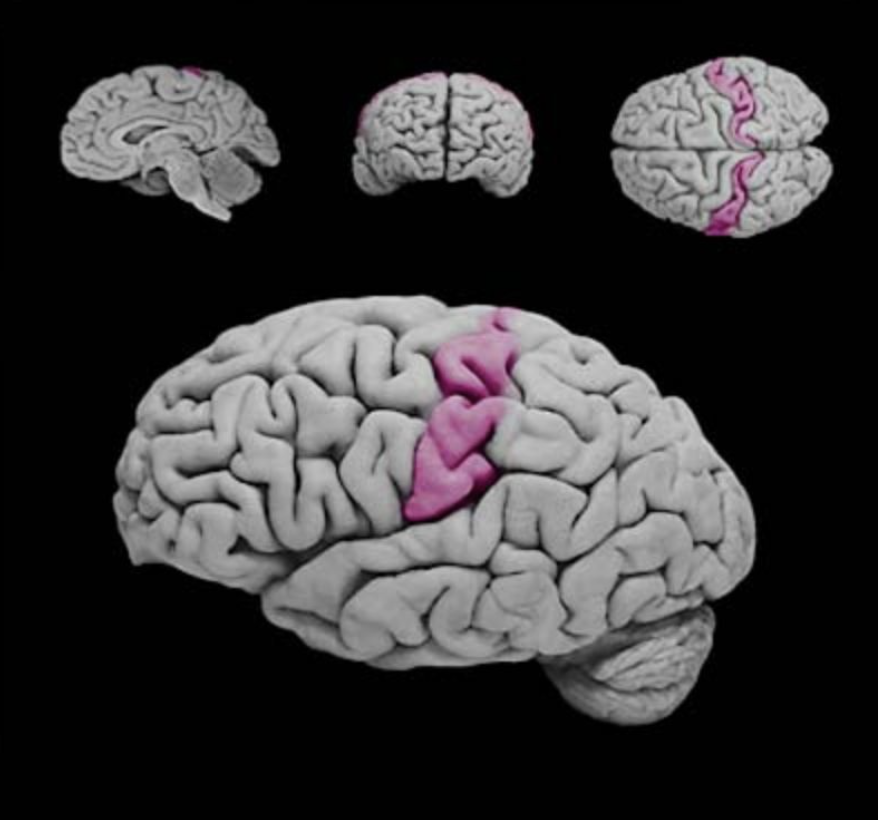
Postcentral gyrus
Vertically oriented gyrus that forms the posterior bank of the central sulcus; this gyrus contains the primary somatosensory cortex.
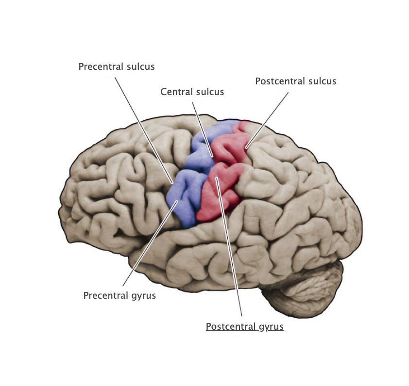
Postcentral sulcus
Vertically oriented sulcus at the posterior margin of the postcentral gyrus.
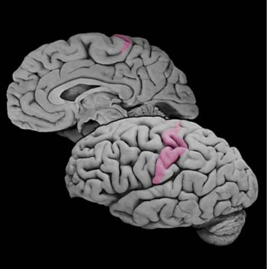
Primary somatic sensory cortex
Cortical areas (Brodmann's areas 3, 1 and 2) in the postcentral gyrus that first receive somatosensory signals from the relevant thalamic nuclei (ventral posterior complex). Also called primary “somatosensory” cortex.
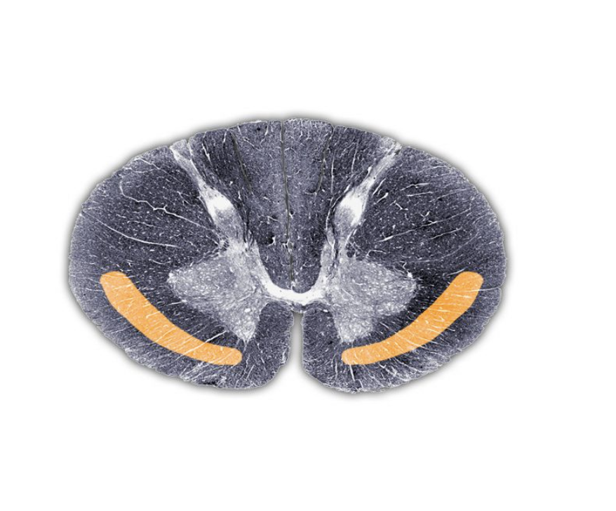
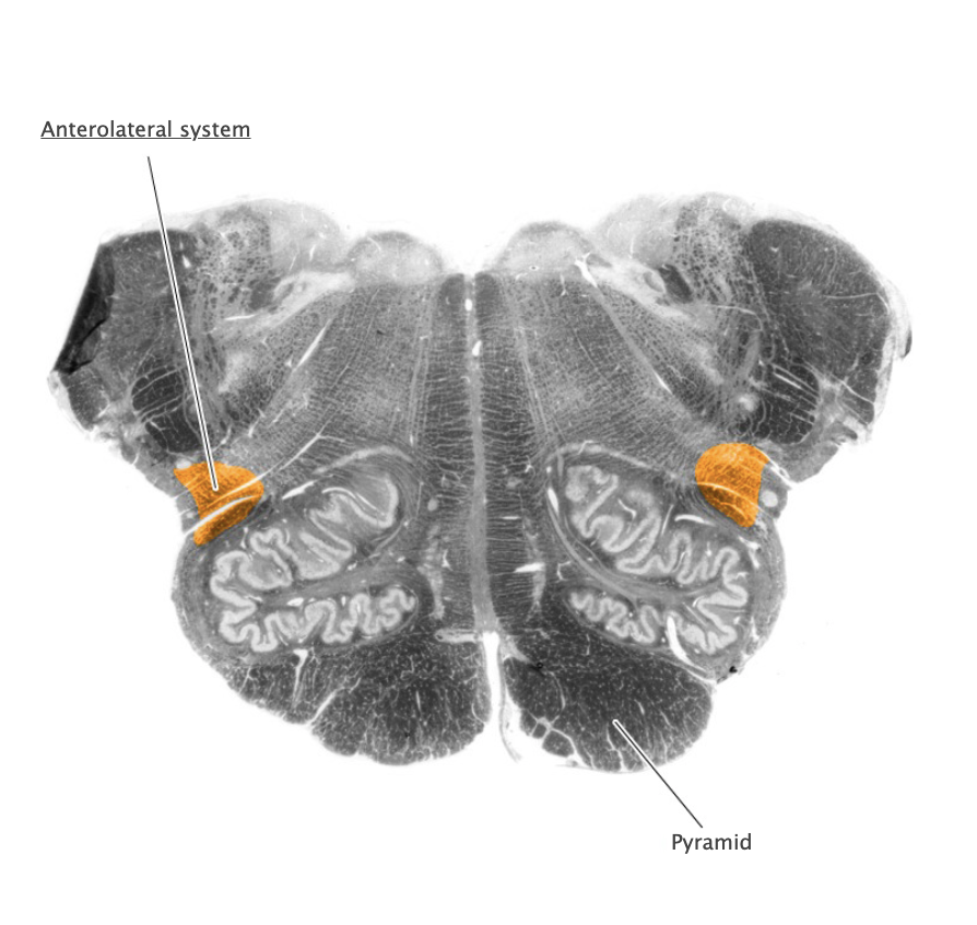
Spinothalamic tract
nerve pathway from the spine to the thalamus along which pain impulses are carried to the brain, major component of the anterolateral system
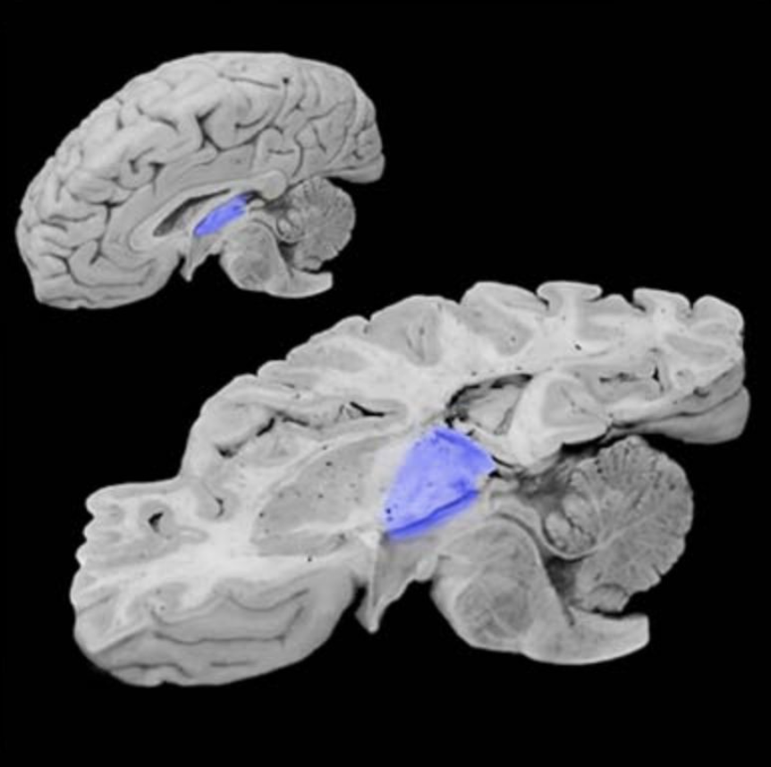
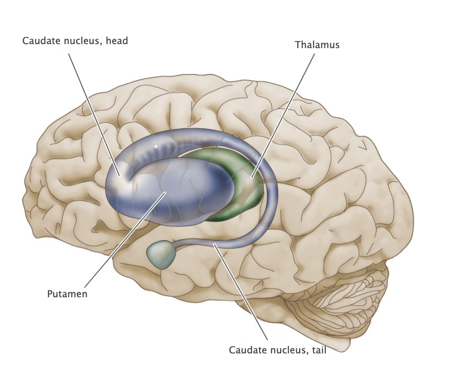
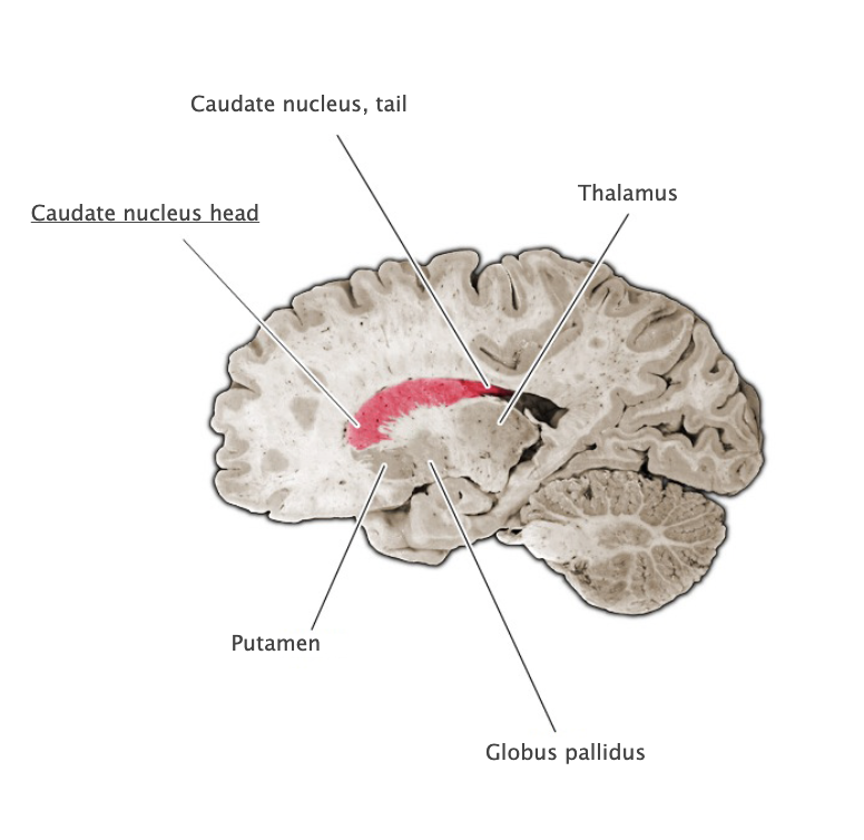
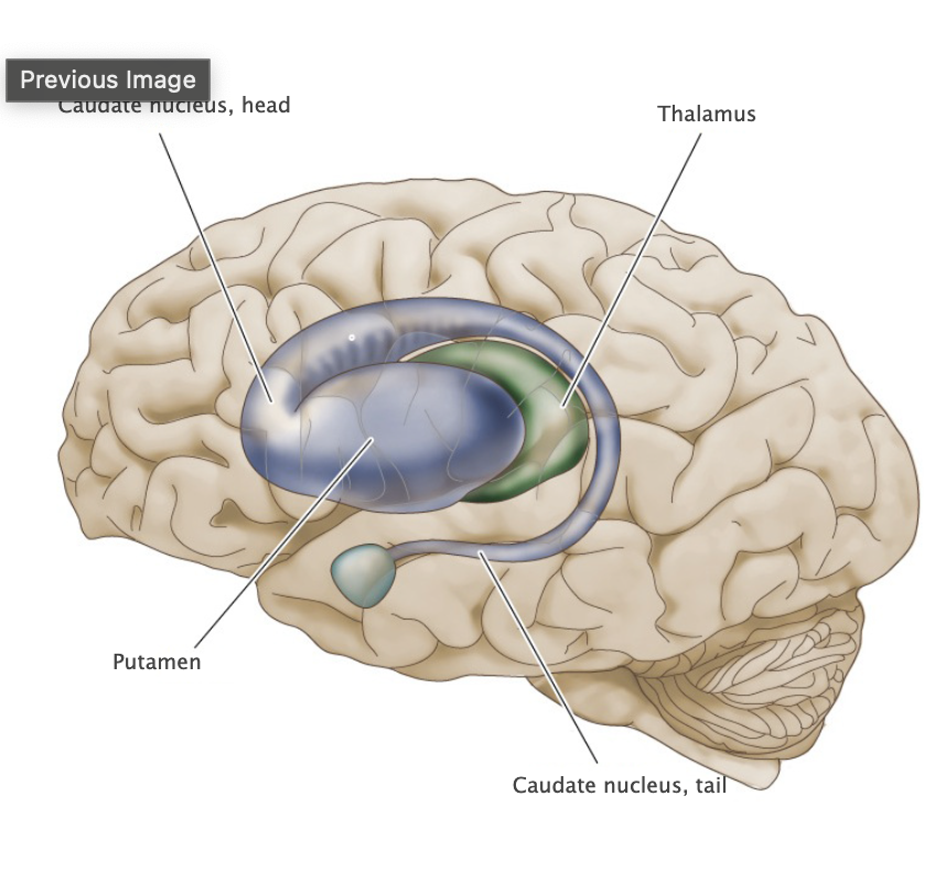
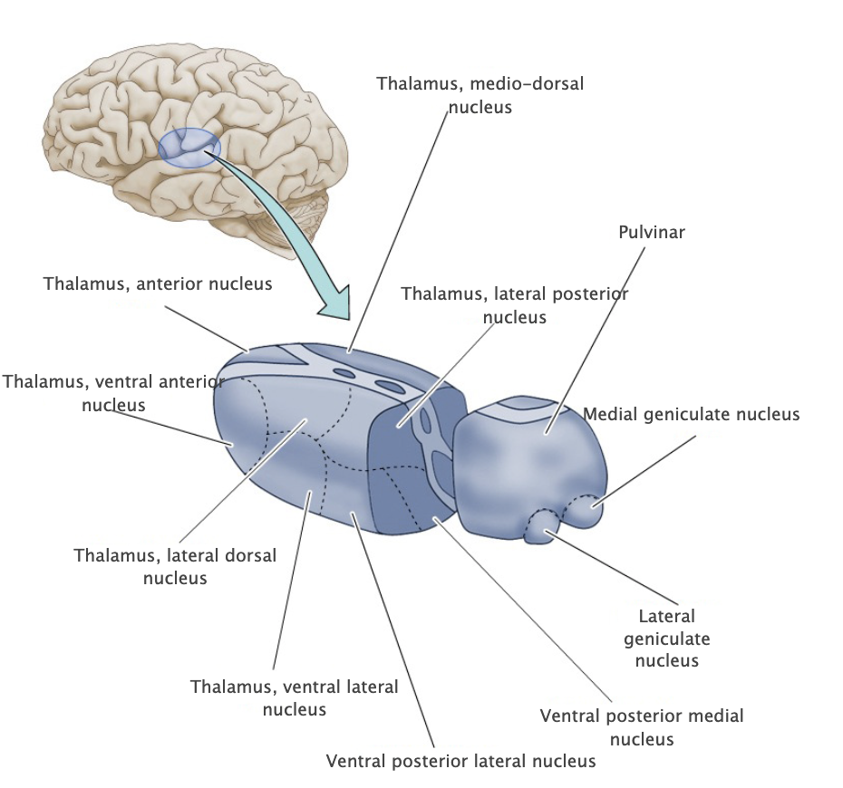
Thalamus
Major division of the diencephalon comprising numerous nuclei that are heavily interconnected to the cerebral cortex. Major functional roles of the thalamus include relaying sensory information from brainstem and spinal cord regions to specific areas of the cerebral cortex, and relaying motor signals from the basal ganglia and cerebellum to the motor cortex.
Basal Ganglia
A group of prominent nuclei lying deep in the subcortical white matter of the cerebral hemispheres that are involved in motor and cognitive functions. The caudate nucleus, putamen, and the globus pallidus are the major components of the basal ganglia; however, additional structures, including the subthalamic nucleus and substantia nigra, modulate the processing of signals in the intrinsic circuitry of the basal ganglia. The basal ganglia serve to regulate the expression of bodily movements, affective behavior and certain cognitive processes by integrating widespread inputs from the cerebral cortex and modulating the activity of relevant thalamic relay nuclei.
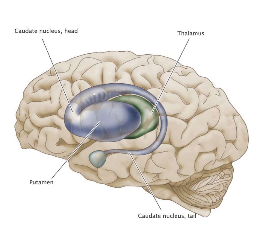

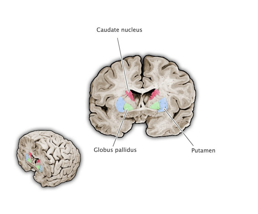
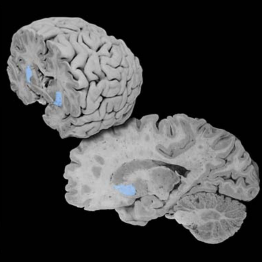
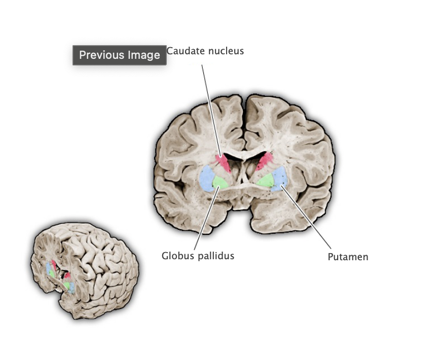
Caudate nucleus head
Rostral portion of the caudate nucleus.
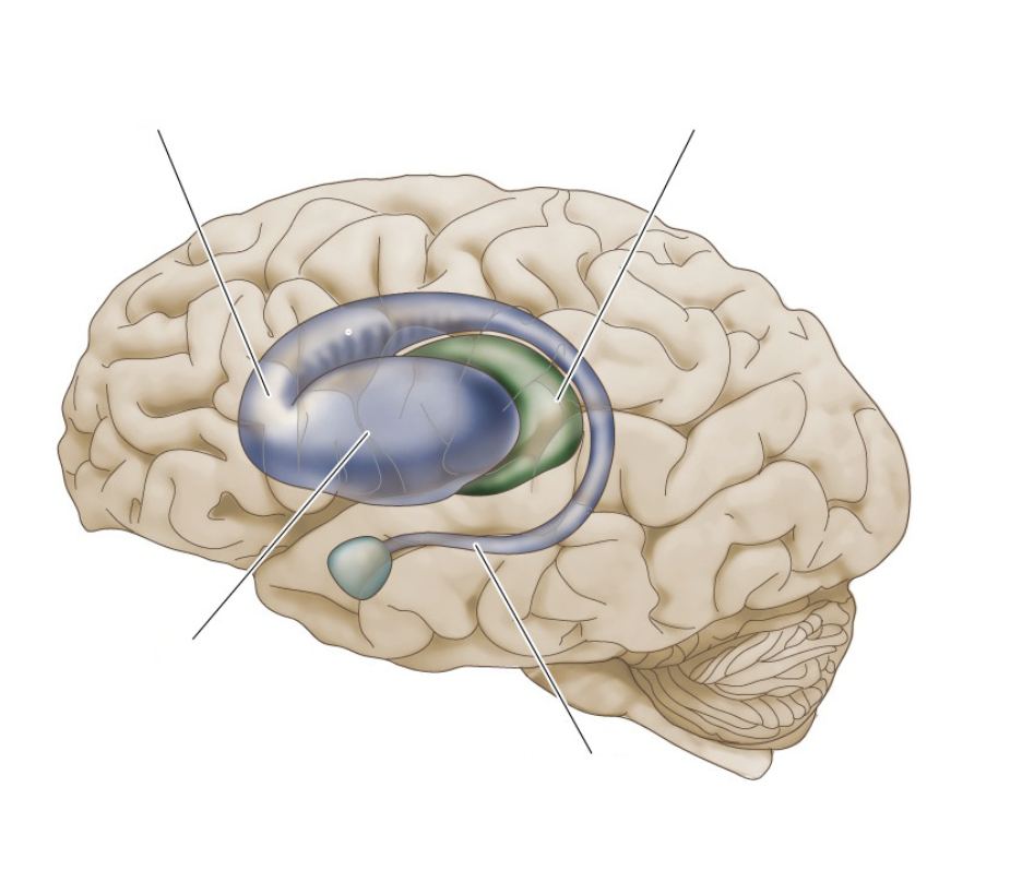
Caudate nucleus tail
Caudal and ventral portion of caudate nucleus that extends into the temporal lobe near the wall of the lateral ventricles.

Globus pallidus
Nucleus of the basal ganglia whose medial division (internal segment) provides the principal output of the basal ganglia to the motor nuclei of the thalamus; the lateral division (external segment) participates in intrinsic circuits that modulate the activity of the medial division.
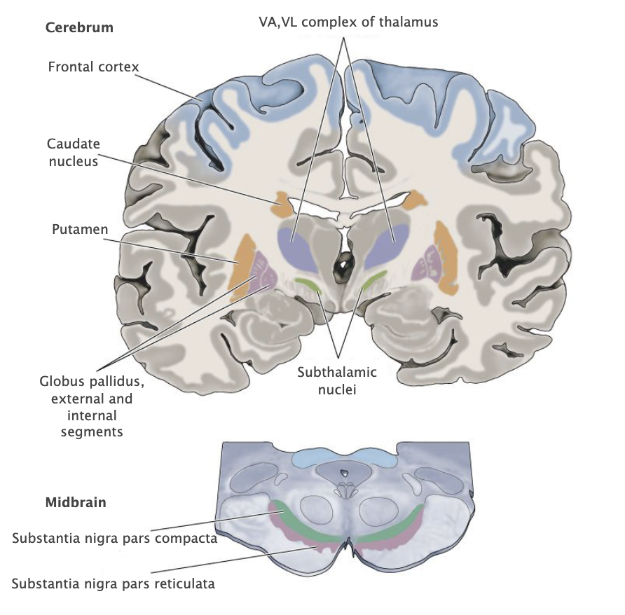
Putamen
A major gray matter component of the basal ganglia representing the lateral aspect of the striatum; located deep in the frontal lobe, just lateral to the internal capsule, caudate nucleus and globus pallidus. Together with other structures in the basal ganglia, the putamen is involved in regulating the expression of bodily movements by integrating widespread inputs from the cerebral cortex and modulating the activity of relevant thalamic relay nuclei.
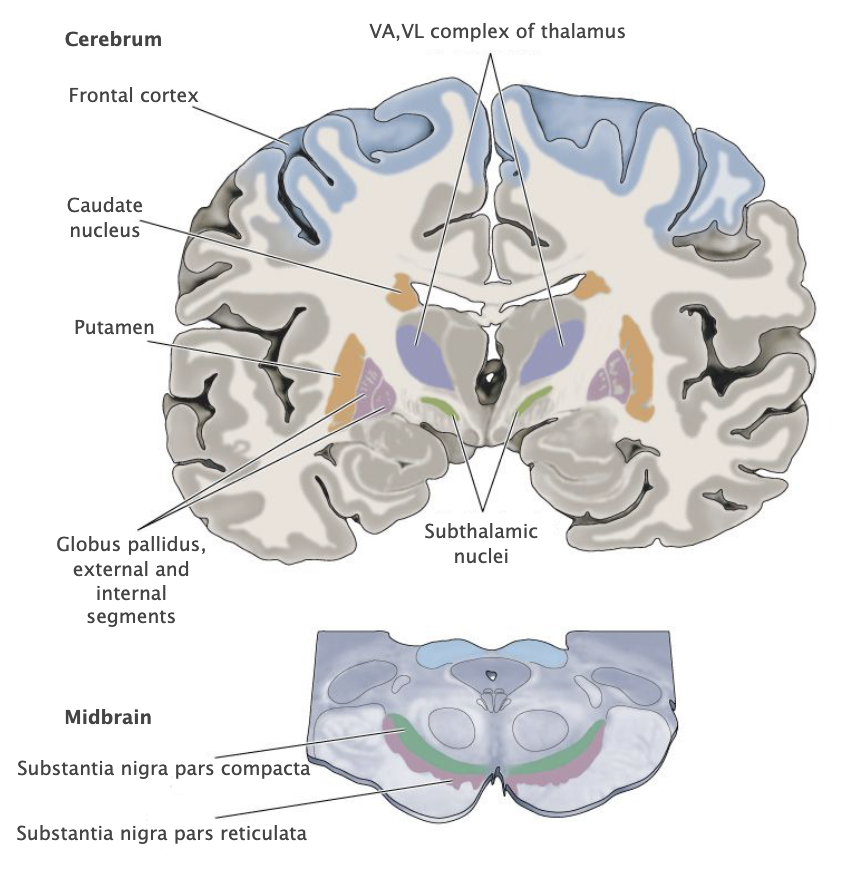
Striatum
Grey matter structures of the basil ganglia that receive command signals from the cerebral cortex and modulatory signals from dopaminergic neurons in the midbrain; provides phasic inhibitory input to pallidal structures. The striatum comprises two major components, the caudate nucleus and the putamen, plus additional ventral structures, including the nucleus accumbens and the olfactory tubercle.

Internal capsule
Large white matter structure that lies between the diencephalon and the basal ganglia comprising numerous cortical and thalamic projection fibers, including sensory axons that run from the thalamus to the cortex and motor axons that run from the cortex to the brainstem and spinal cord.
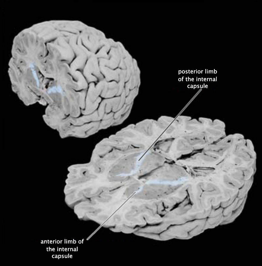
Internal capsule, anterior limb
Anterior portion of the internal capsule that lies between the caudate nucleus and the putamen.

Internal capsule, posterior limb
Posterior portion of the internal capsule that lies between the putamen and the thalamus.
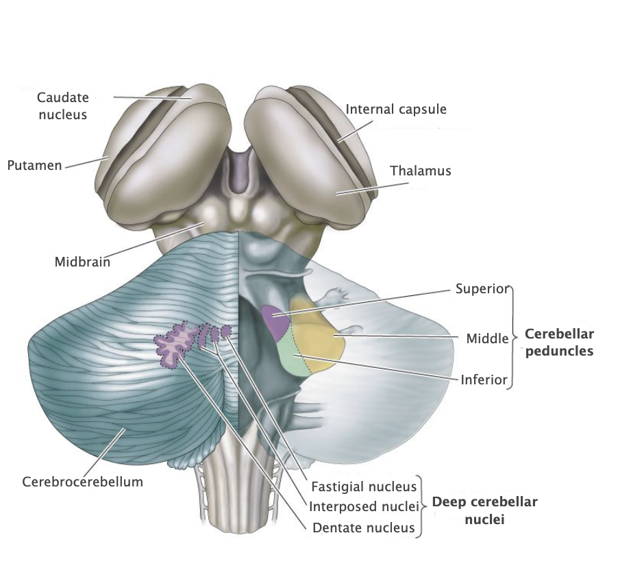
Cerebral peduncles
Three major white matter structures that contain efferent and afferent axons of the cerebellum (peduncle means stalk). These three peduncles are named the inferior, middle, and superior cerebellar peduncles.
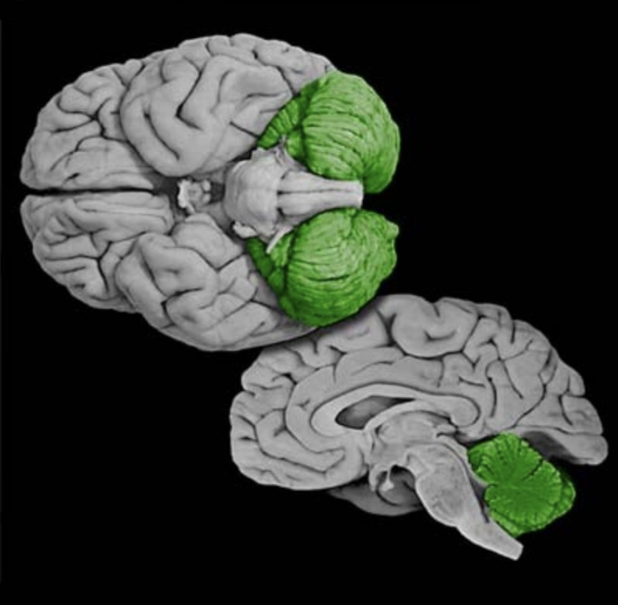
Cerebellum
Prominent convoluted hindbrain structure (Latin for "little brain") consisting of a three-layered cortex, subcortical white matter, deep nuclei, and three peduncles that connect the cerebellum and brainstem. The cerebellum is concerned with the coordination of ongoing movement, motor planning, and motor learning; it also plays a role in coordinating and sequencing cognitive processing.
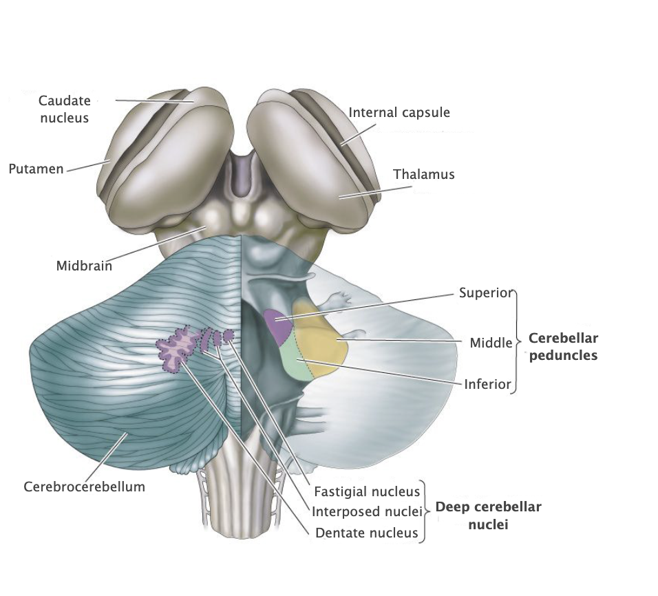

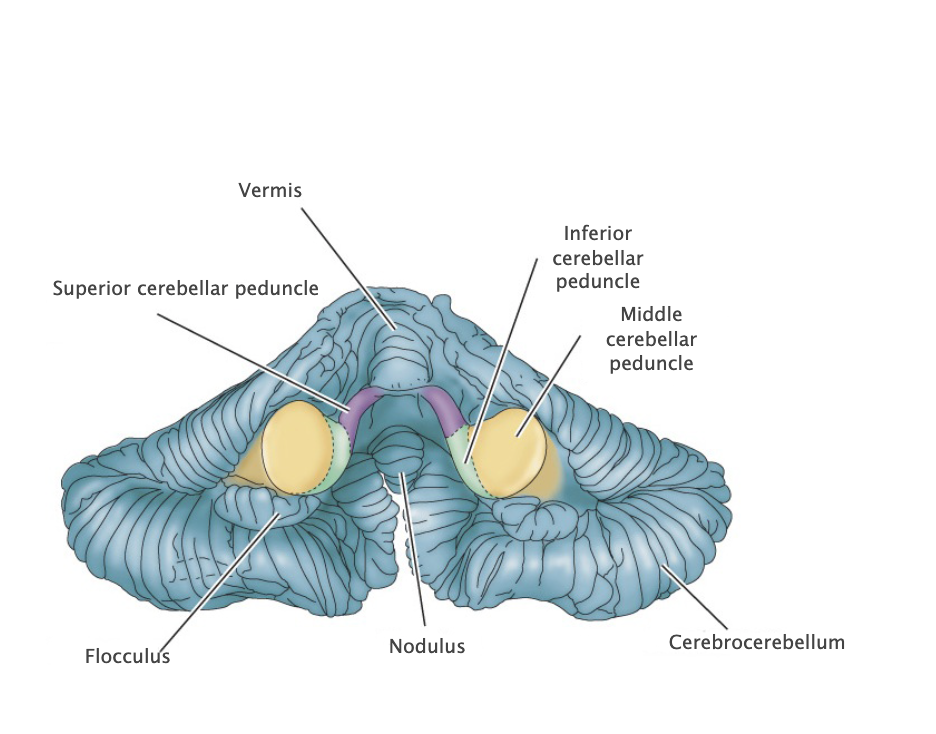
Cerebellar peduncles (superior, middle, inferior)
Three major white matter structures that contain efferent and afferent axons of the cerebellum (peduncle means stalk). These three peduncles are named the inferior, middle, and superior cerebellar peduncles. Prominent structures (peduncle means "stalk") observed on the ventral surface of the midbrain comprising the tegmentum and the more ventrally located white matter tracts (basis pedunculi). In common usage, the term cerebral peduncle denotes these white matter tracts, which contain the efferent axons of the cerebral cortex that project to the brainstem and spinal cord.
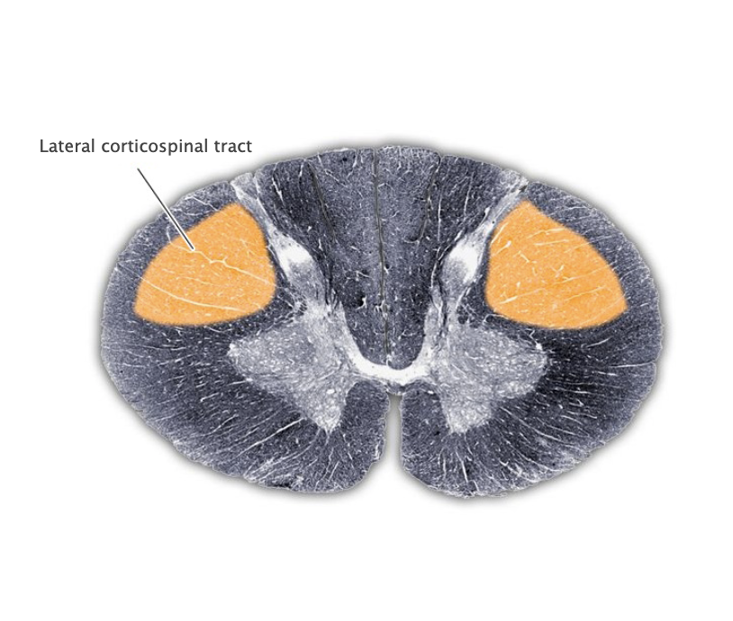
Lateral corticospinal tract
Decussating component (about 90% of total) of the corticospinal projection that originates in the motor cortical areas of the posterior frontal lobe and the postcentral gyrus. The lateral corticospinal tract is located in the lateral white column of the spinal cord and terminates in the lateral portion of the ventral horn, where it governs the lower motor neuronal pools that control the distal extremities.
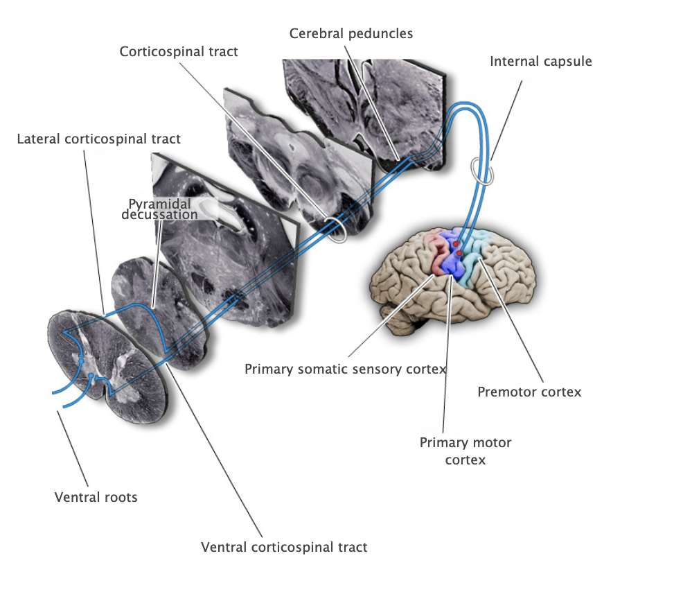

Ventral corticospinal tract
Non-decussating component (about 10% of total) of the corticospinal projection that originates in the posterior frontal lobe, terminates bilaterally in the medial aspect of the spinal cord ventral horn, where it is involved in the control of axial and proximal limb musculature (also called the "anterior corticospinal tract").

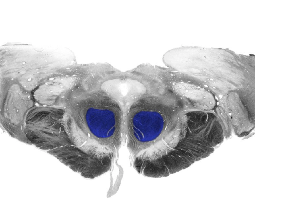
Red nucleus
Prominent nucleus located in the tegmentum of the rostral midbrain. Its major parvocellular (small-celled) division conveys signals from the cerebral cortex to the inferior olivary nucleus, which in turn modulates activity in the cerebellum. A minor magnocellular (large-celled) division may contribute to the control of the distal upper extremities via projections to the lateral ventral horn of the spinal cord, projections that form the rubrospinal tract; however, this projection is vestigial and variable in humans and its functional significance is uncertain.
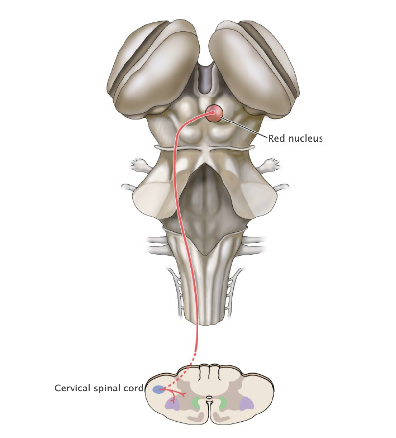
Rubrospinal tract
Descending motor pathway by which upper motor neurons in the red nucleus project to lower motor neurons in the lateral ventral horn of the cervical spinal cord; however, this projection is vestigial and variable in humans and its functional significance is uncertain.
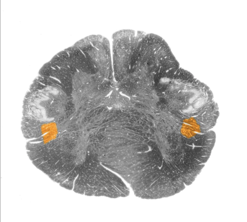
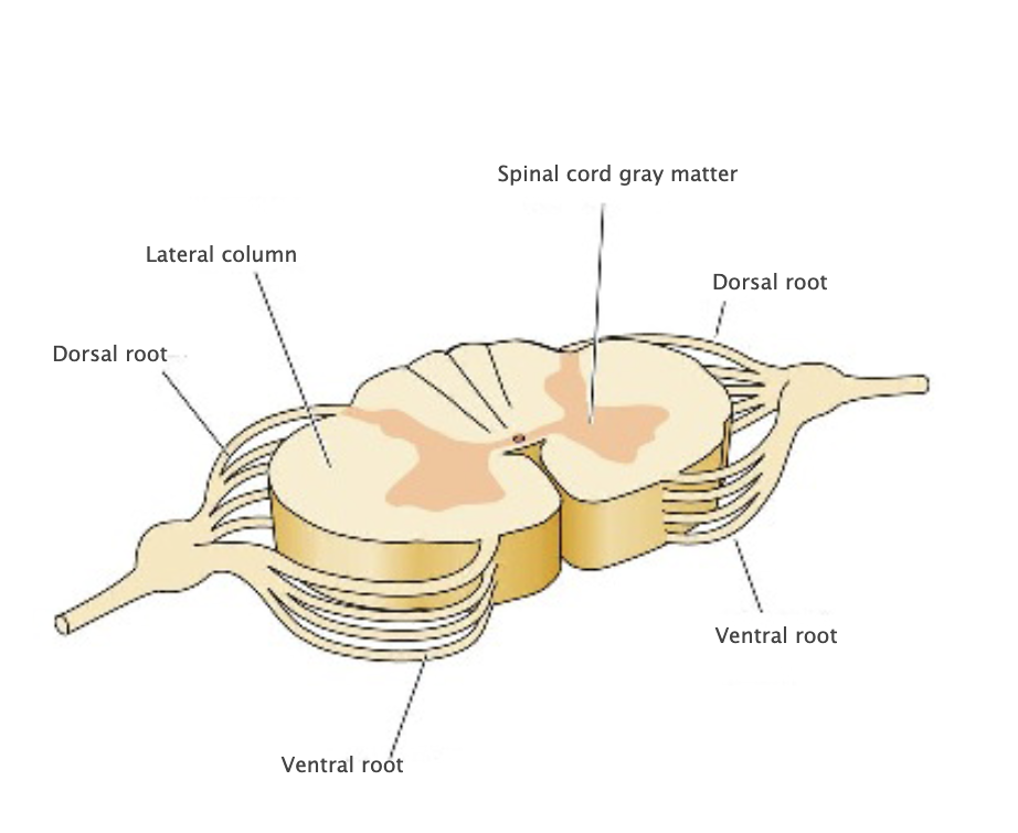
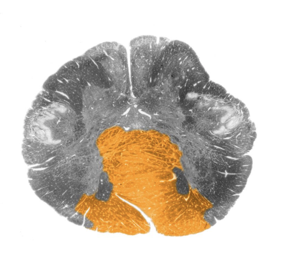
Lateral columns
White matter in the lateral portion of the spinal cord, bounded by the dorsal and ventral nerve roots and the gray matter.
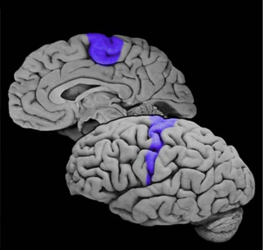
Primary motor cortex
Cortical area (Brodmann's area 4) in the precentral gyrus that contributes to the governance of lower motor neurons in the brainstem and spinal cord and the execution of volitional movements. This area is termed "primary" to indicate that this cortical area, among all other divisions of the motor cortex, has the most intimate functional and anatomical relation to lower motor neurons.
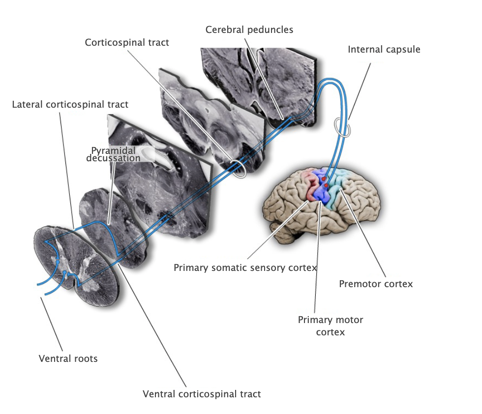
Pyramidal decussation
Prominent structure in the caudal medulla formed by the midline crossing (i.e., decussation) of corticospinal tract axons; the approximately 90% of the corticospinal tract axons that decussate in the caudal medulla then form the lateral corticospinal tract, and the 10% of the axons that do not cross the midline form the ventral (or anterior) corticospinal tract.
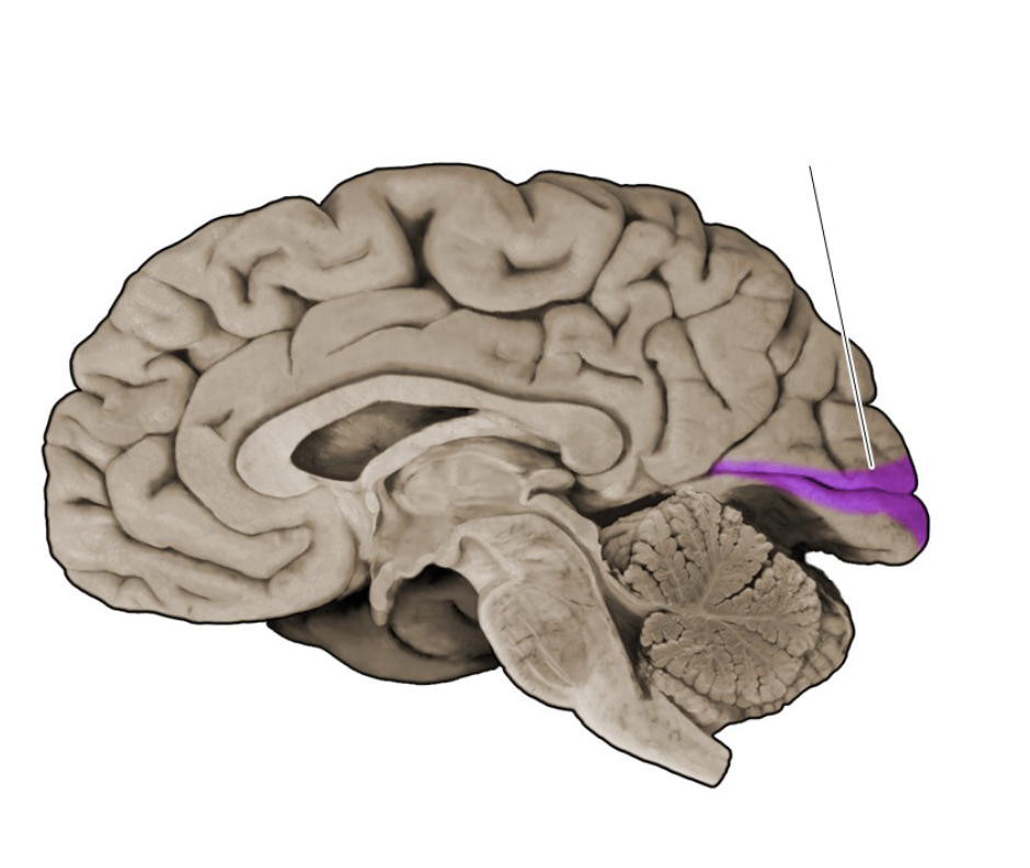
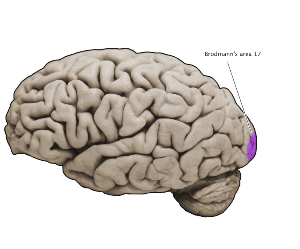
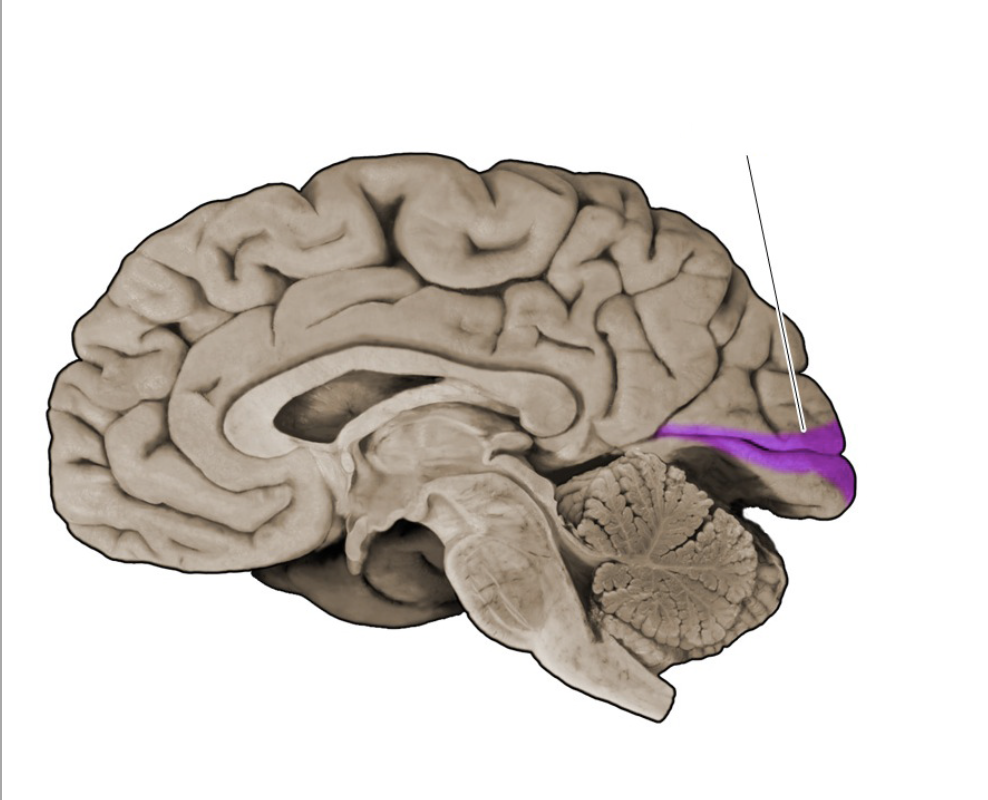
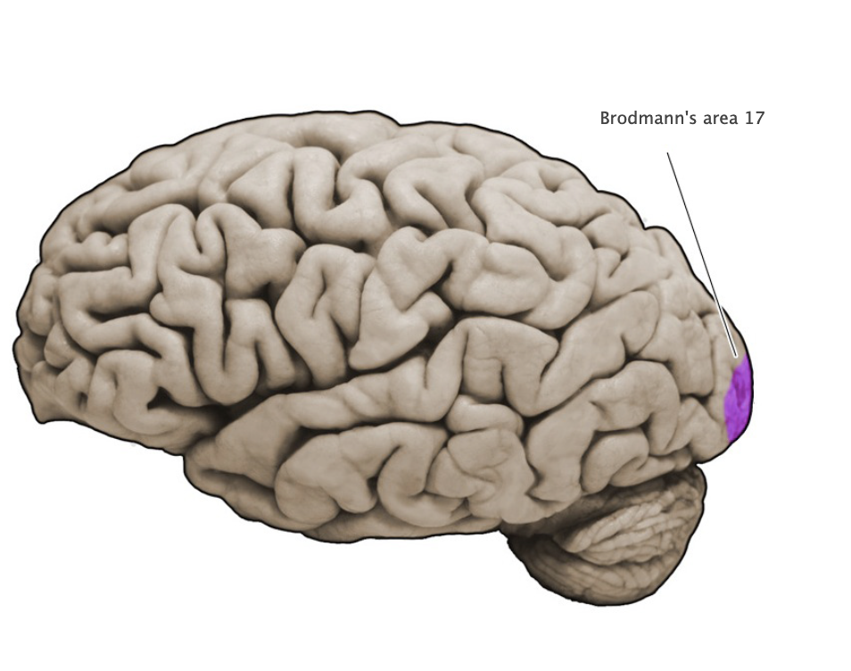
Brodmann's area 17
Sensory cortical area in the banks of the calcarine sulcus (lingual and cuneus gyral formations of the medial occipital lobe); this area corresponds to the primary visual cortex (also known as "striate cortex").
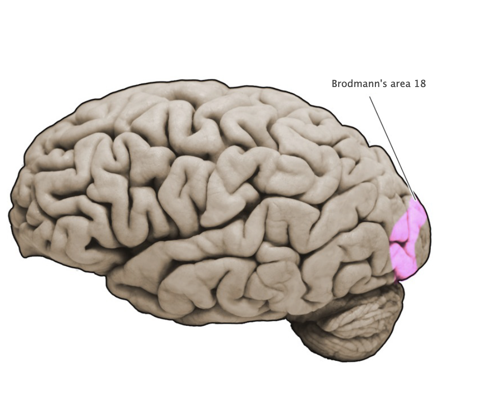
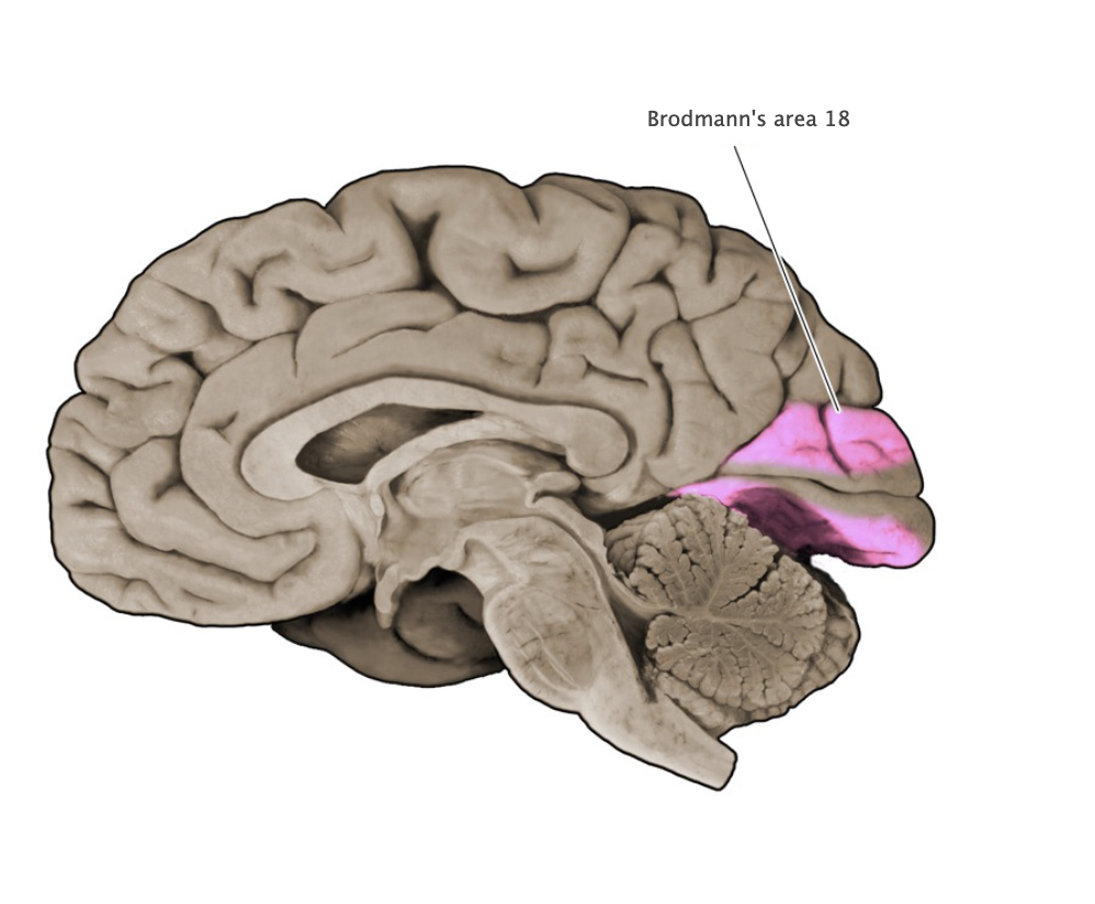
Brodmann's area 18
Sensory cortical area in the medial and lateral aspect of the occipital lobe; this area is part of the extrastriate visual cortex that surrounds the primary visual cortex (area 17 is also known as "striate cortex").
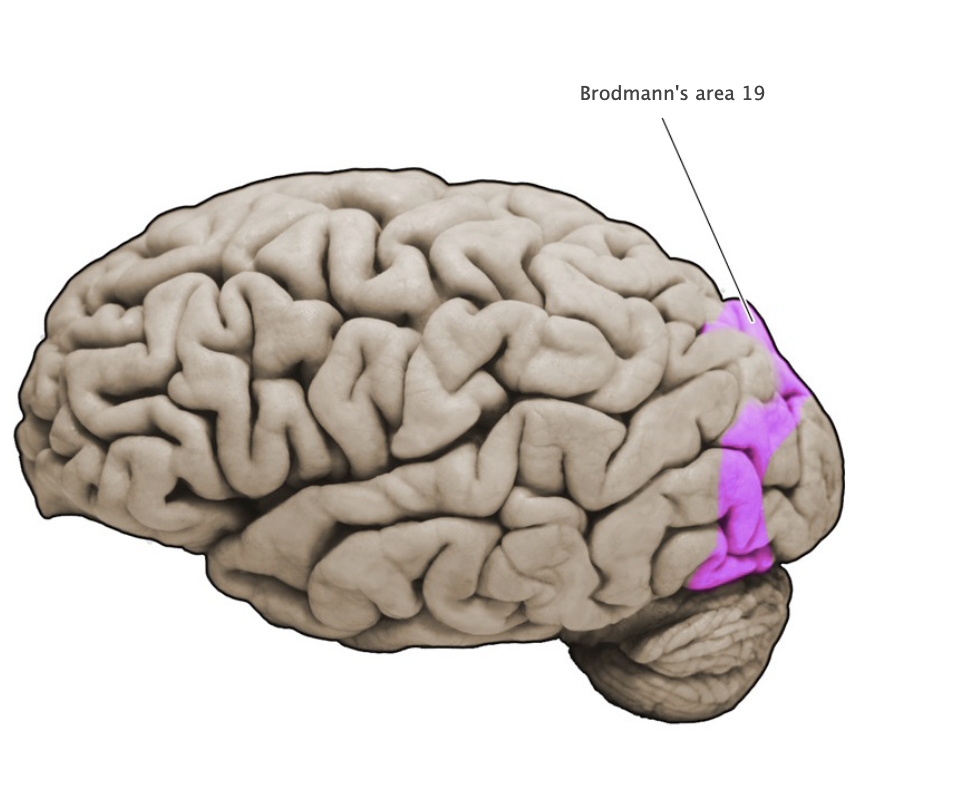
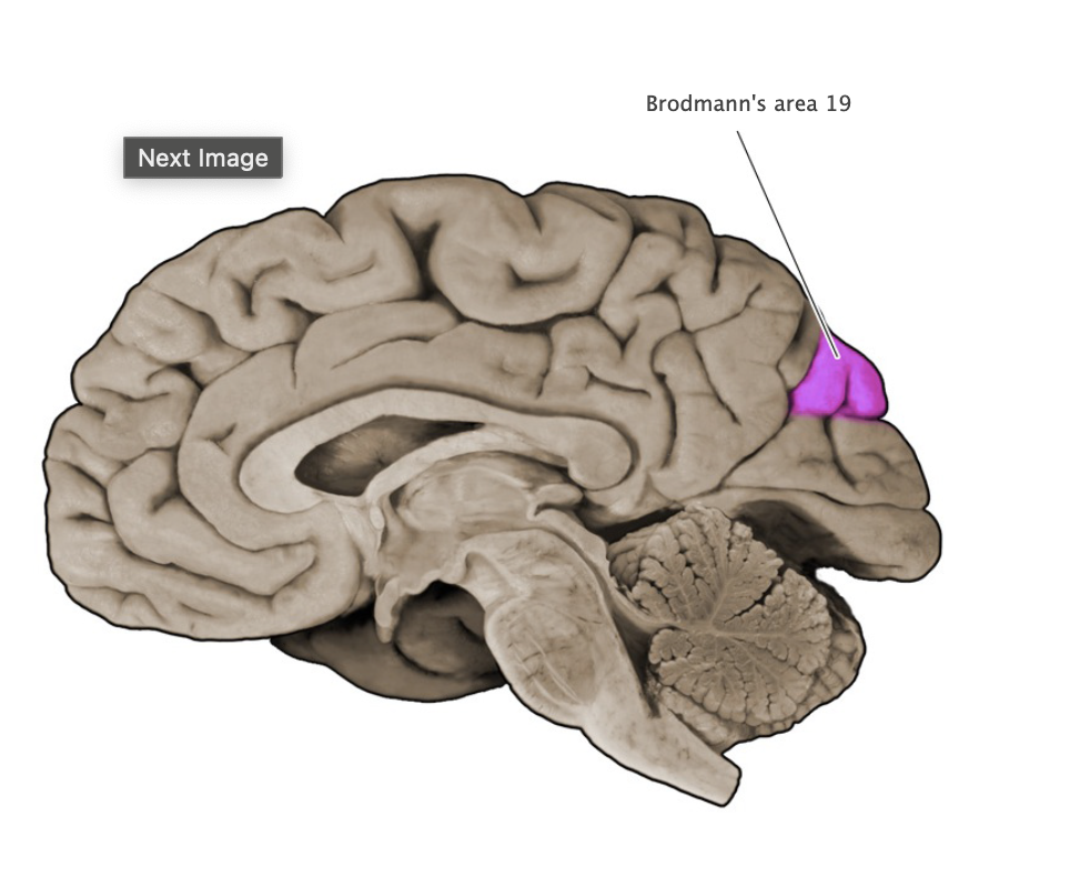
Brodmann's area 19
Sensory cortical area in the medial and lateral aspect of the occipital lobe; this area is part of the extrastriale visual cortex that surrounds the primary visual cortex (area 17 is also known as "striate cortex").
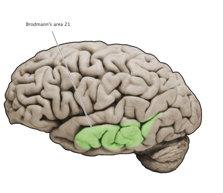
Brodmann's area 21
Associational cortical area in the middle temporal gyrus; this area participates in the analysis of visual signals related to object form and motion.
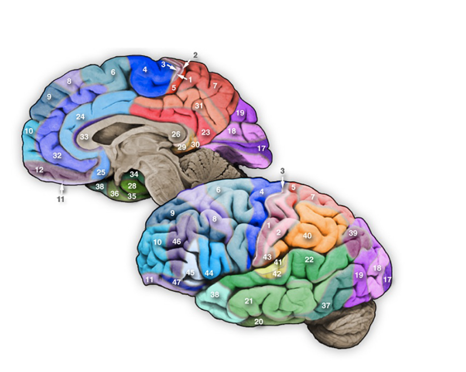
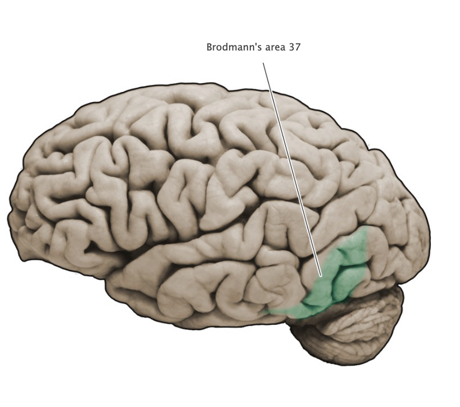
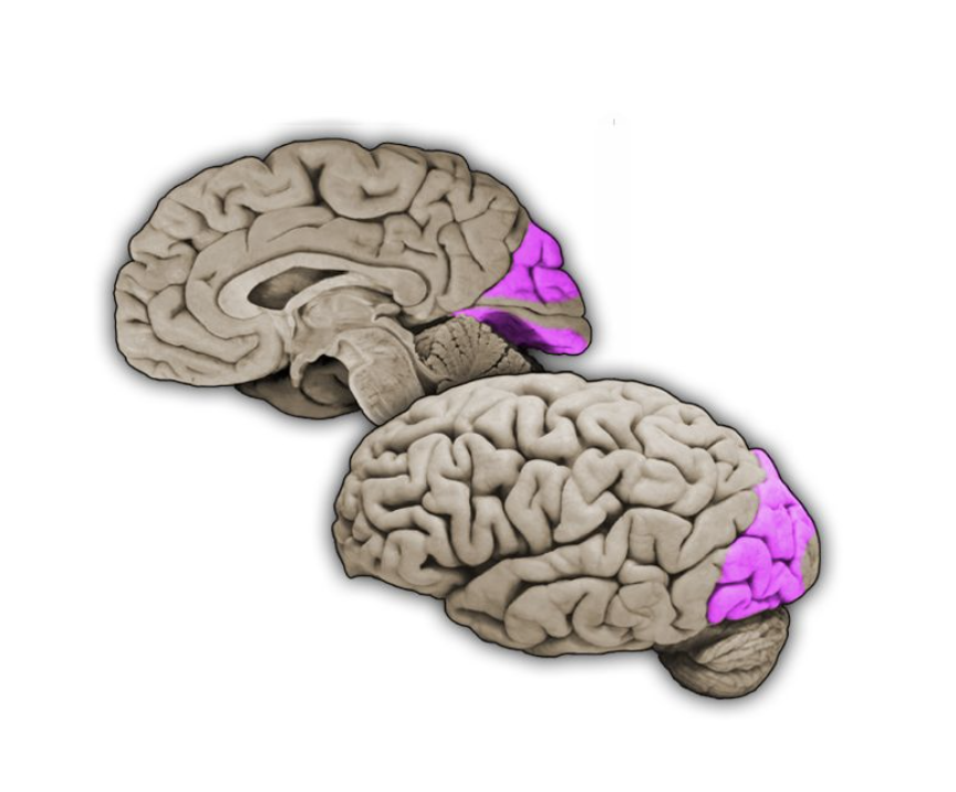
Brodmann's area 37
Associational cortical area in the temporal lobe that extends from the medial to the lateral sides of this lobe: this area participates in the analysis of visual form. motion, and the representation of objects.
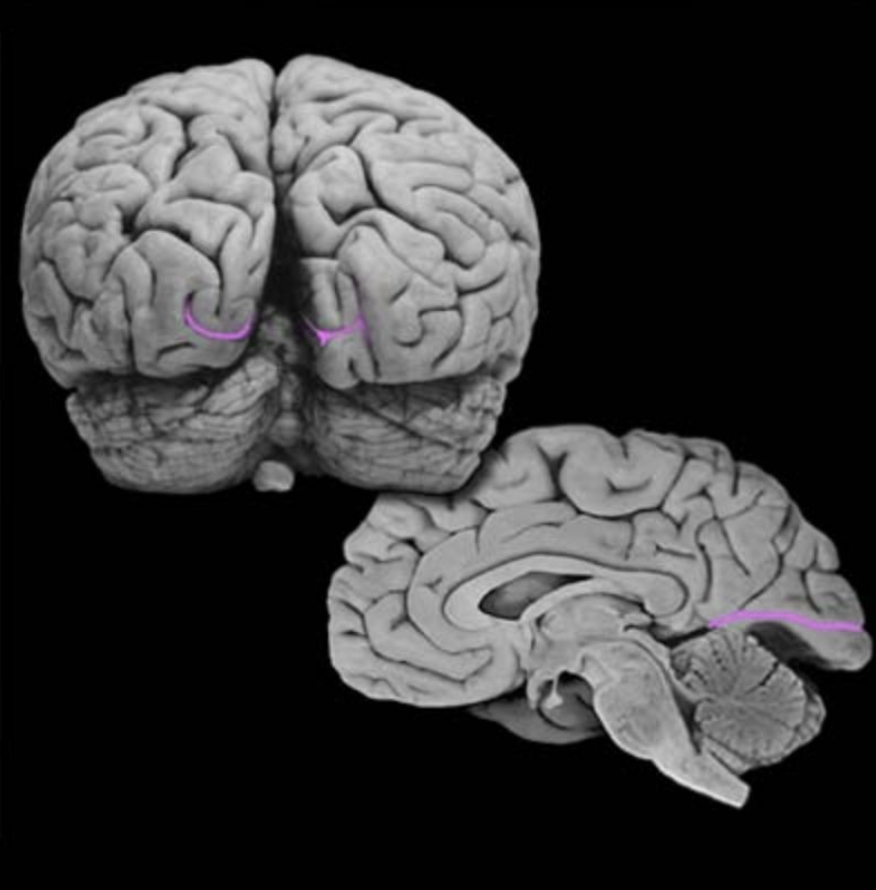
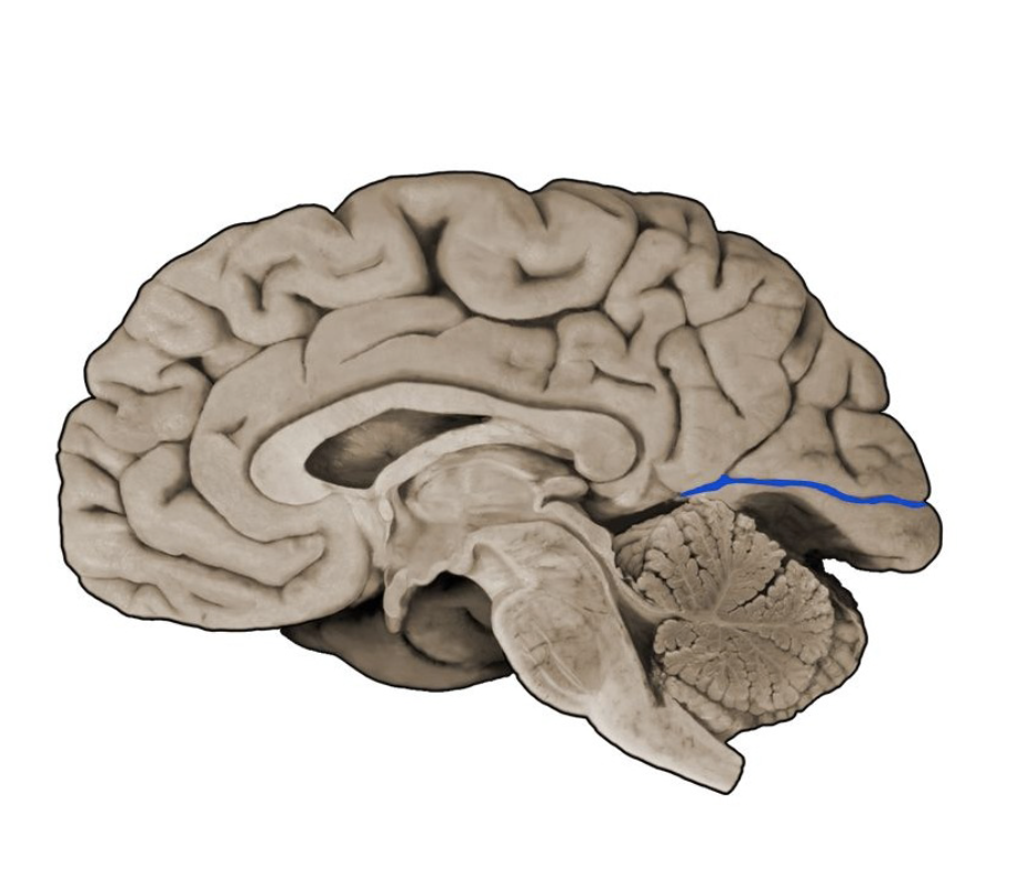
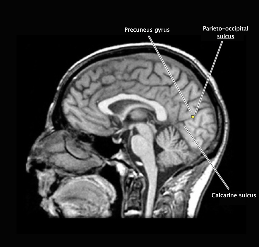
Calcarine sulcus
A prominent sulcus in the medial occipital lobe formed by the cuneus and lingual gyral structures; this sulcus extends roughly horizontally from the parieto-occipital sulcus to its termination in the occipital pole. The calcarine sulcus contains the primary visual cortex (also called the "striate cortex").
Extrastriate cortex
Cortical regions outside of the primary visual cortex (area 17) that participate in visual processing.
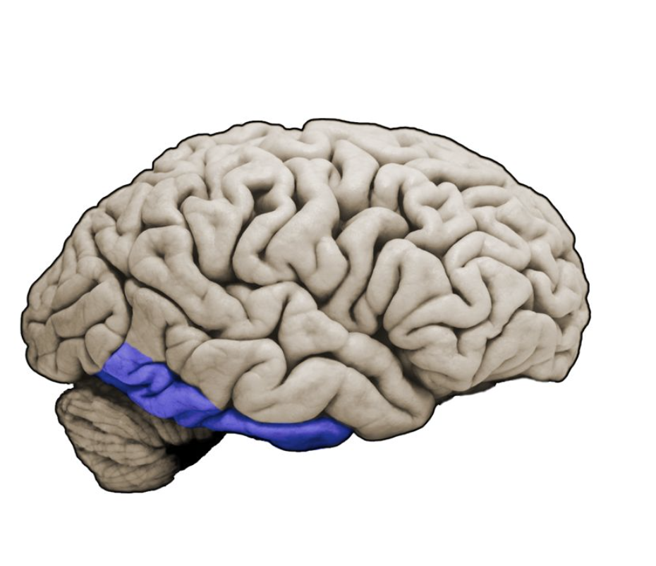
Inferior temporal gyrus
Longitudinal gyrus on the inferior, ventrolateral margin of the temporal lobe.
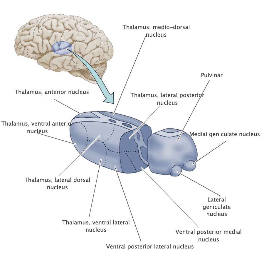
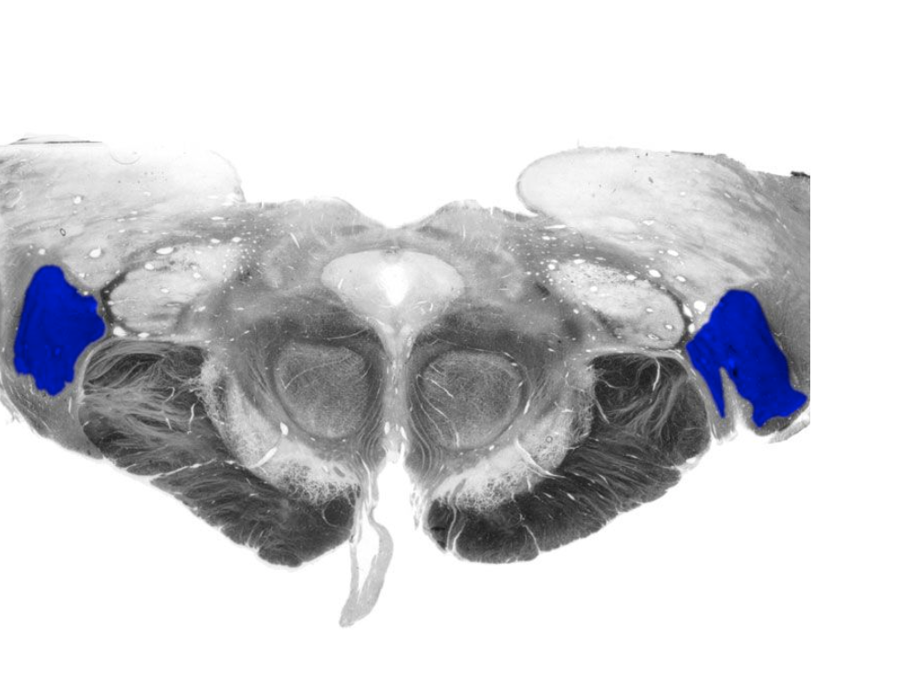
Lateral geniculate nucleus
Nucleus of the posterior, dorsal thalamus that receives the axonal projections of retinal ganglion cells via the optic tract, projects to the primary visual cortex (Brodmann's area 17) in the calcarine sulcus of the occipital lobe.
Occipital association cortex
Cortical regions of the occipital lobe beyond the primary visual cortex that are involved in higher-order visual processing.
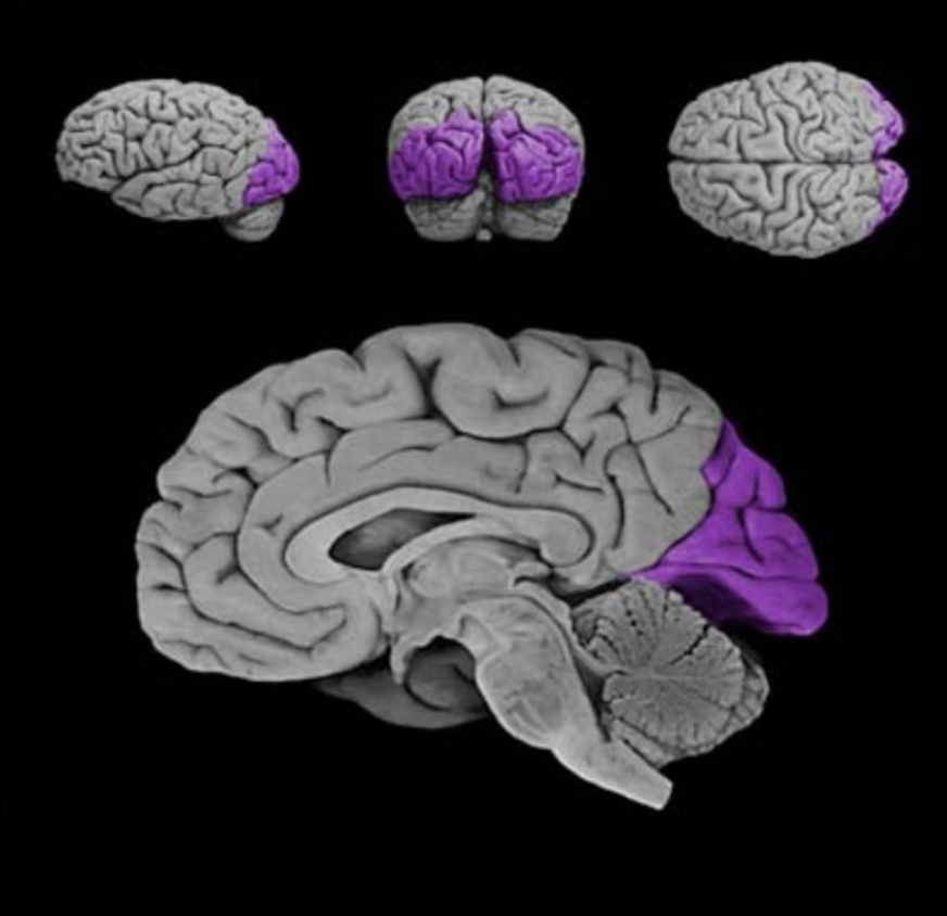
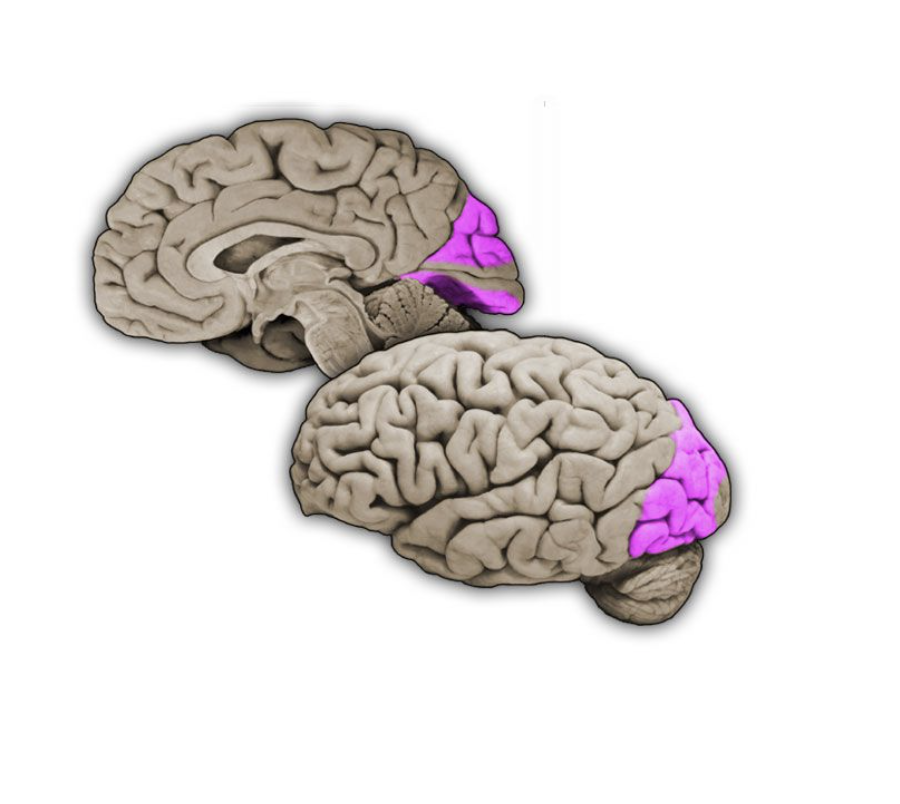
Occipital lobe
Most posterior of the four lobes of the cerebral hemisphere and is primarily involved in visual functions.
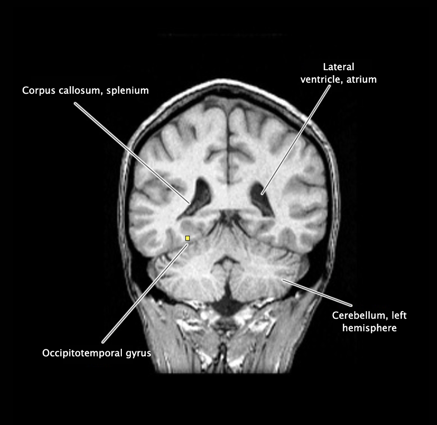
Occipitotemporal gyrus
Longitudinal gyrus on the inferior surface of the temporal and occipital lobes, bounded by the collateral sulcus medially and the lateral occipitotemporal suicus laterally.

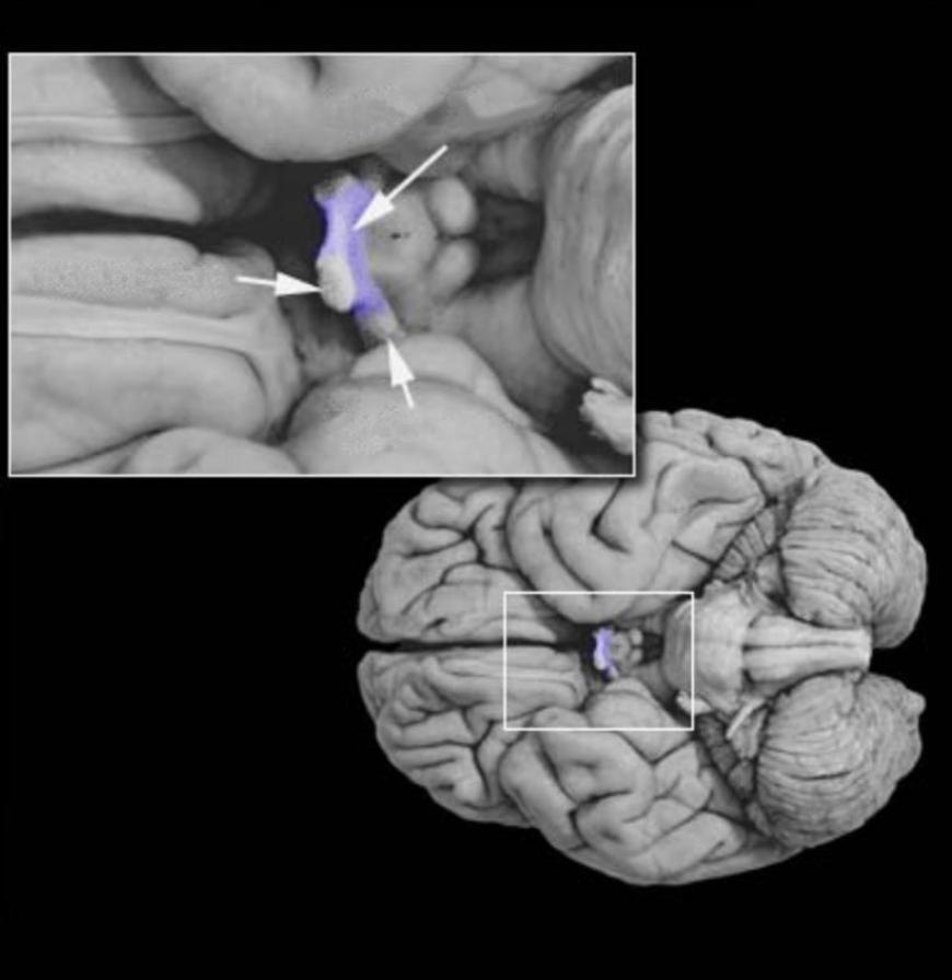
Optic chiasm
White matter structure formed by the partial decussation of the optic nerves. contains the axons of retinal ganglion cells in the nasal parts of each retina that project across the midline to contralateral diencephalic and mesencephalic targets. Caudal to the optic chiasm, these axons join other axons from the temporal retina to form the optic tract.
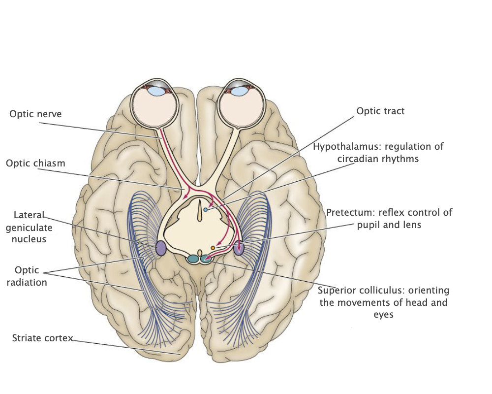
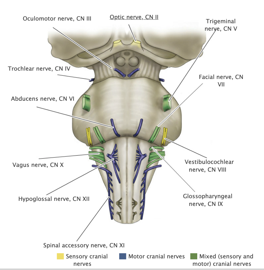
Optic nerve, Il
Cranial nerve ll formed by the axons of retinal ganlgion cells that project to targets in the diencephalon and mesencephalon.
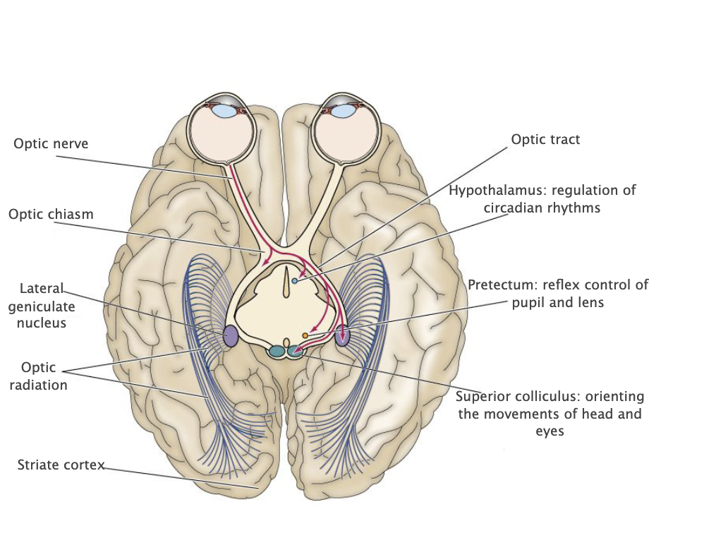
Optic radiations
Well-defined fiber bundles in the subcortical white matter of the parietal, temporal, and occipital lobes that contain the axons of lateral geniculate neurons, which carry visual information to the primary visual cortex (Brodmann's area 17); the temporal portion of these bundles that loop around the inferior horn of the lateral ventricle is known as "Meyer's loop."
Optic tract
The axons of retinal ganglion cells caudal to the optic chiasm which project to targets in the diencephalon and mesencephalon.

Parietal association cortex
Cthortical areas of the parietal lobe, excluding the primary somatosensory cortex; involved in numerous complex functions, including multimodal proprioception, language comprehension, attention and spatial awareness.
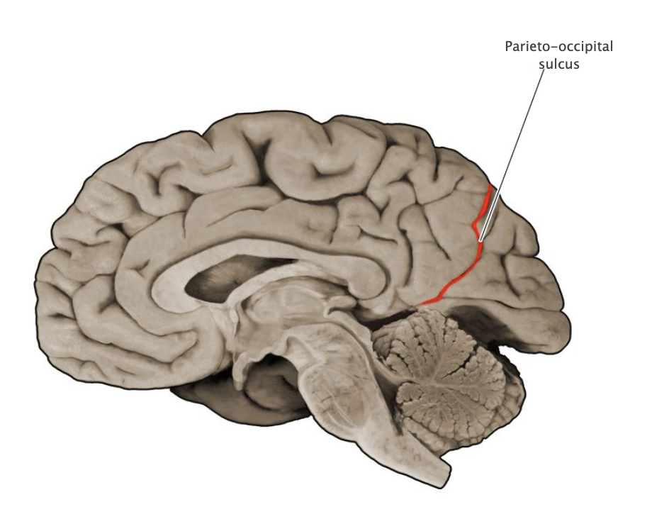
Parieto-occipital sulcus
Prominent vertically oriented sulcus on the medial aspect of the hemisphere that divides the parietal and occipital lobes; intersects the anterior terminus of the calcarine sulcus at nearly a right angle.
Primary visual cortex
Cortical area (Brodmann's area 17; also referred to as the "striate cortex") in the banks of the calcarine sulcus that first receives visual signals from the relevant thalamic nucleus (lateral geniculate nucleus).
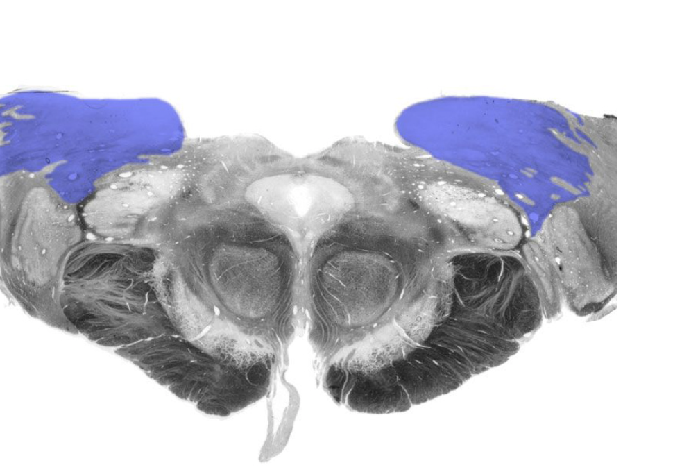
Pulvinar
Nucleus in the posterior-dorsal thalamus that, together with the lateral posterior nucleus, projects to higher-order visual and associational areas in the occipital, parietal and temporal lobes.
Superior colliculus
Paired hillocks on the dorsal surface of the midbrain overlying the cerebral aqueduct, which are involved in orienting movements of the head and eyes. Together with the inferior colliculi, these structures are also known as the "tectum" (which means roof) and the "quadrageminal bodies".
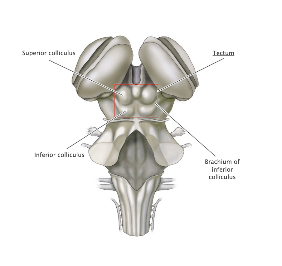
Tectum
Structure in the dorsal midbrain that forms a roof over the cerebral aqueduct (tectum means roof); comprises the superior and inferior colliculi (also called the "quadrageminal bodies").
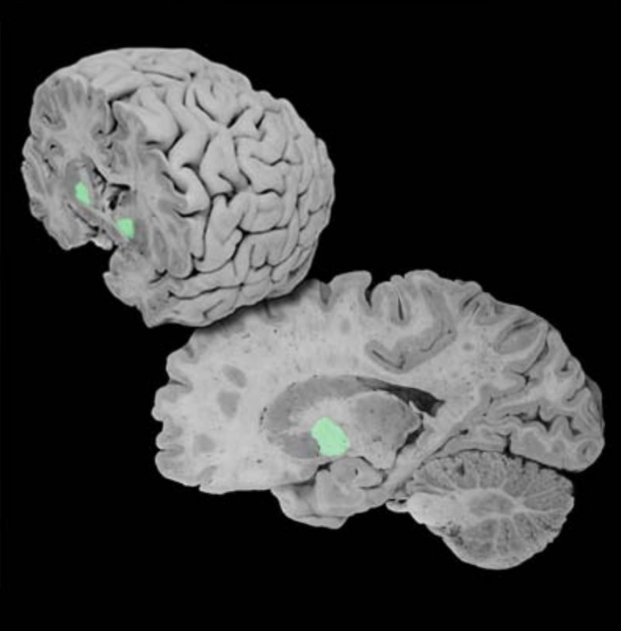
Caudate nucleus
Major gray matter component of the basal ganglia located deep in the frontal lobe. Involved in regulating the expression of certain bodily movements, affective behavior and cognitive processes by integrating widespread inputs from the cerebral cortex and modulating the activity of relevant thalamic relay nuclei.