Chromosomes and Cellular Reproduction Overview
1/174
There's no tags or description
Looks like no tags are added yet.
Name | Mastery | Learn | Test | Matching | Spaced |
|---|
No study sessions yet.
175 Terms
Prokaryotic cell
No nucleus, no paired chromosomes (haploid), typically single circular chromosome.
Eukaryotic cell
Nucleus present, paired chromosomes common (diploid), typically multiple linear chromosomes.
Cell reproduction fundamental events
A cell's genetic information must be copied, the copies of the genetic information must be separated from one another, and the cell must divide into two cells.
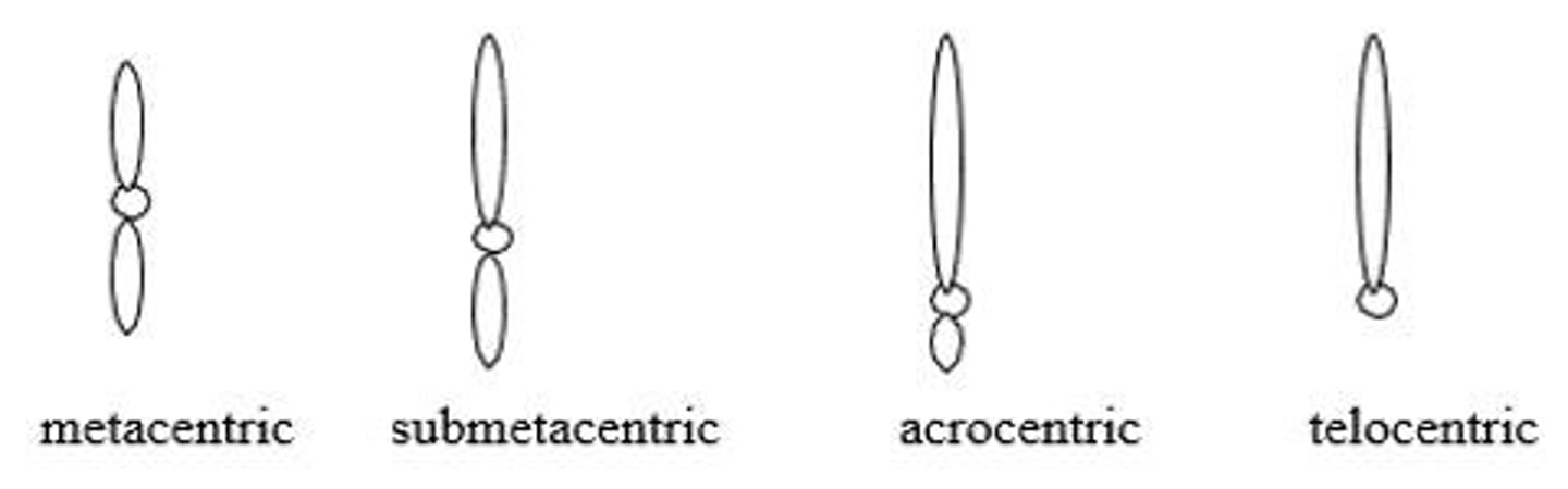
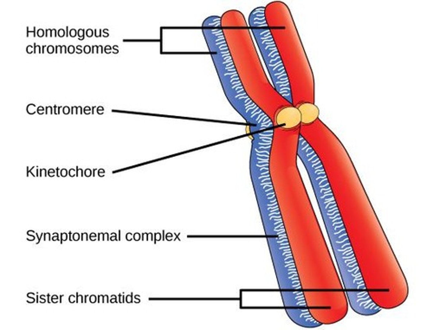
Centromere
Serves as the point of attachment for the kinetochore to which spindle fibers (microtubules) attach.
Telomeres
Serve to stabilize the ends of the chromosome and limit cell division.
Origins of replication
Serve as the starting place for DNA synthesis.
G1 (Gap 1)
In this phase, the cell grows and synthesizes proteins necessary for cell division. During G1, the G1/S checkpoint takes place.
S phase
During S phase, DNA replication takes place.
G2 (Gap 2)
In G2, additional biochemical reactions take place that prepare the cell for mitosis. Near the end of G2 is the G2/M checkpoint.
G0 phase
A nondividing stage some cells may exit the active cell cycle.
Checkpoints
Function to ensure that all the cellular components are present and functioning before the cell moves to the next stage of the cell cycle.
G1/S checkpoint
Occurs during G1 prior to the S phase.
G2/M checkpoint
Occurs in G2 prior to mitosis.
Spindle-assembly checkpoint
Occurs during mitosis.
Prophase
The chromosomes condense and become visible. The mitotic spindle forms. The centrosomes move apart and microtubules form from the centrosomes.
Prometaphase
The nuclear membrane disintegrates. Spindle microtubules enter the nuclear region and attach to the chromosomes.
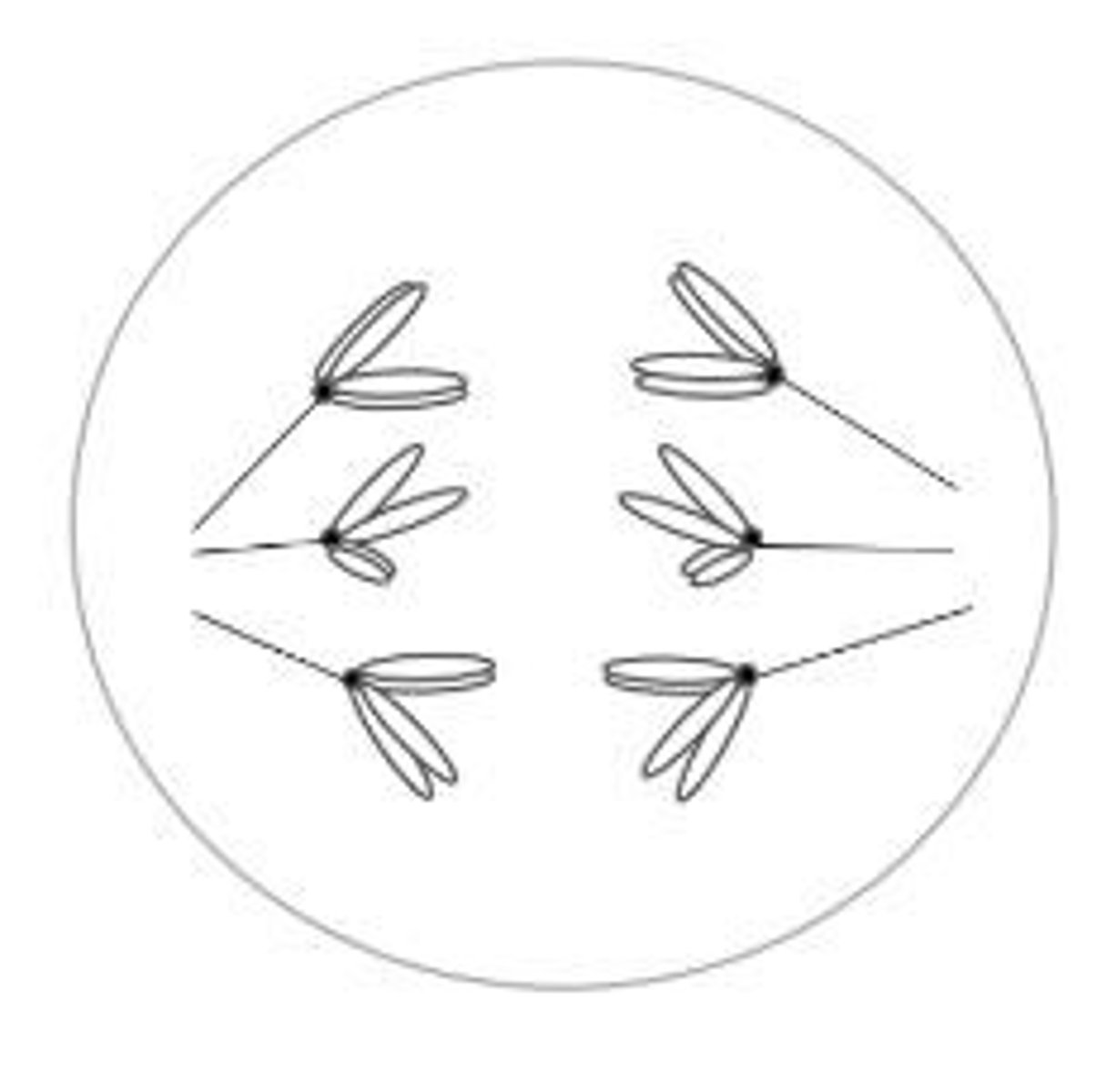
Metaphase
The chromosomes become arranged on the metaphase plate of the cell.
Anaphase
The sister chromatids separate, and the resulting chromosomes move to the opposite poles of the cell.
Telophase
The chromosomes arrive at the spindle poles. The nuclear membrane reforms around each set of chromosomes.
Genetically important results of the cell cycle and mitosis
The outcomes of these processes contribute to genetic diversity and cell function.
Cell Cycle
The cell cycle produces two cells that are genetically identical and that contain a full complement of chromosomes; there is no net reduction or increase in chromosome number.
Genetic Identity of Cells
The two cells are genetically identical because an exact copy of each DNA molecule was created during S phase.
Sister Chromatids
These exact copies give rise to the two identical sister chromatids.
Mitosis
Mitosis ensures that each new cell receives one of the identical sister chromatids.
Meiosis I
Separation of homologous chromosomes.
Prophase I
The chromosomes condense and homologous pairs of chromosomes undergo synapsis. While the chromosomes are synapsed, crossing over occurs.
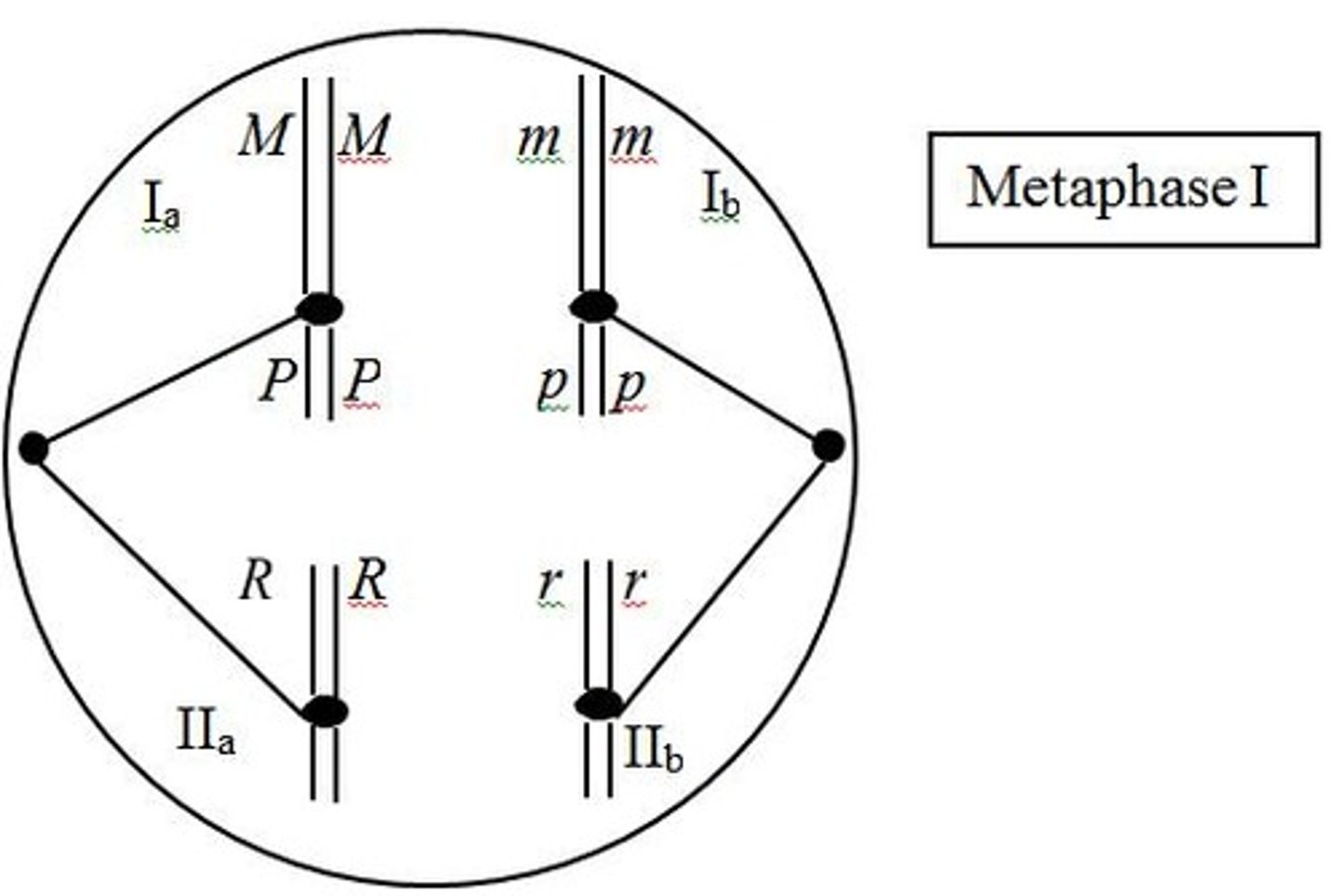
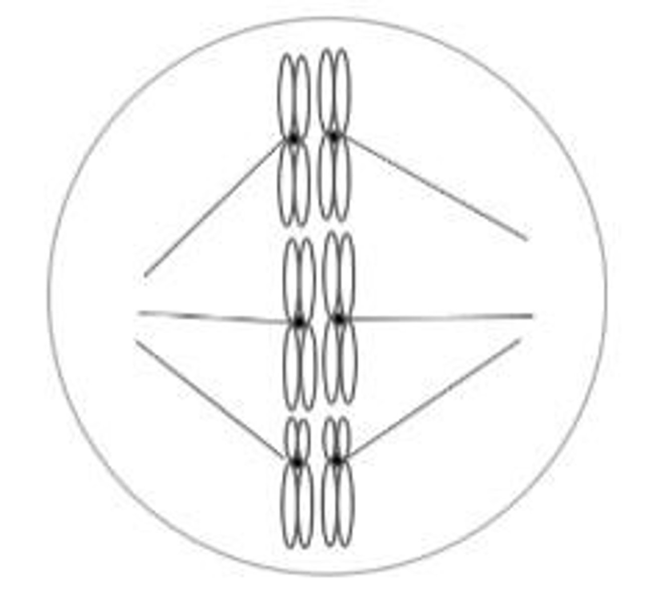
Metaphase I
The homologous pairs of chromosomes line up on the metaphase plate.
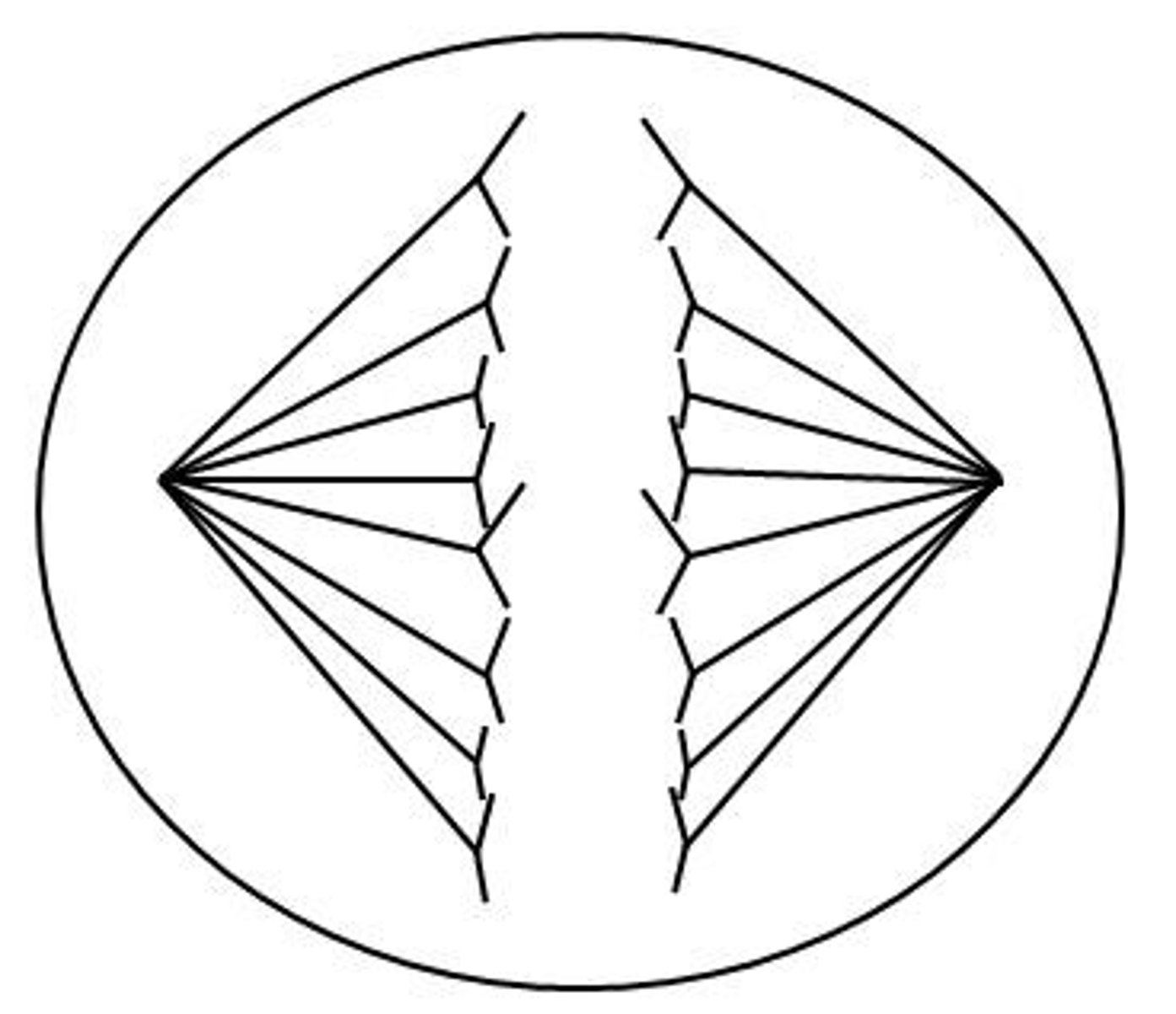
Anaphase I
Homologous chromosomes separate and move to opposite poles of the cell.
Telophase I
The separated homologous chromosomes reach the spindle poles and are at opposite ends of the cell.
Cytokinesis after Meiosis I
Results in the division of the cytoplasm and the production of two haploid cells.
Meiosis II
Separation of sister chromatids.
Prophase II
Chromosomes condense, the nuclear envelope breaks down, and the spindle fibers form.
Metaphase II
Chromosomes line up at the metaphase plate.
Anaphase II
The sister chromatids separate and are pulled to opposite poles.
Telophase II
The chromosomes arrive at the spindle poles. The nuclear membrane reforms, and the spindle fibers break down.
Cytokinesis after Meiosis II
Takes place following meiosis II.
Results of Meiosis
Meiosis involves two cell divisions, thus producing four new cells. The chromosome number is reduced by half.
Genetic Variation in Meiosis
Crossing over occurs in prophase I and the random separation of homologous chromosomes takes place in anaphase I.
Anaphase I vs. Anaphase of Mitosis
In anaphase I of meiosis, homologous chromosomes separate, whereas in anaphase of mitosis, sister chromatids separate.
Spermatogenesis
Occurs in the testes; primordial diploid germ cells divide mitotically to produce diploid spermatogonia.
Primary Spermatocytes
Diploid cells that enter prophase I and complete meiosis I to become two secondary spermatocytes.
Secondary Spermatocytes
Haploid cells that complete meiosis II, producing a total of four haploid spermatids.
Oogenesis
The process by which female animals produce eggs.
Primary Oocyte
A diploid cell that has entered prophase I during oogenesis.
Secondary Oocyte
The haploid cell that receives most of the cytoplasm after meiosis I.
First Polar Body
The smaller haploid cell that receives only a small portion of the cytoplasm after meiosis I.
Ovum
The cell that receives most of the cytoplasm from the secondary oocyte after meiosis II.
Second Polar Body
The smaller haploid cell produced during oogenesis that does not develop into an ovum.
Polar Bodies
Typically, the polar bodies disintegrate, and only the ovum is capable of being fertilized.
Meiosis II in Humans
In humans and many other mammals, meiosis II is not completed until the sperm penetrates the secondary oocyte.
Gametophyte
A multicellular haploid stage in plants.
Sporophyte
A multicellular diploid stage in plants.
Meiosis in Plants
Meiosis in the diploid sporophyte stage of plants produces haploid spores that develop into the gametophyte.
Microsporocytes
Found in the stamen of the flower, they undergo meiosis to produce four haploid microspores.
Pollen Grain
Each microspore divides by mitosis to produce the pollen grain, which contains two haploid nuclei.
Sperm Cells
One of the haploid nuclei in the pollen grain divides by mitosis to produce two sperm cells.
Pollen Tube
The other haploid nucleus in the pollen grain directs the formation of the pollen tube.
Megasporocytes
Diploid megasporocytes found within the ovary divide by meiosis to produce four megaspores.
Embryo Sac
The remaining megaspore divides mitotically three times to produce eight haploid nuclei that form the embryo sac (or female gametophyte).
Egg Cell
Of the eight nuclei formed in the embryo sac, one will become the egg.
Homologous Pair
The two pairs (four socks in all) of each color represent the two chromosomes of a homologous pair, each with two sister chromatids.
Cohesin
The thread that connects the two socks of a pair represents cohesin.
Archaea
This cell is most likely an archaea because it lacks a nuclear membrane and has a single circular chromosome.
Viruses
Viruses are neither prokaryotes nor eukaryotes, because they do not possess a cellular structure.
Chromosome 1
The centromere in chromosome 1 is centrally located, so it is metacentric.
Chromosome 4
The centromere of chromosome 4 is located between the center and the end of the chromosome, so it is submetacentric.
Cell Cycle Duration
If the complete cell cycle requires 24 hours, the average duration of the M phase and metaphase can be calculated based on the proportion of cells in each stage.
Average Duration Calculation
To calculate the time required for a given stage, multiply 24 hours by the proportion of cells at that stage.
Interphase
Stage of the cell cycle where the cell prepares for division, with 160 cells counted, 0.80 proportion of cells, and an average duration of 19.2 hours.
M phase
The phase of the cell cycle that lasts 4.8 hours, determined by adding up the hours spent in each stage of mitosis.
Metaphase duration
Metaphase requires 0.24 hours, or 14.4 minutes.
Preformationism
Theory proposing that a tiny, fully formed adult (the homunculus) exists in the egg or sperm, predicting all traits are inherited from one parent.
Homunculus
The tiny, fully formed adult proposed by preformationism that enlarges during development.
Meiosis
A type of cell division that reduces the chromosome number by half, producing haploid cells.
G1 phase
The first gap phase of the cell cycle where the cell grows and prepares for DNA replication, with 4 chromosomes.
G2 phase
The second gap phase of the cell cycle where the cell prepares for mitosis, containing 12 chromosomes and 24 DNA molecules.
Prophase of mitosis
Stage of mitosis where the cell contains 12 chromosomes and 24 DNA molecules.
Anaphase I of meiosis
Stage of meiosis where homologous chromosomes separate, with 12 chromosomes and 24 DNA molecules.
Anaphase II of meiosis
Stage of meiosis where sister chromatids separate, resulting in 12 chromosomes and 12 DNA molecules.
Prophase II of meiosis
Stage of meiosis where haploid cells contain 6 chromosomes and 12 DNA molecules.
Cytokinesis after mitosis
Each cell will contain 12 chromosomes and 12 DNA molecules.
Diploid number of chromosomes in the plant
There are six chromosomes in this species.
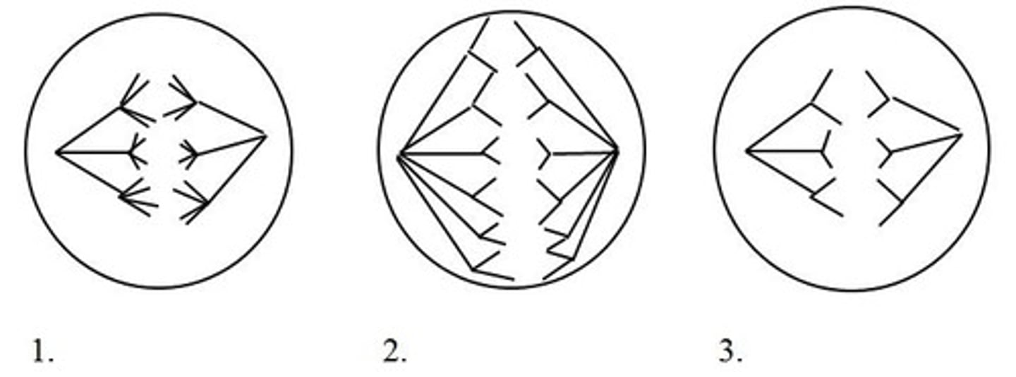
Cell 1 during meiosis
Undergoing anaphase of meiosis I, with six chromosomes, each with two chromatids.
Cell 2 during mitosis
In anaphase of mitosis, where six chromosomes have separated into 12 chromosomes, each with a single chromatid.
Cell 3 during meiosis
In anaphase II of meiosis, with six chromosomes present.
DNA molecules in Cell 1
There are 12 DNA molecules, as there are six chromosomes each with two chromatids.
DNA molecules in Cell 2
There are 12 DNA molecules, as there are 12 chromosomes and sister chromatids are not present.
DNA molecules in Cell 3
There are six DNA molecules, as there are six chromosomes and sister chromatids are not present.
Amount of DNA in G1
7.3 pg, occurring prior to S phase and the doubling of DNA.
Amount of DNA in Prophase I
14.6 pg, as the amount of DNA is doubled during S phase.
Amount of DNA in G2
14.6 pg, as it takes place directly after S phase where the amount of DNA is doubled.
Amount of DNA following telophase II and cytokinesis
3.7 pg, as the chromosome number is reduced by half in meiosis I and then again in telophase II.
Amount of DNA in Anaphase I
14.6 pg, as the amount of DNA does not change from G2 to anaphase I.
Cytokinesis in spermatogenesis
Cytokinesis is equal, resulting in haploid cells of similar sizes.
Cytokinesis in oogenesis
Cytokinesis is unequal, resulting in a larger secondary oocyte and smaller polar bodies.
Homologous pairs of chromosomes
Pairs of chromosomes that are similar in shape, size, and genetic content.
7.3 pg
The amount of DNA present in metaphase II, which is half that in G2.
Oculocutaneous type 2 albinism
A recessive condition where a person has a copy of the DNA sequence that causes albinism on each of two homologous chromosomes.
Anaphase of mitosis
During this stage, there will be four copies of the DNA sequence causing albinism present in the cell.
G1 of interphase
In this stage, there will be two copies of the DNA sequence causing albinism.