BPK 241 Lecture 7
1/44
There's no tags or description
Looks like no tags are added yet.
Name | Mastery | Learn | Test | Matching | Spaced | Call with Kai |
|---|
No analytics yet
Send a link to your students to track their progress
45 Terms
Knee Joint types
Anatomically = synovial (fluid, sacs)
Functionally = hinge (one phase of movement)
Physiologically = Rotation/ rolling & gliding
Knee Bones
Femur
Tibia
Patella
Knee Articulations
Tibiofemoral - Femur & tibia
Patellofemoral - Patella & femur
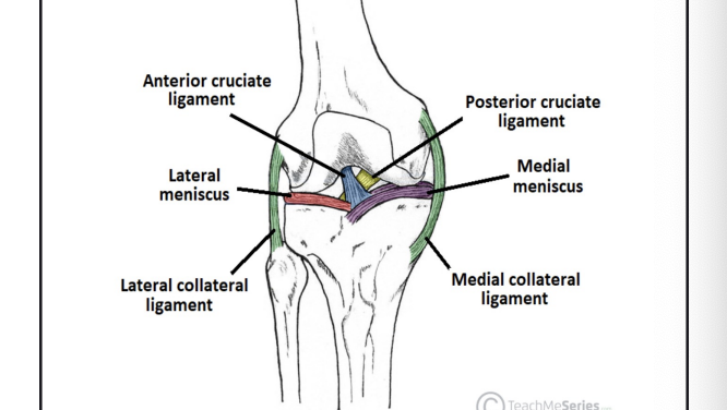
Knee Stability
Capsule
Ligaments
Menisci
Muscles
Tendon
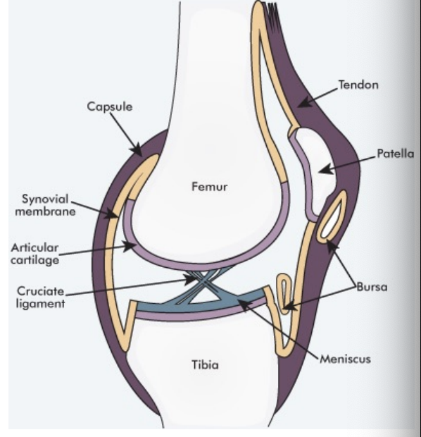
Knee Capsule
Extensive & Redundant
Resists hyperextension
Provides rotational stability
Swelling accumulate at front of knee from capsule space
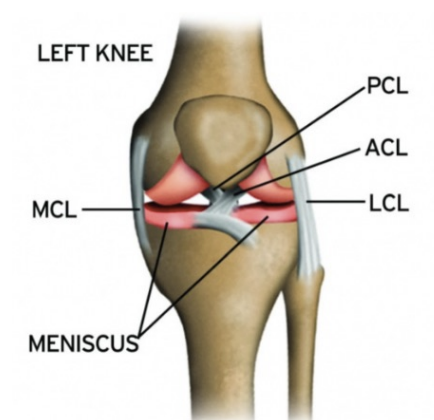
Extracapsular ligaments
Medial collateral (MCL)
Short inner, long outer fibres
Medial epicondyle (push side of knee); where is originates
prevent knee from force going inward
Lateral collateral (LCL)
Attaches to femur and head of fibula
prevent knee outward
Intracapsular ligament
Anterior cruciate (ACL)
Anterior tibia, then up, back, out to medial surface of lateral epicondyle
Prevents anterior displacement of tibia on fixed femur, hyperextension
prevent femur posteriorly, prevent tibia anteriorly
Posterior cruciate (PCL)
Posterior tibia, then up, forward, in to lateral surface of medial condyle
Prevents posterior displacement of tibia on fixed femur
prevent femur forward, prevent tibia backward
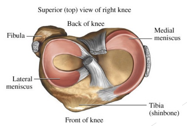
Knee menisci
Fibrocartilage
Medial and tibial
Attached to tibial plateau
Tough fibre material
cushion for shock observes
acts as a sponge, soaks synovial fluid and releases it for blood supplly
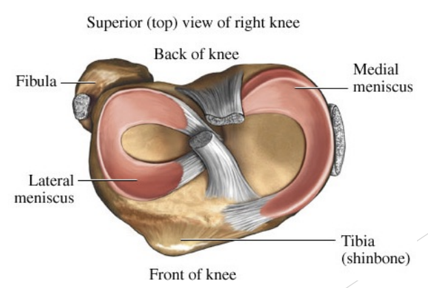
Red zone of Menisci
1/3 Outer
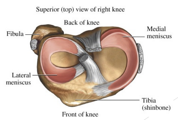
Pink zone of Menisci
Middle 1/3 - terrible blood supply
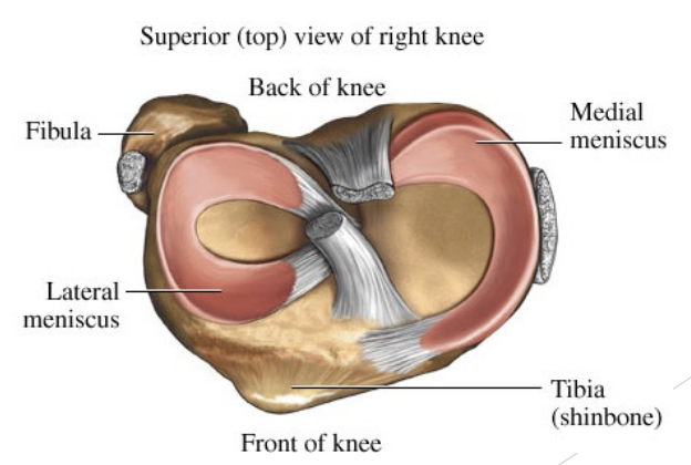
White zone of Menisci
Inner 1/3 - No blood supply, can’t heal itself
Menisci & Coronary ligament
Attached to tibial plateau
Attached to capsule by coronary ligaments
Provides cushioning & stability
Increases synovial fluid circulation
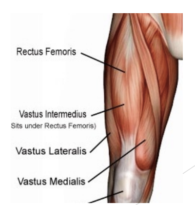
Knee Muscles
Quadriceps femoris (extend the knee, helps PCL from tibia moving backwards)
Rectus femoris (o=ilium at AIIS)
Vastus medialis (o=femur)
Vastus intermedias (o=femur)
Vastus lateralis (o=femur)
All insert by quadriceps tendon and patellar tendon (patellar ligament) on tibial tuberosity
Function: extension of leg at knee
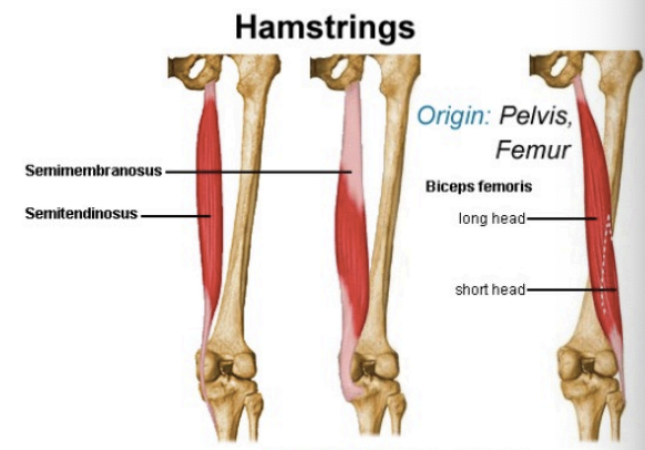
Hamstrings
o = ischuim
Biceps femoris
Semimebranosis
Semitendinosis
insert onto medial tibia
Bicep femoris inserts on head of fibula
flex lower leg on thigh at knee
Extend thigh on trunk at hip
Help a little with hip extension and help ACL from tibia moving forward
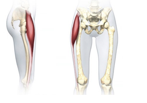
Tensor Fascia Latae (TFL)
o = ischium
Inserts on tibia and into fascia of the thigh (via iliotibial band)
Helps flex and abduct thigh on trunk at hip
Adds to lateral stability of knee
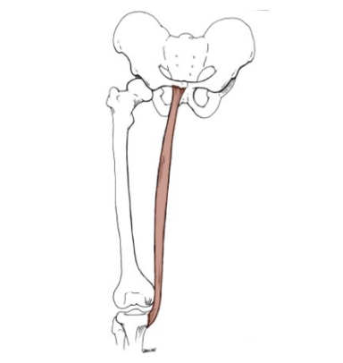
Gracilis
Same origin and insertion as semimebranosis and semitendinosis
Assists with knee flexion
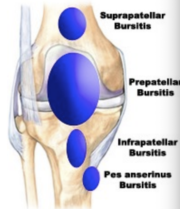
Knee Bursae
Prepatellar
Suprapatellar (and fat pad)
Infrapatellar (and fat pad)
Decrease friction over the bone
most common in chronic onset injury
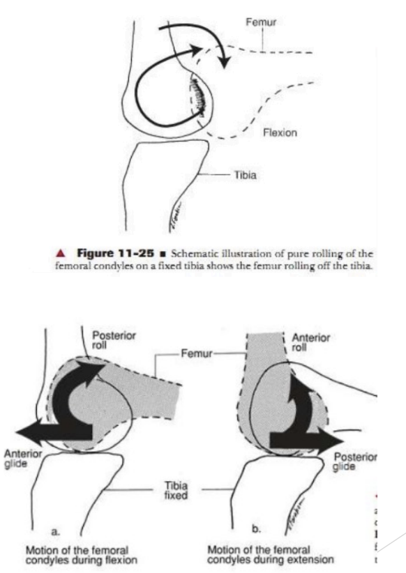
Knee movements
Flexion & extension
Gliding of condyles on plateau and menisci
Rolling (posterior) & gliding (anterior) happening at the same time when flexion and extension occurs
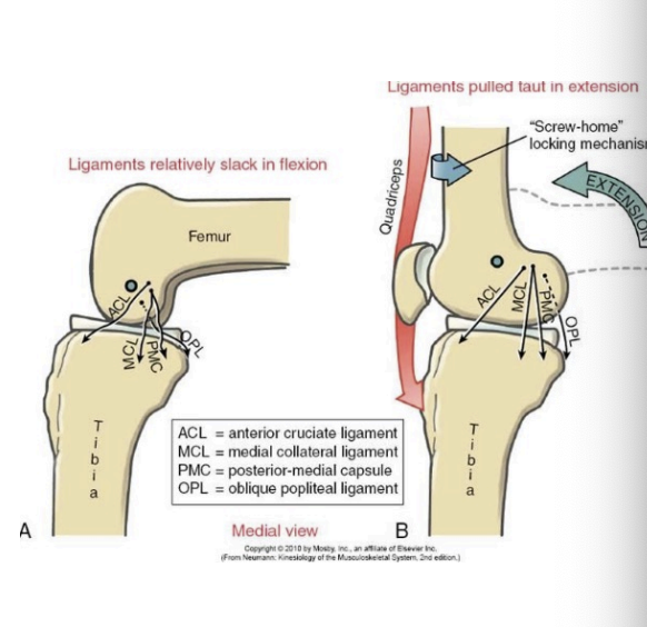
Locking Mechanism
Medial condyle is larger than lateral
Internal rotation of thigh AND/OR rotation of lower leg completes full extension at knee (locking)
Possible medial meniscal distortion
Locked knee: ACL taught, PCL taught, knee joint taught - forces femur in internal rotation & tibia in external rotation
Unlocked knees: Pull femur in external rotation, tibia in internal rotation
Knee Assessment
Hx: What happened, where
PHx: priory injury? Rehab? Ongoing?
SSx:
Unstable on knee
“snap”, “pop”
Examination
Observation
Limp, instability
Swelling (acute or delayed - 24?)
Gait analyses
ROM:
Functional tests - compare sides
Thigh circumference - serial
Sends signal to thigh to shut off if severe
Palpation - be systematic
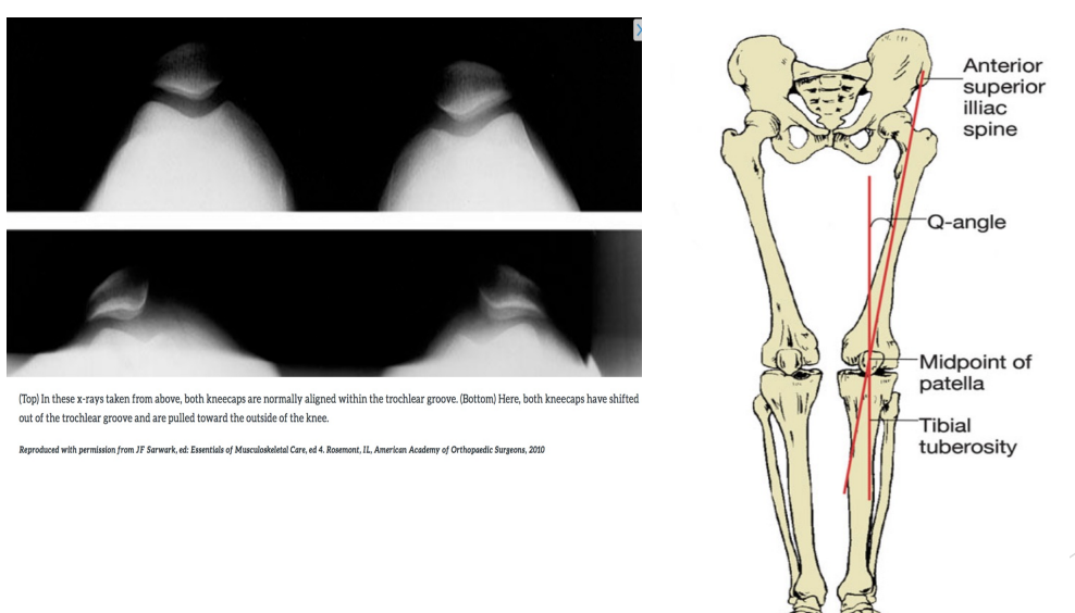
Knee Alignment Deviations
Patellar Malaligments
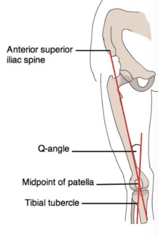
Q- Angle
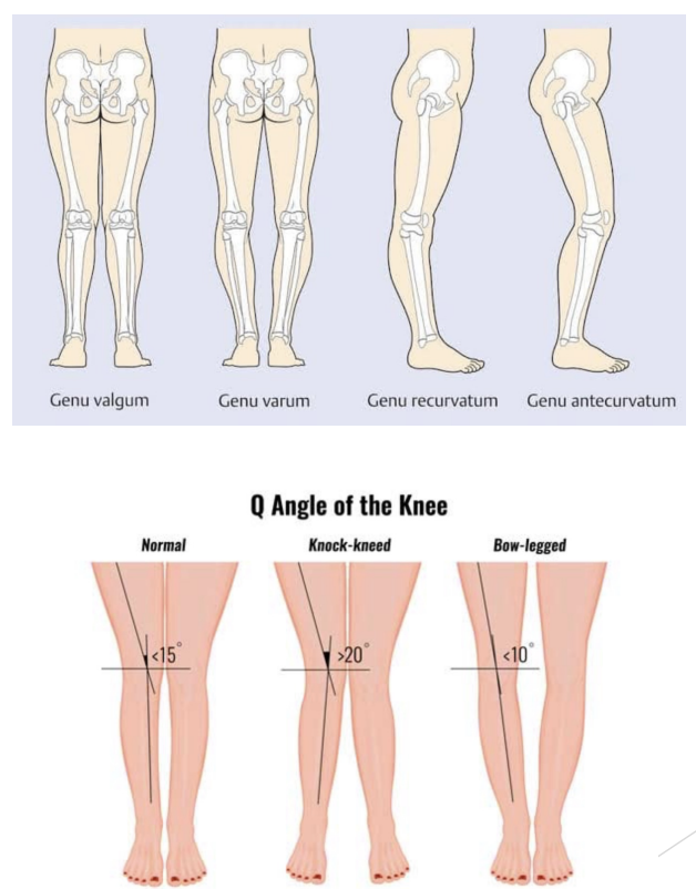
Observation of Knee alignment deviations
Genu Valgrum (knocked kneed)
Increase Q-angle
Genu Varum (bow legged)
Decrease Q-angle
Genu recurvatum
Hyperextension
Genu antecurvatum
Weight Bearing Functional Assessment
Gait
Cycle
Single leg squat
Thessaly - menisci
Duck walk (Childress Test) meniscus
2 legged hop
Single leg hop
Test on strength, distance
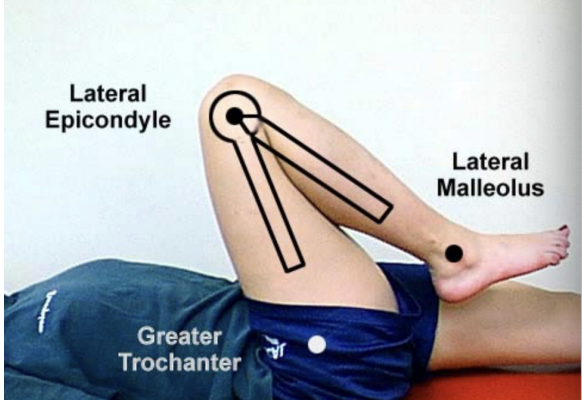
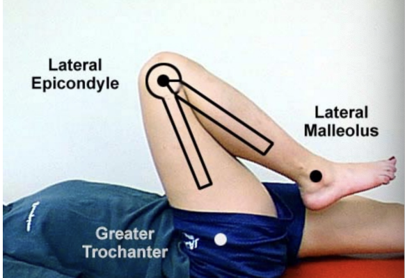
Knee Assessment ROM
Flexion/Extension
Active
Passive
Resisted (strength)
Extacapsular ligaments: LCL/ MCL
Valgus stress test
Varus stress test
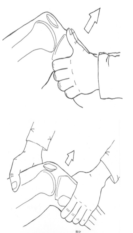
Intracapsullar Swelling Tests
ACL, MCL, Meniscus (medial)
Patellar compression - Ballottement
Swipe (sweep) test
Pushing down on patella
force swelling of knee from medial to lateral side of knee
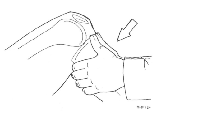
Knee Assessment - ACL
Anterior drawer (injured vs uninjured)
Lachman’s (pulling tibia forward)
Pivot shift
MRI accuracy for diagnosis of ACL injury 95% or >
Knee Assessment - PCL
Posterior sag
Posterior drawer
Knee Assessment - Meniscus
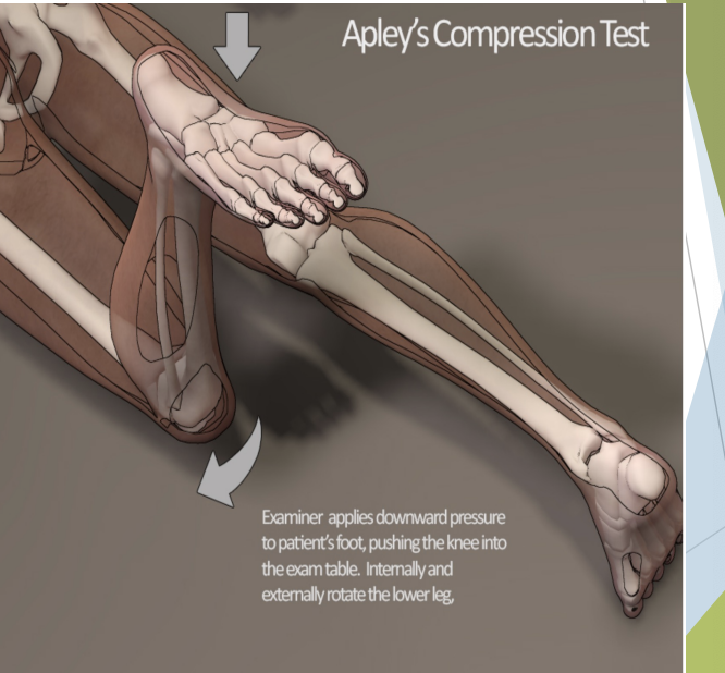
Apley’s compression (bent knee position “forced knee rotation”)
McMurray’s
Joint line tenderness
Knee Contusions
Direct blow
Worse if muscle relaxed
SSx:
Pain, tenderness
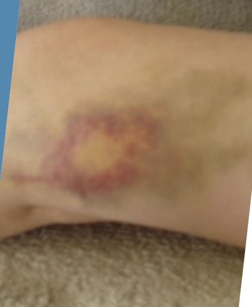
Swelling (circumscribed)
Discolouration
Limp
Tx:
Crutches, ice
POLICE for 2 days
ROM, padding - return to sport
Physiotherapy and rehabilitation
N.B. No massage, no heat directly over bruise (myositis ossificans - surplus of calcium forced)
Bursitis
Direct blow or kneeling
Overuse
SSx:
Inflammation (five stages)
Tenderness
Painful on knee extension if infrapatellar or suprapatellar
Tx:
POLICE, padding, NSAIDs
Heat, physiotherapy, rehabilitation
Early aspiration if acute, traumatic, drain fluid from bursae
Complications
Chronicity, recurrence, infection
Sprains
Direct blow (e.g. valgus anterior)
Torsion or hyperextension
Worse if foot is fixed (planted)
1st degree Sprain
Mild pain, mild swelling
NO Snap or Pop
NO limp, NO effusion, NO increased laxity
Tx: POLICE, physiotherapy, brace
2nd Degree Sprain
Pain, tenderness, “snap”
Swelling (and effusion if intraarticular)
Limp
Increased laxity with firm endpoint
Tx: as for 1st degree, plus see MD. Longer recovery period
3rd Degrees
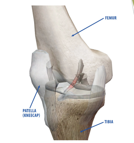
Complete rupture of ligament(s)
SSx:
More pain, tenderness, “snap”
Marked swelling, effusion
Unstable or won’t bear weight
Marked increased laxity, soft endpoint
Tx:
NPO
Stabilize and transport to hospital
Will need brace ± or surgery
Follow with extensive physio, rehab
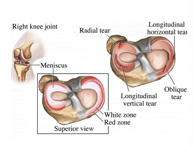
Meniscal tears
Mechanisms
Torsion
Hyperextension
SSx:
Acute
Pain, tenderness
Effusion
Chronic or acute
Locking or buckling
Intermittent pain, swelling, effusion
Positive McMurray’s, Apley’s tests
Tx:
Physiotherapy
Arthroscopic repair or excision
Exercise
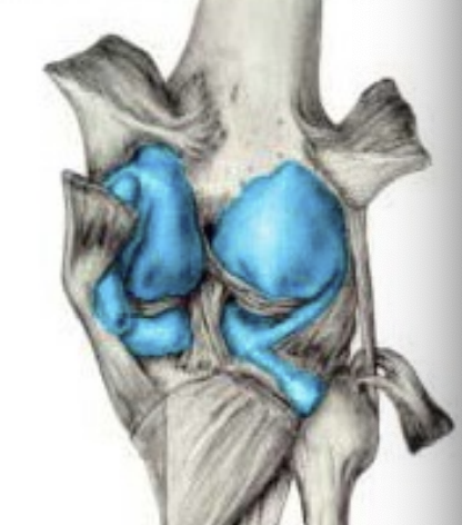
Capsular tears
Mechanisms
Torsion
Hyperextension
SSx:
Pain
Swelling?
Tenderness
May cause rotary instability
Mimics torn meniscus or tendon injury
Tx:
Rest, physiotherapy, rehabilitation
Surgery if symptoms persist
Patellofemoral Pain Syndrome
Pain around patellofemoral joint
Related to abnormal tracking of patellae in femoral groove
Causes of abnormal patellar tracking can include: genu valgrum, external tibial torsion, overpronation, greater than normal Q angle
Chondromalacia Patellae
Softening and deterioration of cartilage on back of patellae
SSx: Pain walking up or down, running, squating
Tx: Conservative (POLICE, NSAIDs, activity modification, strengthening, orthotics)
Surgery is last resort
Patellofemoral Stress Syndrome
Lateral tracking of patellae in femoral groove
Tight lateral musculature, weak hip abductors/ stabilizers
SSx: Pain lateral patellae, crepitis with patellar compression
Tx: POLICE, avoiding aggravating activities, strengthening VMO over VL. Stretching lateral soft tissues, McConnell taping/bracing
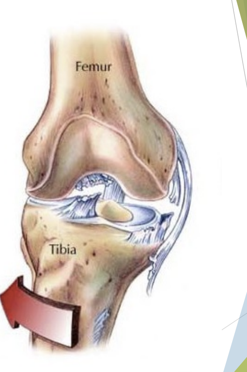
Unhappy triad
Structures damaged
ACL Tear
Positive anterior drawer Lachmann’s test, pivot shift
Effusion
“snap”
MCL tear
Valgus instability, point tenderness
Meniscal tear
Tenderness
Locking or buckling
Capsular tear
Tx:
Reconstructive surgery
Brace, physio, rehab
NB. Effusion? To hospital ASAP
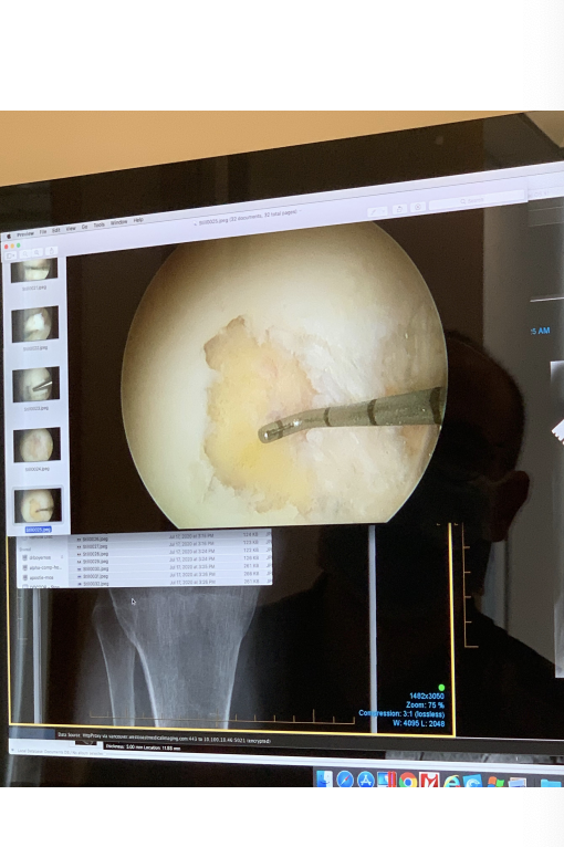
Osteochondritis dissecans
Damage to cartilage and subchondral bone
Most commonly in knee, medial femoral condyle 70% of time
Causes: hereditary, traumatic, vascular
Progression: softening of cartilage
Early cartilage separation from bone
Partial detachment of cartilage
Osteochondral separation with loose bodies
ACL - Post Surgical Rehab
Quadriceps/ hamstrings activation
ROM focusing on full extension
3 to 4 months post surgery - proceed with caution
Closed kinetic chain → Open kinetic chain
Strengthening - hamstrings and quadriceps balanced
6 months in - increase loading, jogging, running
Neuromuscular training
Return to sport - education/ technique modification
Return to sport - functional testing
Brace?
Prevention of ACL injury: FIFA 11
ROM
Agility
Strength test
Dynamic warm up
Screening for Risk of Knee Injuries
Drop vertical jump test