Lab Quiz 1 Bones: pelvic girdle, lower limb, ankle, foot
 Call Kai
 Learn
Learn Practice Test
 Spaced Repetition
 Match
Match Flashcards
 Knowt Play
1/27

There's no tags or description
Looks like no tags are added yet.
Name | Mastery | Learn | Test | Matching | Spaced | Call with Kai | Chat |
|---|
No analytics yet
Send a link to your students to track their progress
28 Terms
What is the pelvic girdle composed of?
Hips and the sacrum
Male pelvic girdle
v shape
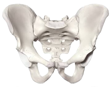
female pelvic girdle
u shape

coxal bone
also known as the hip bone, is comprised of three fused elements: the ilium, ischium, and pubis. It forms a major part of the pelvic girdle.

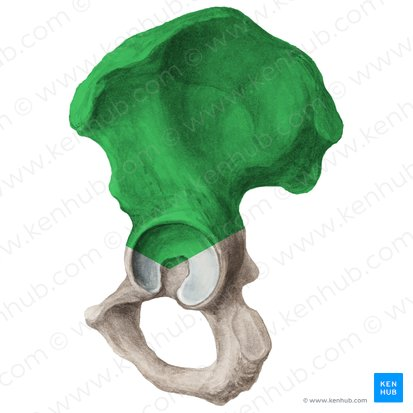
Ilium
the uppermost and largest part of the coxal bone, contributing to the structure of the pelvis.
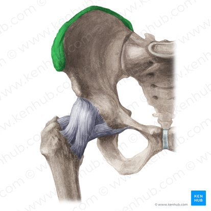
Iliac Crest
the prominent ridge on the superior border of the ilium, serving as an attachment site for muscles and ligaments.
Greater sciatic notch
the large indentation located on the posterior aspect of the ilium, which allows for the passage of nerves and blood vessels from the pelvis to the lower limb.

ischium (THINK ABOUT ANATOMIC POSITION)
the curved, lower part of the coxal bone that forms the posterior portion of the pelvis, providing support during sitting and serving as an attachment point for muscles.
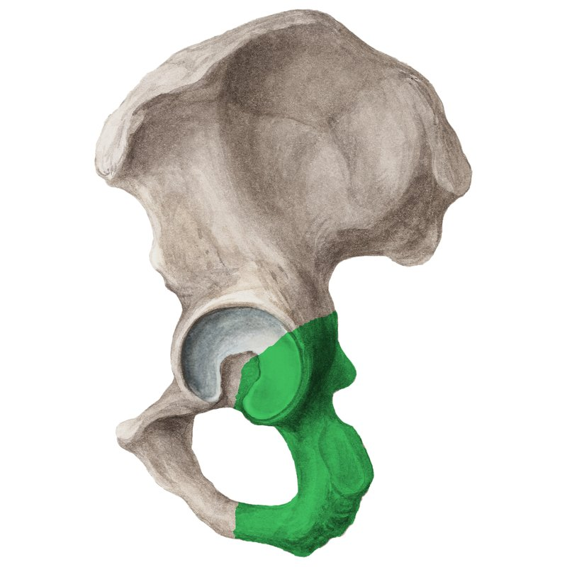
ischial tuberosity
the bony prominence on the ischium, providing support when sitting and serving as an attachment site for muscles and ligaments.

Pubis (THINK ABOUT ANATOMIC POSITION)
the medial and anterior portion of the coxal bone, contributing to the pelvic girdle and forming part of the pubic symphysis.

Auricular surface
the articular surface of the ilium that forms the joint with the sacrum, facilitating the connection between the pelvis and the vertebral column.

obturator foramen
the large opening in the pelvis formed by the pubis and ischium, allowing passage for nerves and blood vessels to the thigh.

acetabulum
The cup-shaped socket of the hip bone that receives the head of the femur, enabling hip joint articulation. “oil cup”

pubic symphysis
The fibrocartilaginous joint located between the left and right pubic bones, providing slight mobility and stability to the pelvis.

pubic arch
ormed by the inferior rami of the pubic bones, contributing to the pelvic inlet and shaping the pelvic cavity.

Femur
The longest and strongest bone in the human body, located in the thigh, articulating with the hip joint at its proximal end and the knee joint at its distal end.

femur head
The rounded proximal end of the femur that fits into the acetabulum of the pelvis, crucial for hip joint articulation.
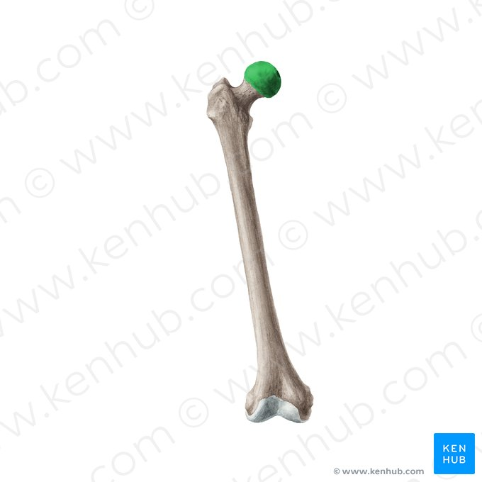
femur fovea capitis
A small pit on the femur head where the ligamentum teres attaches, helping stabilize the hip joint.

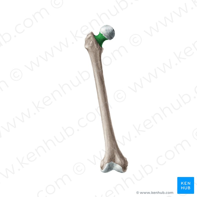
Femur neck
The constricted region just below the femur head, serving as a pivotal connection between the head and the shaft of the femur.
femur greater trochanter
A large, bony prominence located on the lateral aspect of the femur, serving as an important site for muscle attachment and leverage.
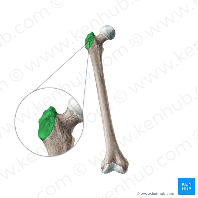
femur lesser trochanter
A small, bony projection located on the medial aspect of the femur, serving as a key site for muscle attachment, particularly for the iliopsoas muscle.

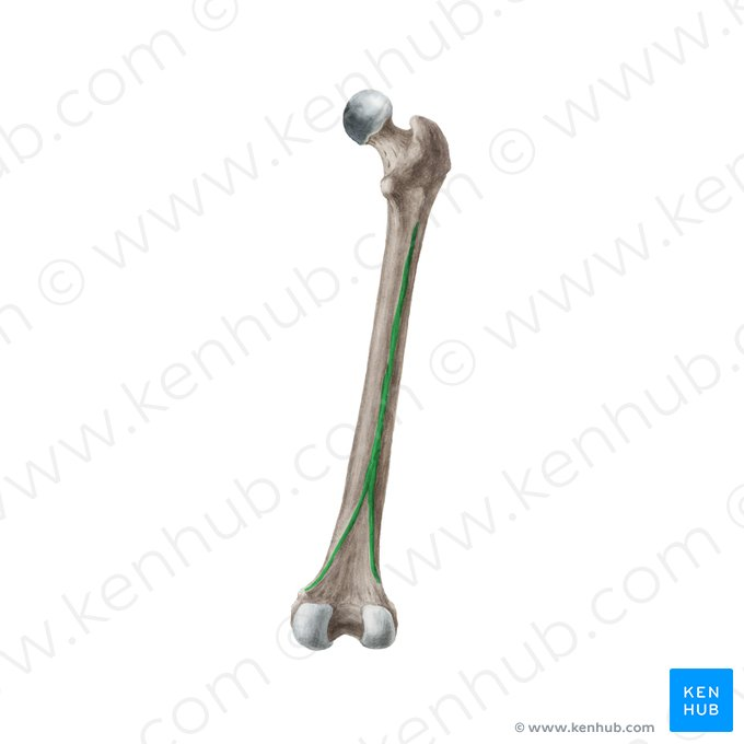
femur linea aspera
A prominent longitudinal ridge located on the posterior surface of the femur shaft, providing attachment for muscles and contributing to the femur's structural strength.
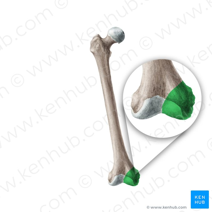
femur medial condyle
rounded, articular surface located on the distal end of the femur, which articulates with the tibia at the knee joint.
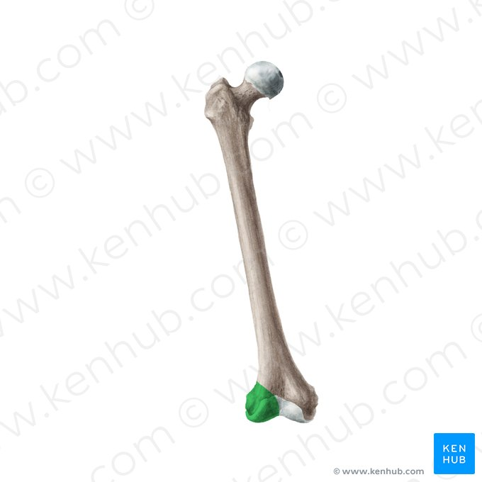
femur lateral condyle
rounded, articular surface located on the distal end of the femur, which articulates with the tibia and fibula at the knee joint.
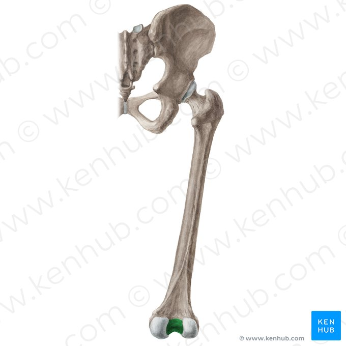
femur intercondylar fossa
deep notch located between the medial and lateral condyles of the femur, allowing for the attachment of ligaments and facilitating movement at the knee joint.
femur medial epicondyle
bony prominence on the medial side of the femur, providing attachment for ligaments and muscles, particularly the adductor muscles.
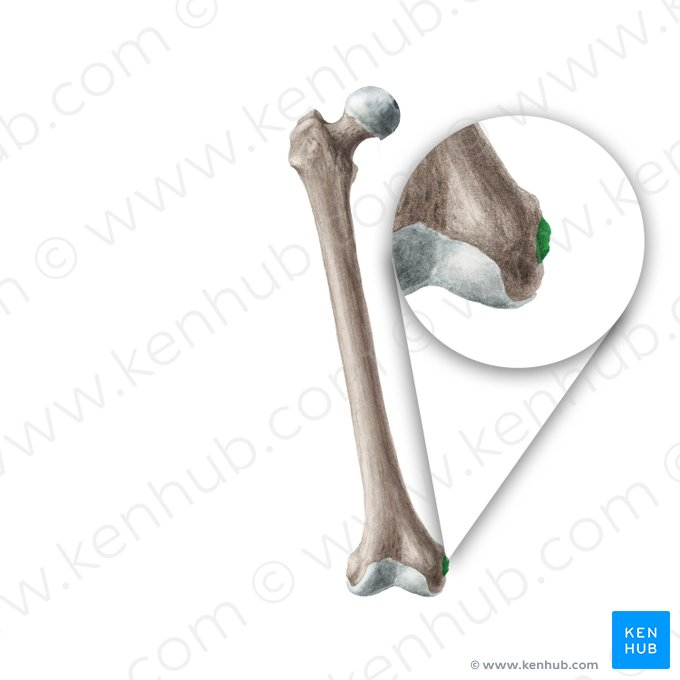
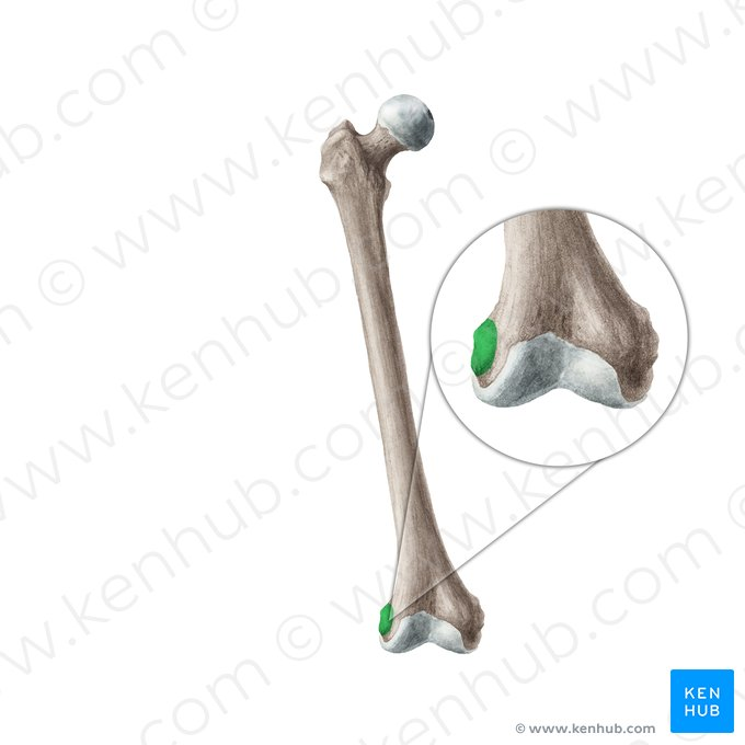
femur lateral epicondyle
bony prominence on the lateral side of the femur, serving as an attachment point for ligaments and muscles, including those involved in knee stabilization and movement.