NRSE 210 Exam #2 part 2
 Call Kai
 Learn
Learn Practice Test
 Spaced Repetition
 Match
Match Flashcards
 Knowt Play
1/104

There's no tags or description
Looks like no tags are added yet.
Name | Mastery | Learn | Test | Matching | Spaced | Call with Kai | Chat |
|---|
No analytics yet
Send a link to your students to track their progress
105 Terms
external ear (function, structures)
funnel sound waves
auricle (pinna), external auditory canal, tympanic membrane
Auricle (pinna)- (structure)
cartilage structure, includes orkle at top
External auditory canal (structure)
3 cm, lined with glands that secrete cerumen
cerumen
ear wax
keeps foreign objects out, lubricates ear
Tympanic membrane (description, location)
eardrum
pearly grey, oval slightly concave
separates external and middle ear
Middle ear (description, function)
small air- filled cavity
contains ossicles (malleus, incus, Staples)
Function: transmit sound vibrations from eardrum to inner ear
Inner ear: bony labyrinth
containing sensory organs for hearing and equilibrium
Inner ear: cochlea
central hearing apparatus
CN VII
vestibulocochiear nerve (inner ear is connected to)
Pathways of hearing: air conduction
normal, efficient pathway]
external and middle ear
Pathways of hearing: bone conduction
skull bones vibrate
vibrations transmits to inner ear
hearing loss: conductive loss (cause, examples)
mechanical dysfunction of external/ middle ear
partial loss, amplifying may help
examples: foreign body, perforated eardrum, wax, fluid
hearing loss: senseorineural loss
pathology of inner ear, CN VII, or cerebral cortex
amplifying sound does not improve hearing
examples: presbycusis (age related progression)
hearing loss: mixed loss
combination of sensorineural and conductive
vertigo (cause)
cause by labyrinth inflammation, sending wrong locations to the brain
Tinnitus
ringing, buzzing (caused by lound noises)
Tx- asorin, lasix, antibiotics, NSAIDS
Otoscope exam positioning
adults: pull pinna up and back
young children: pull pinnadown and back
Otitis externa (definition and affect)
swimmers ear
infection, pain with pinna/ tragus movement, swelling, drainage
external ear
Darwin’s tubercle
small bump on helix, harmless
external ear
Tophi
uric acid nodules, sign of gout
external ear
Chondrodermatits
painful red bump from pressure/ sun
external ear
keloid
overgrowth of scar tissue
carcinoma
non- healing lesion, bleeding, often sun exposed
Palperbral fissure
space between eyelids
limbus
border between sclera and cornea
canthus
corners of eyes
caruncle
fleshy mass at inner canthus with sebaceous glands
conjunctiva
palperbral (lines lids)
bulbar (covers eyeballs
What CN’s control extraocular muscles
III, IV, VI
Outer layer of eye (makeup)
sclera
cornea
Middle layer of eye makeup
iris
pupil
lens
anterior chamber
what CN controls the size of the pupil
CN III
Inner layer of eye makeup
retina- conversion light to nerve impulses
optic disc, rerinal vessels, macula
Pupillary light reflex
direct: same eye constricts
consensual: other eye also constricts
visual accommodation
pupils constrict, eye's converge for near vision
cataracts
aging eye changes
lens opacity, surgical removal
glaucoma
aging eye changes
increased intraocular pressure, damage to the optic nerve
macular degeneration
aging eye changes
affects central vision
leading cause to blindness
Physical exam test: Snellen chart
distance vision (20/20 is normal)
Physical exam test: near vision
card is 14 inches from the patients face
normal result is 14/14
Physical exam test: confrontation test
peripheral vision
Physical exam test: corneal light reflex
eye alignment
Physical exam test: cover test
strabismus check, lazy eye
Physical exam test: diagnostic positions test
6 cardinal gazes
eyelid disorders
perior edema, exophthalmos, enophthalmos
ptosis, ectopic, entropin
eye lesions
blepharitis, dacryocystitis, chalazion, hordeolum (stye)
Pupil disorders
anisocoria (unequal pupils)
mydriasis (dilated/ fixed)
external eye disorders
conjunctivitis (pink eye)
Thoracic cage structure
bony structure made of sternum
12 pairs of ribs
12 thoracic vertebrae
Diaphragm structure
forms the floor
separates abdomen from thoracic cavity
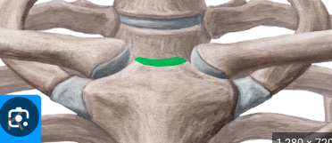
suprasternal notch structure
U- shaped, abover sternum, between clavicles
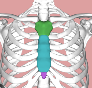
sternum structure
breast bone with three parts:
manubrium
body
xiphoid
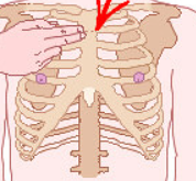
Manubriosternal angle
AKA angle of louis
between manubrium and body
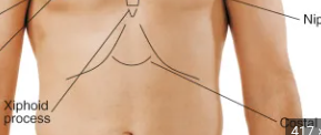
costal angle
right and left of xiphoid process
normal= 90 degrees
angle increased when rib cage is chronically overinflated (ex.COPD)
vertebra prominens structure
C7
flex head to chin
bony protrusion at the back of the spine
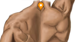
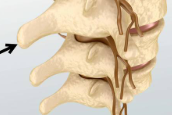
Spinous processes structure
T1- T2 (top to bottom of spine
Inferior border of scapula structure
located symmetrically in each hemithorax
aligns with 7th- 8th rib
Twelfth rib structure
midway between spine and side
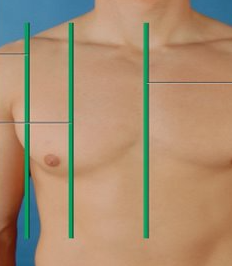
Mid sternal line
exact middle (divides body into left and right)
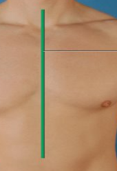
Mid clavicular line
bisects center of each clavicle
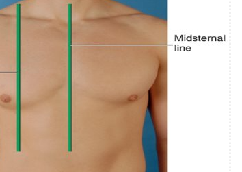
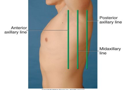
Anterior axillary line
runs down the front side of the body through the armpit
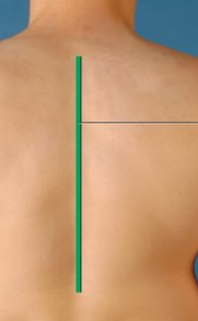
vertebral line
runs down middle of the back (divides left and right(
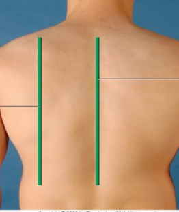
scapular line
posterior version of the mid- clavicular line
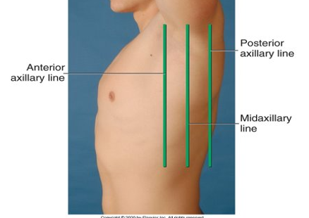
Mid axillary line
lateral reference
from the apex of axilla staright down the body
splits anterior and posterior
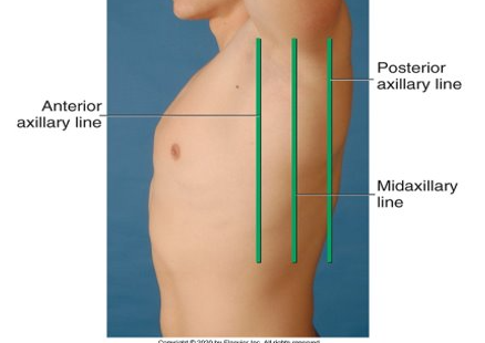
Anterior axillary line
runs down the front side of the lateral position
Posterior axillary line
runs down the back side of the lateral position
Mediastinum cavity
contains esophagus, trachea, heart, and great vessels
Lung borders and descriptions (3)
Apex: highest point of lung tissue
Base: lowest border; sits on diaphragm (around 6th rib)
Posterior: lungs extend from T3 to T10
Right lung description
3 lobes
shorter because of liver
lobes are separated by fissures
left lung description
2 lobes
narrower because of the heart
no middle lobe
lobes are separated by fissures
anterior chest lung POV
mostly upper and middle lobes
posterior chest POV
almost all lower lobe
Pleurae (makeup, cavity, negative pressure)
thin slippery envelope between lungs and chest wall
inside of the envelope contains small amount of lubricating fluid
negative pressure: vacuum holds lungs tightly against the chest wall
Trachea description
anterior to esophagus
10 cm long
splits sternal angle into right and left main bronchi
Bronchi decription
lined with goblet cells (secrete mucus) and cilia (sweep particles upward)
involuntary mechanisms are controlled by what
brain stem
inspiration
air rushes in, diaphragm expands
expiration
air is expelled as chest recoils
AP/ Transverse ratio (description and abnormalities)
The normal anteroposterior (AP) to transverse chest ratio is approximately 1:2, meaning the distance from front to back is half the distance from side to side
barrel chest is a 1:1 ration
pigeon chest description
sternum bulging outward
Funnel chest description
sternum is inward
Symmetrical chest expansion test (normal/ abnormalities during test)
hands on mid back, thumbs should move apart evenly
asymmetry movement = pneumonia, trauma, collapsed lung, deformity
Tactile fremitus test (description and abnormalities)
use ball of hand, have patient say 99
avoid scapula; vibrations should be symmetrical
decreased fremitus= emphysema
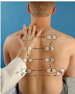
percussion of posterior chest
non dominant hand on patient, strike with dominate hand (avoid scapula)
move side to side, top to bottom
over lung= resonance, scapula= flat, liver= dullness
Abnormal sounds: hyperresonance= too much air. dullness= increased density
ascultate posterior chest procedure
have patient lean forward slightly, arms in lap
breathe slowly through mouth, slightly deeper than normal
Breath sounds: bronchial
inspiration< expiration
loud, over trachea/larynx
normal breath sounds
Breath sounds: bronchovesicular
inspiration= expiration
moderate, over major bronchi/ upper sternum
normal breath sounds
Breath sounds: vesicular
inspiration> expiration
soft pitched, over low lungs (rustling sound)
normal breath sounds
decreased breath sounds means
collapsed lung or obstruction
Adventitious sound discontinuous description (crackles fine/course, rhonchi, pleural friction rub)
Fine crackles- high pitched, collapsed air sacs reopening
Coarse crackles- Velcro sound, coughing doesn’t clear, edema or pneumonia
Rhonchi- low pitched snoring, fluid blocked airways, clear with cough
pleural friction rub- rubbing leather sound, pleural inflammation
Adventitious sound continuous description (wheeze, stridor)
wheeze- musical, high- pitched; mostly expiration (asthma, emphysema)
stridor- high- pitched inspiration; swollen upper airway or obstruction (croup, epiglottis)
barrel chest (description, seen with..)
AP= transverse (1:1)
horizontal ribs
seen in asthma or emphysema
scoliosis (description, seen in...)
lateral spine curvature
uneven shoulders/ scapula/ hips
can limit lung capacity
common in females
Kyphosis
hunchback; pain, reduced motion
aging/ osteoporosis; may impair breathing
Tachypnea (description, seen in..)
greate than or equal to 24 breaths per minute
anxiety, fever, infection, exercise
Bradypnea (description, seen in..)
less than 12 breaths per minute
CNS depression, coma, overdose
Cheyne- strokes (description, seen in..)
alternating fast and slow breathing with apnea
heart/renal failure, overdose, Increase ICP (intracranial pressure)
Asthma (definition, cause, symptoms, treatment)
definition: chronic inflammatory airway disorder, causing airway construction and mucus production
causes: allergens, exercise, cold air, infections, stress
symptoms: wheezing, coughing, shortness of breath, chest tightness
treatment: bronchodilators (Albuterol), avoiding triggers
COPD (definition, cause, symptoms, treatment)
chronic obstructive pulmonary disease
definition: progressive airway obstruction due to chronic bronchitis or emphysema
cause: smoking, air pollutants, genetic factors
symptoms: chronic cough, sputum production, fatigue
treatment: smoking cessation, oxygen therapy, bronchodilators
Pneumonia (definition, cause, symptoms, treatment)
definition: infection that inflates air sacs in one or both lungs
cause: bacteria, virus, fungi
symptoms: fever, cough, phlegm, chest pain, fatigue
treatment: antibiotics (for bacterial), rest, fluid, oxygen therapy if needed