Nervous System- Anatomy/Physiology
1/92
Earn XP
Description and Tags
Dr. Breslin Anatomy/Physiology H
Name | Mastery | Learn | Test | Matching | Spaced | Call with Kai |
|---|
No analytics yet
Send a link to your students to track their progress
93 Terms
Main Functions of the Nervous System
Detection of internal and external changes of the body
Integration of information from each sense
Coordination of muscles, organs, and glands
Central Nervous System
Includes the brain and spinal cord.
Integrates and coordinates sensory information
Transmission of motor commands (motor output)
Peripheral Nervous System
All of the nervous tissue outside of the brain and spinal cord. Includes the afferent division and the efferent division.
Afferent Division
Subdivision of the PNS. Received by receptors. Transmits information from the senses towards the brain
Efferent Division
Subdivision of the PNS. Transmits commands to the muscles and glands from the brain. Divided into two systems: Somatic nervous System- and the autonomic nervous System-.
The Somatic Nervous System
Subdivision of the efferent division. Controls skeletal muscles.
Autonomic Nervous System
Subdivision of the efferent division. Regulates involuntary smooth and cardiac muscles as well as glands. Divided into two divisions: Sympathetic division and Parasympathetic division
Sympathetic Division
Subdivision of the Autonomic nervous System. Includes “fight or flight” response.
Parasympathetic Division
Subdivision of the Autonomic nervous System. Includes “rest and digest” responses.
Neurons
The basic units of the nervous System. Able to communicate with other cells. They specialize in transmitting and receiving messages. Neurons cannot divide.
Neuroglia
Support neurons by regulating their surrounding environment
Dendrites
Recieves, processes, and transfers information from other neurons and the environment. Transfers information towards the cell body.
Axons
Conduct impulses away from cell body. They transmit electrical impulses (messages) to the brain.
Cell Body
The main part of a neuron that contains the neurons organelles. Contains ER and free ribosomes known as nissel bodies (grey color-grey matter).
Axon Hillock
Thickened region of the cell body where action potential originates.
Myelin
Thin pads of lipids that serve as electrical insulation. Myelinated Neurons can transmit electrical signals much faster and appear white. The speed of the signals can reach up to 250 mph
Demyelination
Progressive destruction of myelin sheaths, causing a loss of sensation and motor control (paralysis). Examples include Multiple Sclerosis (MS) and mercury poisoning.

Unipolar Neurons
Exclusively sensory neurons, except for olfactory and sight (optic). Cell body lies off to one side and is housed in the ganglion. One process, one axon, one dendrite.
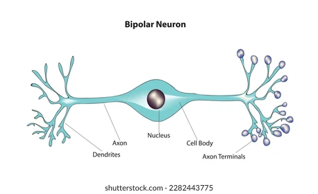
Bipolar Neuron
Olfactory and Optic neurons. Two processes, one axon, and one dendrite with the cell body in-between.
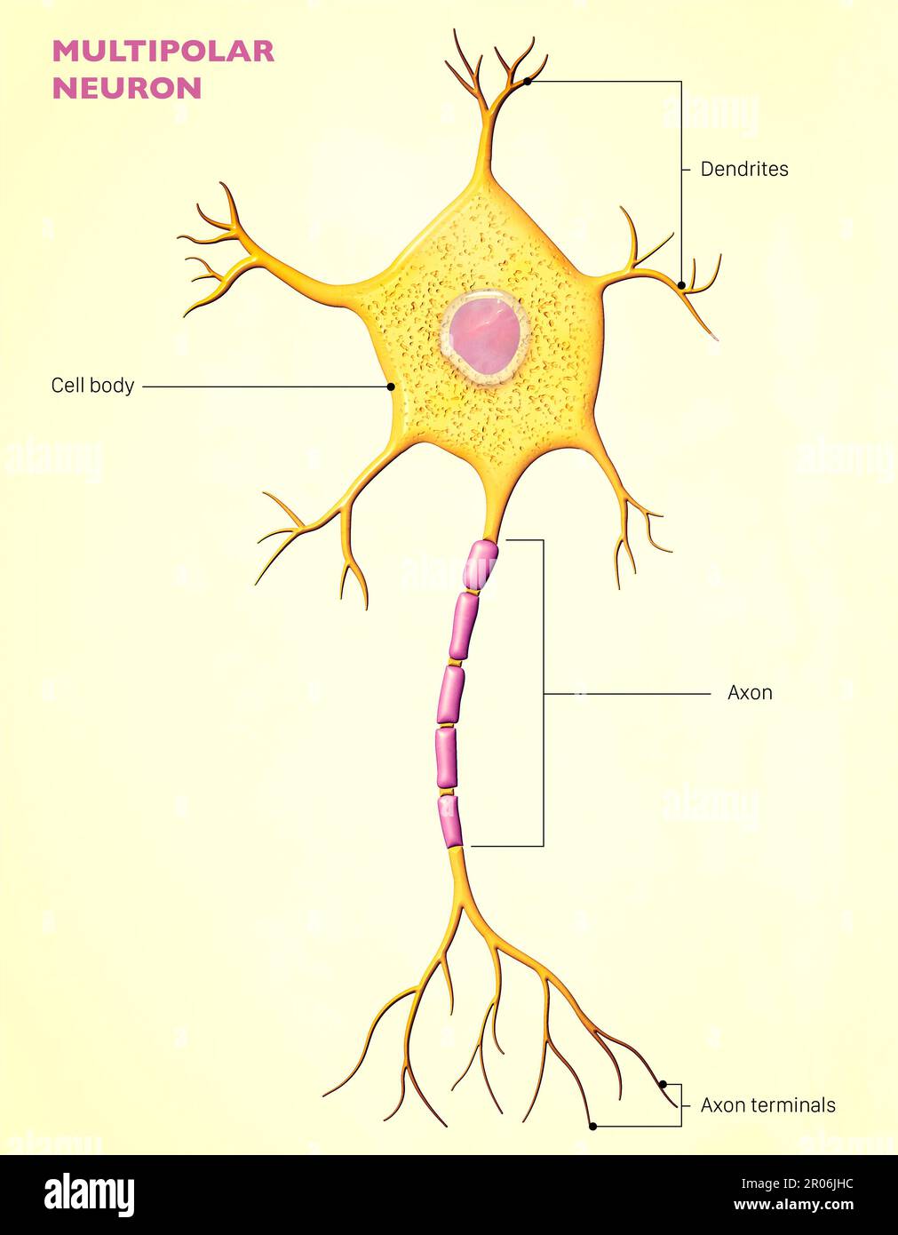
Multipolar Neurons
Multiple processes, two or more dendrites and one axon. Most commonly found in the central nervous System. Make up all motor neurons controlling skeletal muscle.
Sensory Neurons
Part of the efferent division. Brings information from sensory receptors to the brain. We have 10 million of these
Somatic Sensory Receptors
Processes information from the outside world. There are two kinds: External receptors and proprioceptors.
External Receptors
A type of somatic sensory receptors. Send information regarding touch, pressure, and temperature.
Proprioceptor
A type of somatic sensory receptor. Send information regarding the position and movement of skeletal muscles.
Visceral (internal) Receptors
Monitor activity of digestive, respiratory, cardiovascular, urinary, and reproductive systems.
Motor neurons
Part of the efferent division (output). Carry information from the central nervous System to other organs and organ systems. We have half a million of these.
Somatic Motor Neurons
In charge of the skeletal muscles
Visceral Motor Neurons
Part of the Autonomic nervous System. Controls smooth muscle and cardiac muscle.
Interneurons
Only located in the brain and spinal cord (CNS). They are in charge of:
Connecting sensory neurons to motor neurons.
Transferring signals between sensory and motor neurons
Memory, planning, and learning.
The more complex the response, the more interneurons involved. We have around 20 billion of these.
Astrocyte
A type of neuroglia located in the CNS. It is the largest and most numerous. Secretes chemicals that maintain the blood barrier. Also in charge of repairing damaged tissue.
Oligodendrocytes
A type of neuroglia located in the CNS. The tips wrap around axons which creates a sheath of insulation (myelin). One oligodendrocyte can wrap around multiple axons at a time. They myelinate short segments of axon.
Nodes of Ranvier/Internodes
Gaps between myelin sheath. Message travels 250 mph
Saltatory Conduction
Jumps from node to node
Myelinated Neurons
White matter
Unmyelinated Neurons
Grey matter
Microglia
A type of neuroglia found in the CNS. It is the smallest and rarest. Contains phagocytic cells (derived from WBC). Engulfs cellular waste and pathogens.
Ependymal Cells
A type of neuroglia in the CNS. They line the central canal of the spinal cord and ventricles of the brain (contains cerebrovascular fluid). The cilia on the cells help circulate CSF. The ependymal cells also help produce, move, and secrete CSF.
Satellite Cells
A type of neuroglia located in the PNS. They surround and support the neuron cell body.
Schwann Cells
A type of neuroglia in the PNS. They myelinate axons of nerve cells in PNS which forms neurilemma. Can only myelinate 1 axon at a time.
Nerve Pathways
Nerve pathways are always one way. This includes the Sensory and Motor tracts.
Motor Tract
On the central side of the spinal cord. A part of the efferent division. Moves signals away from the brain.
Sensory Tract
On the dorsal side of the spinal cord. A part of the afferent division. Moves signals towards the brain.
Nerves
Always found in the PNS
Tracts
Always found in the CNS
Sensory Nerve Rules
Always go to the CNS
Always have a ganglion to house their nuclei
Always enter the dorsal horn to pass along their message
Message Pathway
Sensory receptors —> Spinal cord- dorsal horn tract —> Brain (cortex) —> Spinal cord (exit ventral tract) —> Motor neuron
Nerve Signal Transmission Process (NST)
Contains five main steps; Action Potential, Resting Stage, Depolarization, Repolarization, and Sodium-Potassium ATP Pump.
Action Potential
First stage of NST. Electrical impulses sent across the membrane of neurons. They are generated by changing amounts of ions inside and outside the neuron. K+, Na+, Cl-, Pr- (regular charged proteins).
Resting Stage
Second stage of NST. A resting neuron has a negative potential of about -70mV.
Depolarization
Third stage of NST. Occurs when a stimulus (such as a neurotransmitter) causes a protein channel to open. Na+ ions diffuse inside the cell membrane, creating a positive charge across the membrane. “Negative charge —> Positive charge”
Repolarization
Fourth stage of NST. The return to the original negative charge as K+ ions diffuse out of the cell. So much K+ leaves the cell that it becomes hyperpolarized (more negative than its original resting state)
Sodium-Potassium ATP Pump
Fifth stage of NST. Restores the original concentrations of K+ and Na+ through active transportation (3 Na out and 2 K in). The neuron is now available to conduct another action potential.
Synapse
A gap between the axon of the neuron and another cell
Neurotransmitter
Chemicals that allow neurons to communicate with each other throughout the body.
Synaptic Cleft
The space after the axon terminal of a neuron between the next target cell. Important for cells to be able to communicate using chemical transmission.
Cholinergic Synapse
Found between motor neurons and muscle cells. Converts a presynaptic electrical signal into a chemical signal (acetylcholine), which diffuses across the synaptic cleft.
Acetylchloline
The neurotransmitter of the cholinergic synapse. Released and binds to receptors on the muscle cell. It is eventually broken down by an enzyme, ending the process.
Botulism
A disease caused by the ingestion of bacterial toxins from rotting food. The toxin prevents the release of acetylchloline at cholinergic receptors, causing paralysis
Tetanus
Caused by a bacterial toxin that inhibits the enzyme that breaks down acetylchloline. This causes prolonged, painful muscle contractions/ Exposure to the bacteria through any wound that punctures the skin. “Prolonged contraction”
Reflex
A rapid, involuntary response to a stimulus. These actions occur more quickly because they occur over a reflex arc (a pathway). These bypass the brain.
Reflex Arc
Begins with a stimulus at a sensory neuron, such as pain or heat. The sensory neuron is depolarized.
Cerebrum (Lobes)
The largest region of the brain. Controls conscious thought, complex movement, and memory. Separated into 2 sides (Left hemisphere and Right hemisphere) by the longitudinal fissure. Four main lobes: Temporal lobe, Parietal lobe, Occipital lobe, and Frontal Lobe.
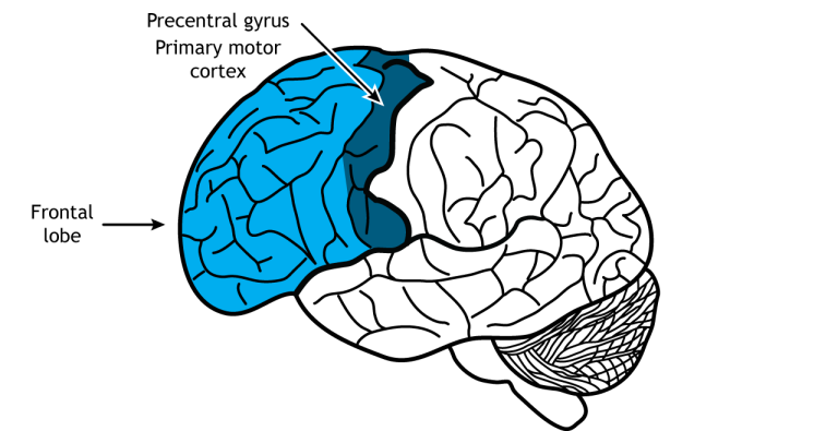
Frontal Lobe
Part of the cerebrum. Considered the output center. This is the largest lobe. It controls skeletal muscle movement, personality, short-term memory, consciousness, Broca’s Area (language; ability to form words), morals, and ethics.
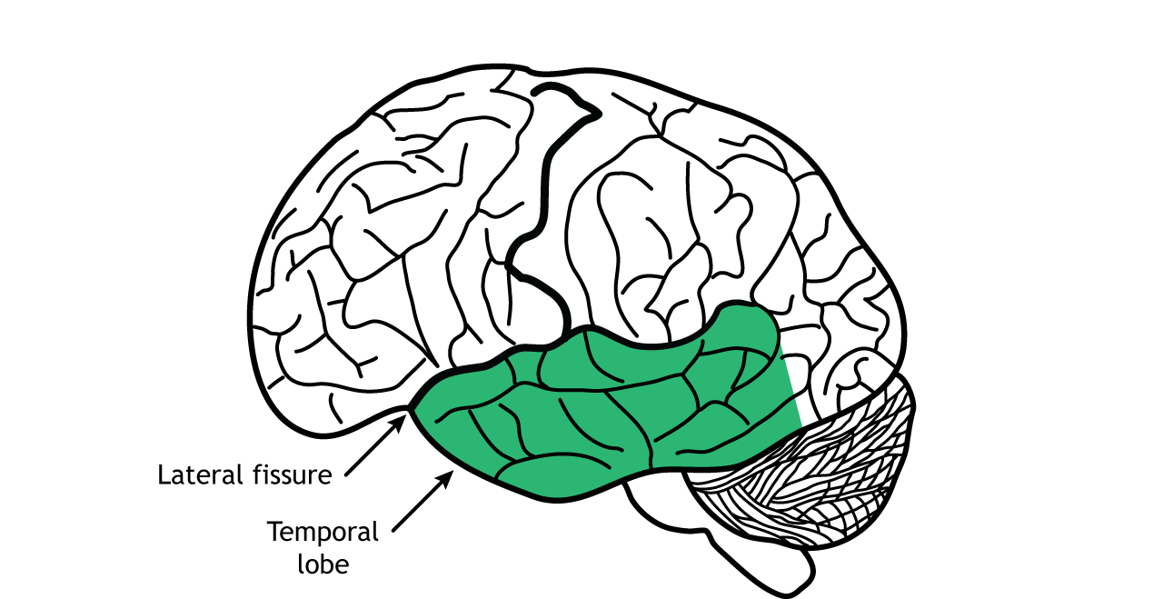
Temporal Lobe
Part of the cerebrum. In charge of hearing, taste (olfactory), balance, and long-term memory.
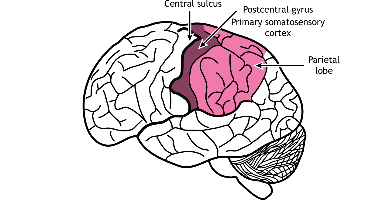
Parietal Lobe
Part of the cerebrum. Considered the Major Input. In charge of integrating sensory information, Broca’s Area (understanding speech), recognition of objects, people, places, and events, and sensation.
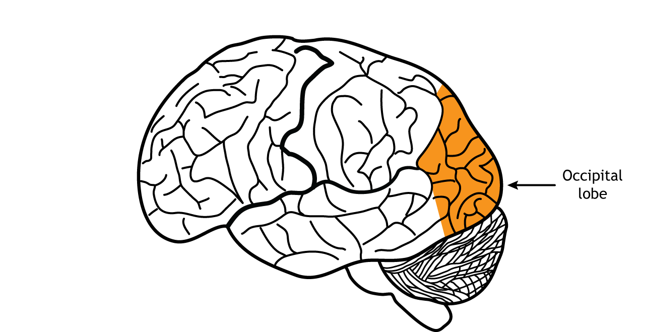
Occipital Lobe
Part of the cerebrum. Contains the vision center. Primary visual cortex.
Cerebral Cortex (Grey Matter)
One layer of the Cerebrum. Considered the grey matter. Unmyelinated, outer grey matter that contains neuron cell bodies (cortex, wrinkled portion).
Corpus Callosum (White Matter)
One layer of the Cerebrum. Considered the white matter. Myelinated, inner white matter that contains axons encased in myelin sheaths. In charge of communication between the Left and Right hemispheres.
Inner Brain Anatomy
Includes: Parietal lobe, central sulcus, frontal lobe, corpus callosum, lateral ventricle, hypothalmus, pituitary gland, thalamus, pons, medulla oblongata, cerebellum, pineal gland, Occipital love, and parieto-occipital sulcus
Diencephalon
Integrates sensory information and motor commands from the brain, spinal cord, and PNS. Collects all senses except for oflaction. Includes: Hypothalmus, Thalmus, and Pineal Gland
Thalmus
A part of the Diencephalon. Transfers (relays) impulses received from sensory neurons to correct region of the cerebrum (cortex).
Hypothalmus
A part of the Diencephalon. Controls many aspects of internal homeostasis, including body temperature, water balance, and overall metabolism (Hunger and Thirst)
Pineal Gland
A part of the Diencephalopn. Releases melatonin, a hormone that regulates day-night cycles (circadian rhythm).
Brain Stem
Directly attaches the brain to the spinal cord. Controls involuntary body functions. The cranial nerves connect through the brain stem. Includes the midbrain, pons, and medulla oblongata.
Pons
Part of the Brain Stem. Contains the neurons responsible for controlling breathing along with the medulla oblongata.
Medulla Oblongata
Part of the Brain Stem. The lowest part of the brain stem. Merges into the spinal cord. Controls heart rate, blood pressure, swallowing, vomiting, and breathing (with the Pons).
Cerebellum
Often called the "“little brain”. Makes up 10% of the mass in the brain. Copies motor commands sent to the spinal cord. When the information is sent correctly, movement is made. Coordinates posture, balance, and body movements.
Protection of the Central Nervous System
Due to the neurons in the CNS being unable to undergo mitosis, the CNS has a lot of protection. It consists of the layers of protection: the bone, the Meninges (dura matter, arachnoid mater, and pia matter), and Cerebrospinal fluid.
Meninges
Below the bone. These are membranes of connective tissue. Made up of three parts: Dura Mater, Arachnoid Mater, and Pia Mater.
Dura Mater
A part of the Meninges. The tough, thick, outermost layer.
Arachnoid Mater
A part of the Meninges. A network of blood vessels that look similar to a web. It contains cerebrospinal fluid.
Pia Mater
A part of the Meninges. The delicate innermost layer that closely adheres to the surface of the brain and spinal cord.
Cerebrospinal Fluid
Similar to plasma. Circulates through a series of cavities (called ventricles) throughout the brain. Provides a watery cushion that protects nervous tissue from trauma.
Blood-Brain Barrier (BBB)
Made of capillaries. Is is a selective semi-permeable membrane that only allows water, glucose, and amino acids into the brain area. It keeps out waste, proteins, random cells, and harmful substances.
Encephalitis
An inflammation of the brain itself caused by a foreign substance or virus. Symptoms include: headache, drowsiness, nausea, and fever.
Meningitis
An inflammation of the Meninges (membranes) of the brain and spinal cord. Symptoms include: fever and stiff neck.
Concussion
A traumatic brain injury caused by an impact that is not absorbed by cerebrospinal fluid and damages the brain itself.
Brain Tumor
Masses of cells that overgrow in specific areas of the brain. Symptoms vary depending on the exact region of the brain that’s affected.