Lecture 7/8 - How neurons communicate
1/125
There's no tags or description
Looks like no tags are added yet.
Name | Mastery | Learn | Test | Matching | Spaced |
|---|
No study sessions yet.
126 Terms
What are the two classic experiments to determine how neurons communicate
Galvani
Loewi
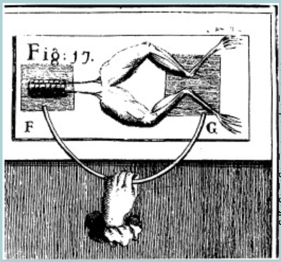
Luigi galvani:
Observed WHAT hanging on a wire would WHAT during an electrical storm
Hypothesized that sparks from the storm activated WHAT
Applied WHAT to a dissected WHAT attached to the leg (the leg WHAT)
Concluded that the WHAT used WHAT to communicate
Luigi galvani:
Observed FROG LEGS hanging on a wire would TWITCH during an electrical storm
Hypothesized that sparks from the storm activated MUSCLES
Applied CURRENT to a dissected NERVE attached to the leg (the leg TWITCHED)
Concluded that the NERVOUS SYSTEM used ELECTRICITY to communicate
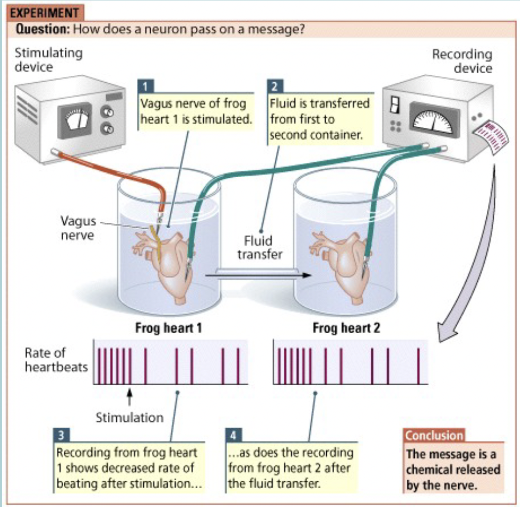
Chemical communication: 1st evidence
Done by Otto loewi WHAT experiment
Stimulated the WHAT
DOES WHAT TO the heart
The two hearts shared fluid and they exhibited the same WHAT even though the other heart was not stimulated
Chemical found to be WHAT
Chemical communication: 1st evidence
Done by Otto Loewi FROG HEART experiment
Stimulated the VAGUS NERVE
SLOWS the heart
The two hearts shared fluid and they exhibited the same RESPONSE even though the other heart was not stimulated
Chemical found to be ACETYLCHOLINE
Movement of ions = WHAT
Movement of ions = CHARGE
Intercellular and extracellular fluid is filled with WHAT
Ions (charged particles)
What are the ions most important for neuron electrical signals
Sodium (Na+)
Potassium (K+)
Chloride (Cl-)
Large negatively charged proteins (A-)
Calcium (Ca 2+)
What contributes to a cells electrical charge:
Outside cell:
WHAT
WHAT
WHAT
Inside cell (leakage channels):
WHAT
WHAT
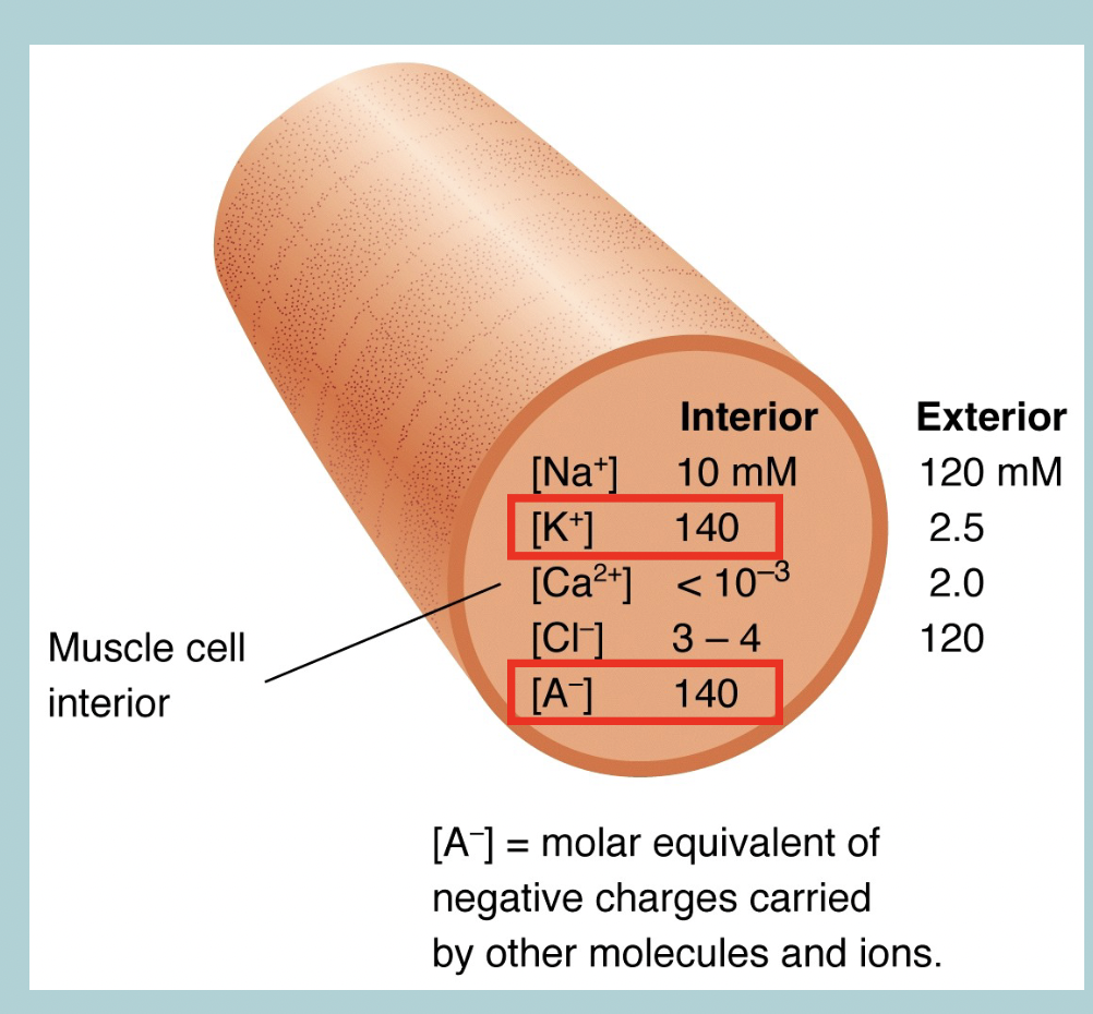
What contributes to a cells electrical charge:
Outside:
Lots of SODIUM (Na+)
Lots of CHLORIDE (Cl-)
Lots of CALCIUM (Ca2+)
Inside:
Lots of POTASSIUM (K+)
Lots of NEGATIVE PROTEINS (A-)
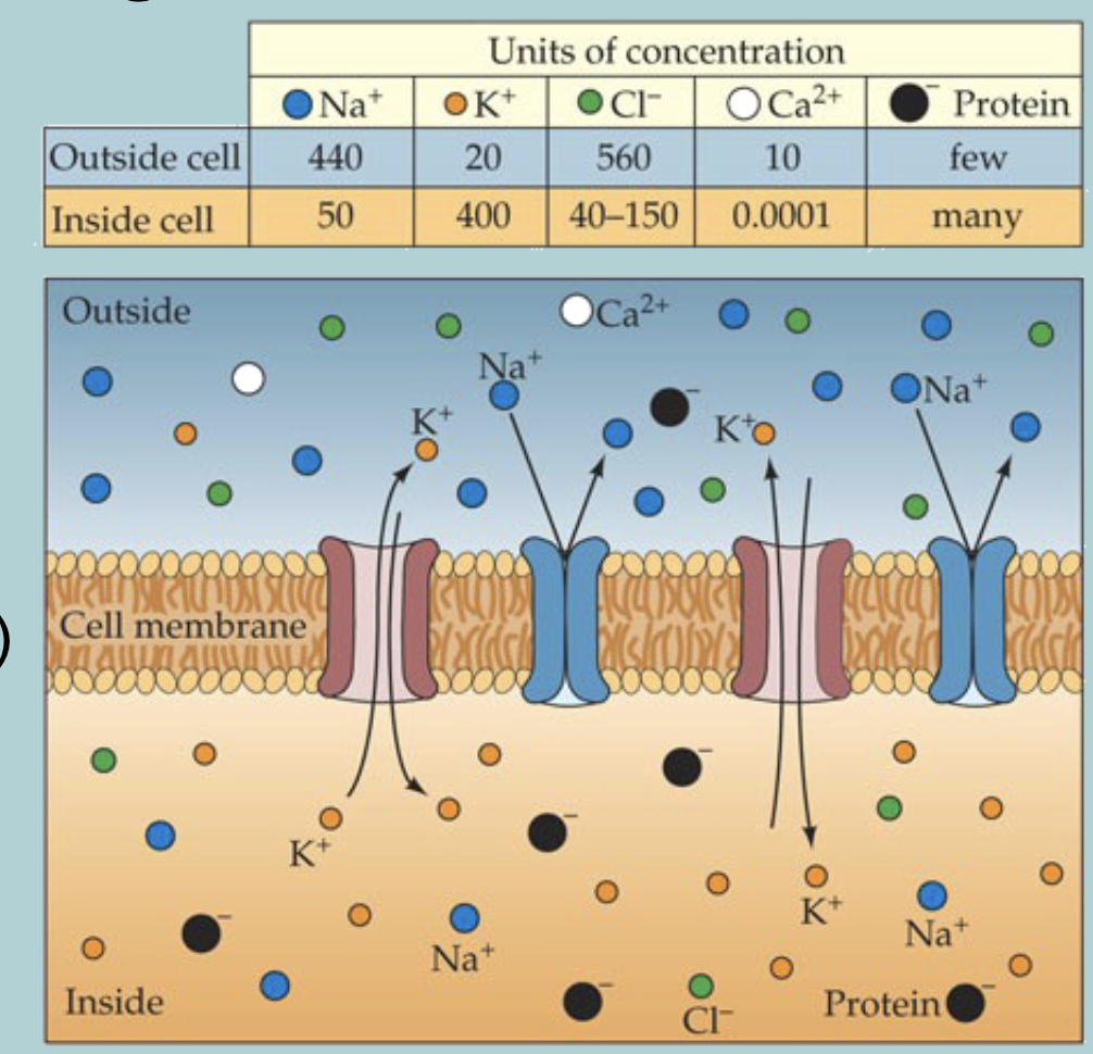
Of all these, only WHAT ions can, to some extent move freely through specific WHAT channels
Of all these, only K (±) ions can, to some extent move freely through specific K (±) channels

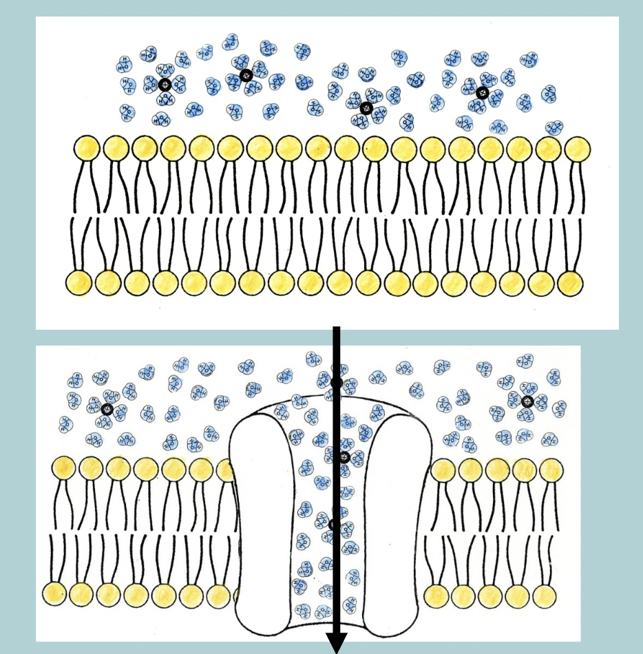
What are ion channels
a HYDROPHILIC pathway that facilitates ion movement across the PLASMAMEMBRANE
What are three features of the ion channel
Permeability
Selectivity
Gating
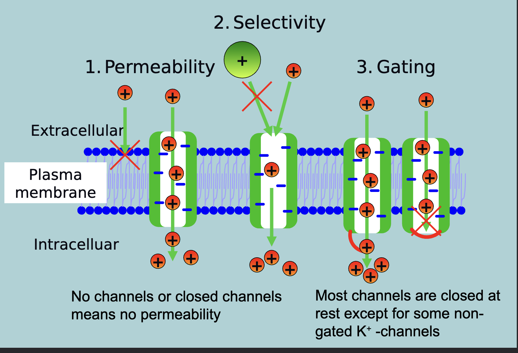
No channels or closed channels means WHAT
No channels or closed channels means NO PERMEABILITY

Most channels are closed at WHAT except for some WHAT
Most channels are closed at REST except for some NON-GATED K (±) channels

The cellular membrane potential is a function of:
WHAT differences mainly in WHAT and WHAT on the inside and outside of the WHAT
Combined with WHAT differences for these WHAT
The cellular membrane potential is a function of:
CONCENTRATION differences mainly in METAL CATIONS and LARGE ORGANIC ANIONS on the inside and outside of the PLASMAMEMBRANE
Combined with SELECTIVE PERMEABILITY differences for these IONS
Movement of ions: Diffusion
All molecules WHAT and therefore will spread from areas where they are more WHAT to areas of WHAT
No WHAT required, due to WHAT
Eventually ions will be distributed WHAT in solution - called WHAT
Movement of ions: Diffusion
All molecules MOVE and therefore will spread from areas where they are more CONCENTRATION to areas of LOW CONCENTRATION
No ENERGY required, due to RANDOM MOVEMENT
Eventually ions will be distributed EVENLY in solution - called DYNAMIC EQUILIBRIUM
Movement of ions: Diffusion:
WHAT selectively restrict the free diffusion of certain molecules
Movement of ions: Diffusion:
SEMI-PERMEABLE MEMBRANES selectively restrict the free diffusion of certain molecules
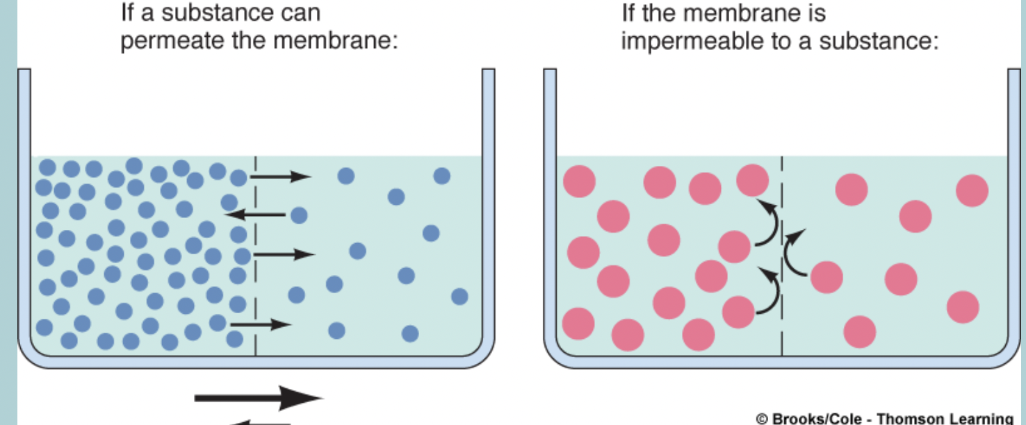
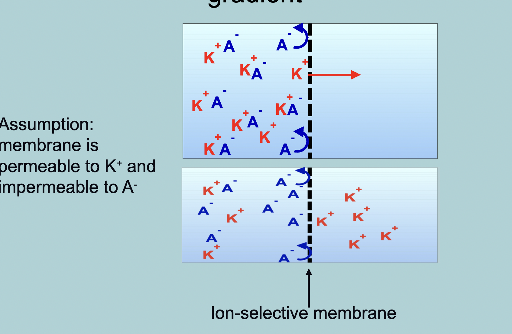
Semipermeable membrane: Concentration gradient
What will the separation look like if the membrane is permeable to K+ and impermeable to A-
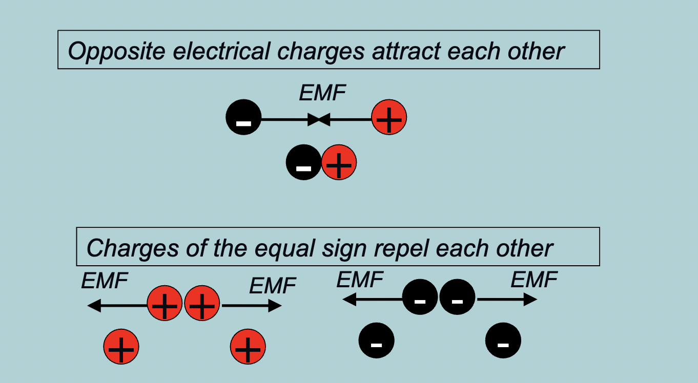
Movement of ions: voltage gradients and electromotive forces:
Move from areas of WHAT to areas of WHAT
Separation of WHAT, which costs WHAT, creates an WHAT
Movement of ions: voltage gradients and electromotive forces:
Move from areas of HIGH CHARGE to areas of LOW CHARGE
Separation of CHARGES, which costs ENERGY, creates an ELECTROMOTIVE FORCE (EMF)
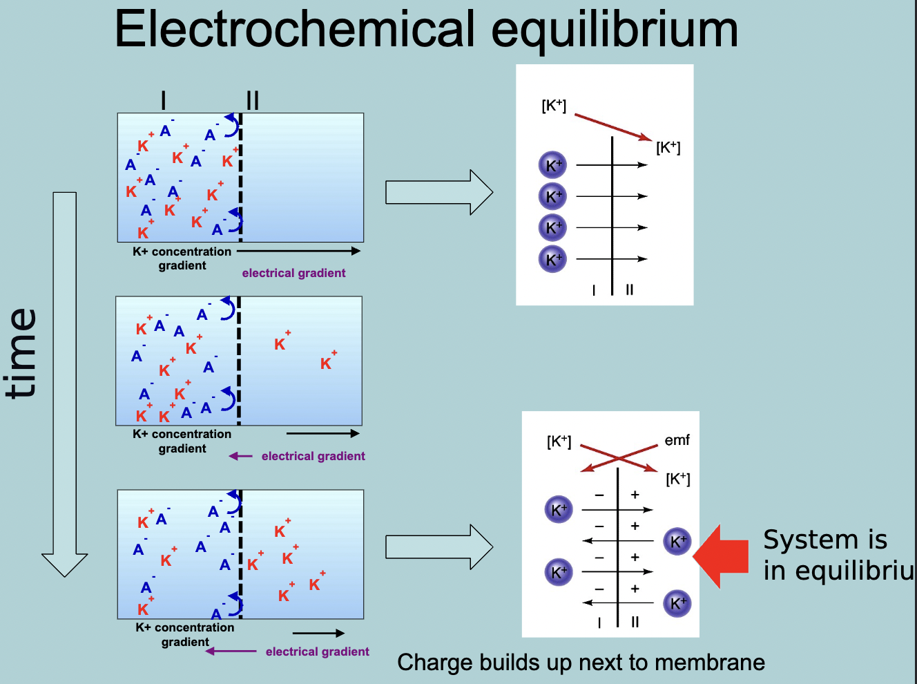
How does electrochemical equilibrium work
Positive charges flow through the semi-permeable membrane till there is an equal amount of charge between the K+ and A-, once equilibrium is reached the K+ will continue to flow back and forth between the semi-permeable to maintain the system of equilibrium
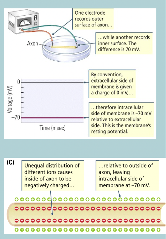
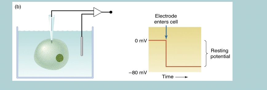
Electrical activity of cell membranes: Resting membrane potential
When undisturbed, there is a WHAT difference across the membrane
Inside the membrane is WHAT relative to WHERE
This voltage difference is called a WHAT “WHAT”
Usually inside is WHAT to WHAT MORE negative than outside (so the inside = WHAT to WHAT)
The inside can range between WHAT and WHAT depending on cell and species
Electrical activity of cell membranes: Resting membrane potential
When undisturbed, there is a STABLE difference across the membrane
Inside the membrane is NEGATIVE relative to OUTSIDE
This voltage difference is called a MEMBRANE “POTENTIAL”
Usually inside is 65mV to 70mV MORE negative than outside (so the inside = -65mV to -70mV)
The inside can range between -40mV and -90mV depending on cell and species
Resting membrane potential mainly depends on the WHAT
Resting membrane potential mainly depends on the CONCENTRATION of K+ IONS
Resting membrane potential is near the WHAT at which the concentration gradient pushes WHAT out of the cell cancels out with the pulling of WHAT into the cell
Resting membrane potential is near the VOLTAGE at which the concentration gradient pushes K+ out of the cell cancels out with the pulling of K+ into the cell
Resting membrane potential varies a little depending on the cells WHAT (remember: most channels are WHAT at rest except for some WHAT channels)
Resting membrane potential varies a little depending on the cells PERMEABILITY to other IONS (remember: most channels are CLOSED at rest except for some K (±) channels)
Resting membrane potential to sodium (Na+):
HOW MUCH more concentrated outside the cell
Membranes is not very WHAT to Na+ (but some leaks in)
Would this slow leak eventually eliminate the charge separation: WHAT
There is a WHAT which is a WHAT pump that reverses the WHAT
Resting membrane potential to sodium (Na+):
10X more concentrated outside the cell
Membranes is not very PERMEABLE to Na+ (but some leaks in)
Would this slow leak eventually eliminate the charge separation: YES
There is a “BILGE PUMP” which is a Na+/K+ pump that reverses the SLOW LEAK

All cells have a WHAT
All cells have a RESTING CELL MEMBRANE
Changes in the membrane WHAT are at the heart of how neurons WHAT to signals and WHAT information
Changes in the membrane VOLTAGE are at the heart of how neurons RESPOND to signals and PROCESS information
What are the three ways the opening state of an ion channel can be changed (change in permeability)
Chemical
Mechanical
Electrical
How do chemicals change the permeability
Neurotranmitter receptors
How do mechanicals change the permeability
For instance, stretch receptors
How do electricals change the permeability
Some channels open when the membrane potential reaches a certain threshold (aka voltage-gated)
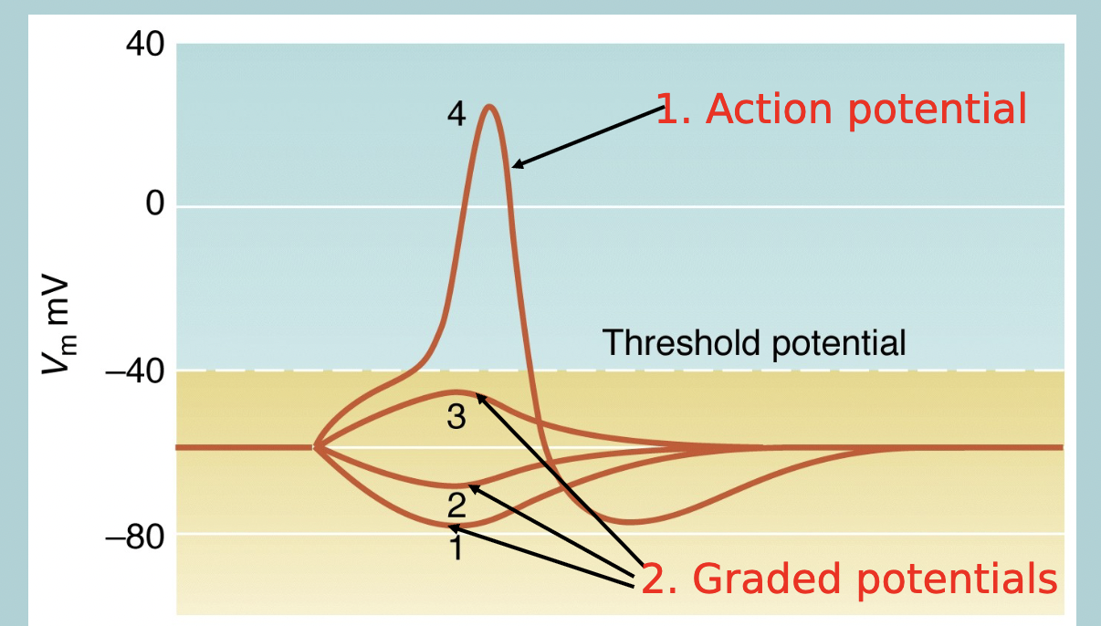
What are the two types of electrical signals
Graded potentials
Action potentials
In the three ways the opening state of an ion channel can be changed (change in permeability) which ones are graded potential and which ones are action potentials
Chemical (graded potential)
Mechanical (graded potential)
Electrical (Action potential)
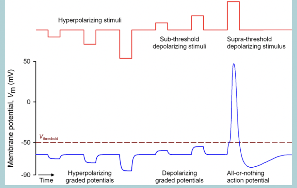
Graded potential:
When the membrane is stimulates a change in WHAT can be produced
WHAT
WHAT
Graded potential:
When the membrane is stimulates a change in MEMBRANE POTENTIAL can be produced
DEPOLARIZATION
HYPERPOLARIZATION

What is depolarization
LESS difference between the inside and outside, thus, neuron is LESS negative
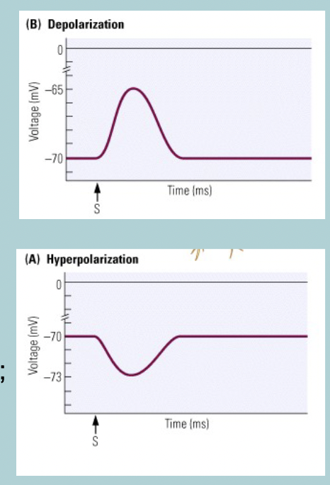
What is hyperpolarization
LARGER difference between the inside and outside, thus, neuron is MORE negative

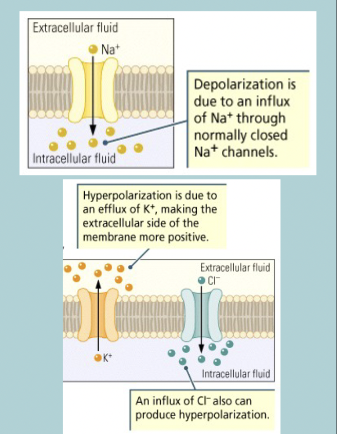
Basis of Graded potentials:
Change in membrane WHAT of certain ions (ie, channels open)
Depolarization:
Increase influx of WHAT (or WHAT)
Hyperpolarization:
Increase influx of WHAT or WHAT
Basis of Graded potentials:
Change in membrane PERMEABILITY of certain ions (ie, channels open)
Depolarization:
Increase influx of Na+ (or Ca 2+)
Hyperpolarization:
Increase influx of K+ or Cl-
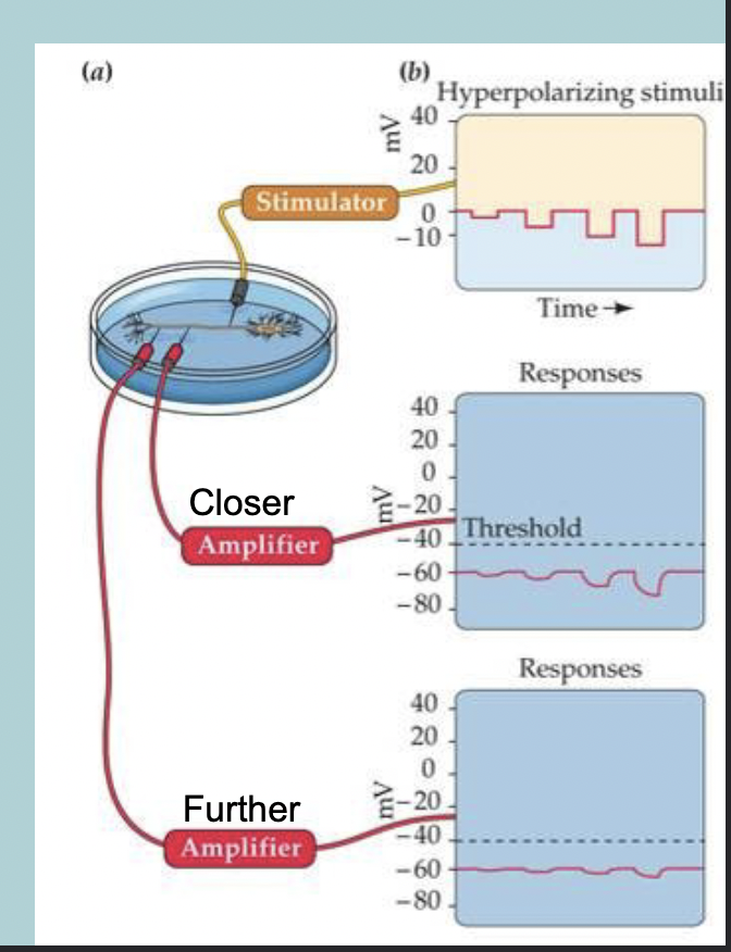
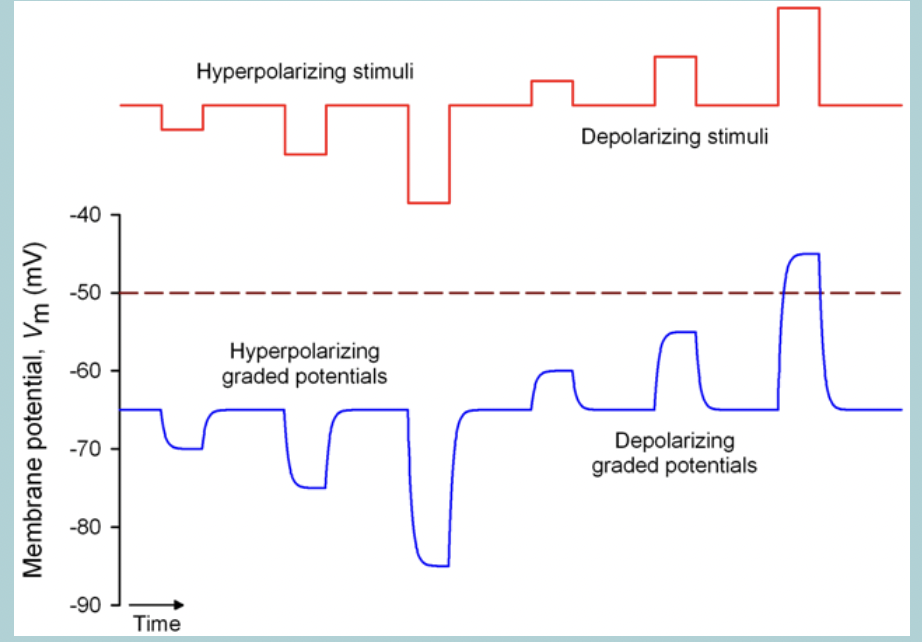
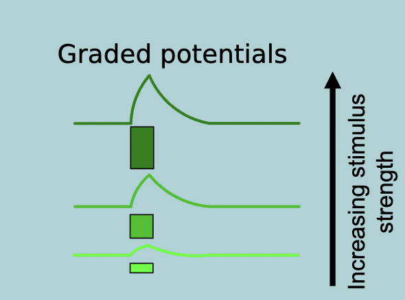
Graded potential - Hyperpolarization:
Graded change in voltage depends on:
WHAT
WHAT
Graded potential - Hyperpolarization:
Graded change in voltage depends on:
Stimulus strength
Distance
Graded potential - Depolarization:
Graded change in voltage depends on:
WHAT
WHAT
WHAT
WHAT is past threshold
Graded potential - Depolarization:
Graded change in voltage depends on:
Stimulus strength
Distance
Threshold
ACTION POTENTIAL is past threshold
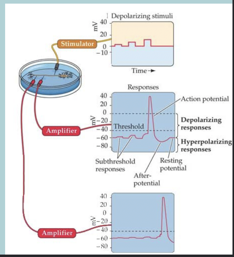
Action potentials are a WHAT response:
The neuron either produces a WHAT and WHAT the electrical signal to the next neuron or no WHAT is generates ( remains “WHAT”)
Action potentials are a ALL-OR-NONE response:
The neuron either produces a FULL ACTION POTENTIAL and TRANSMITS the electrical signal to the next neuron or no ACTION POTENTIAL is generates ( remains “SILENT”)
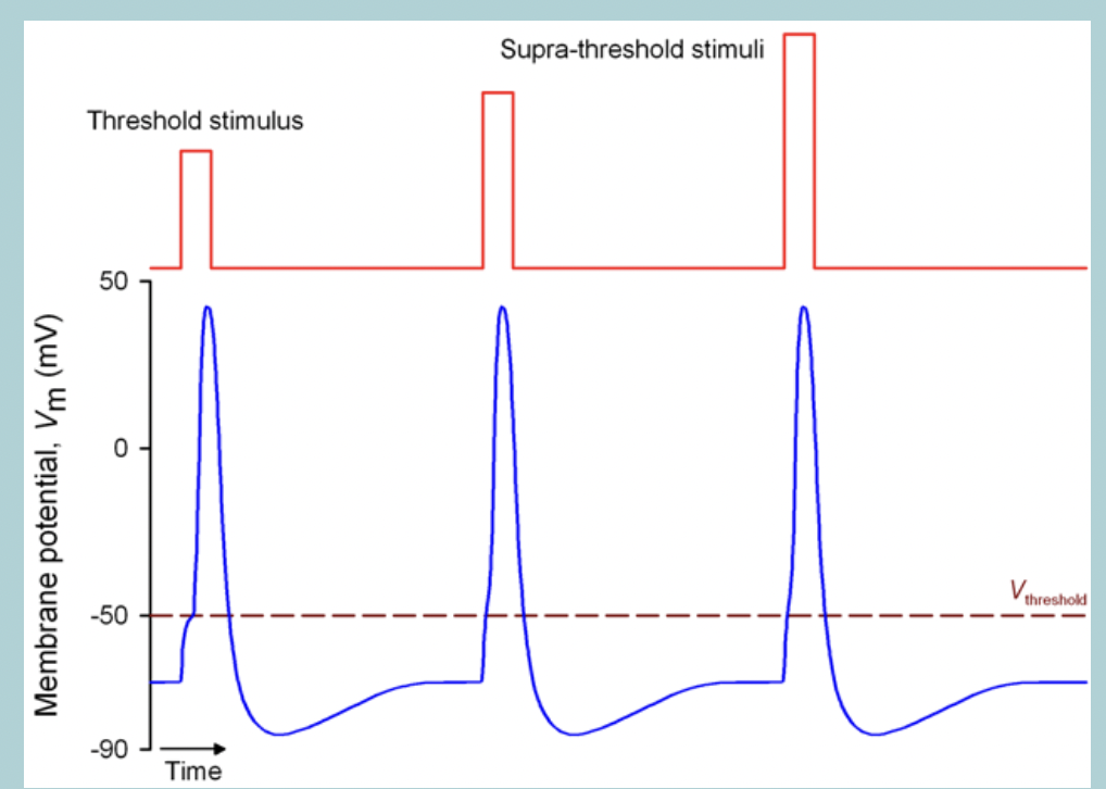
Action potentials are always the same WHAT no matter how strong the WHAT is
Action potentials are always the same SIZE no matter how strong the DEPOLARIZING STIMULUS is
Strength of stimulus is coded in the WHAT and WHAT of action potentials
Strength of stimulus is coded in the PATTERN and FREQUENCY of action potentials
ie, number of action potentials per second (Hz) (think of how many action potential go off when a feather brushes your arm vs someone pinching you)
The Hodgkin-Huxley model:
Hodgkin and Huxley described the ionic basis of the WHAT
This is considered the most important single achievement in WHAT
They derived a relatively simple but detailed mathematical and biophysical model of the WHAT
1980 the scientist were able to identify and record WHAT
1990 cloning of WHAT (to discover their WHAT)
2000 first 3-dimensional structure and function of a WHAT
The Hodgkin-Huxley model:
Hodgkin and Huxley described the ionic basis of the ACTION POTENTIAL
This is considered the most important single achievement in CELLULAR NEUROPHYSIOLOGY
They derived a relatively simple but detailed mathematical and biophysical model of the ACTION POTENTIAL
1980 the scientist were able to identify and record SINGLE ION CHANNELS
1990 cloning of ION CHANNELS (to discover their AMINO ACID SEQUENCE)
2000 first 3-dimensional structure and function of a ION CHANNEL
All cells maintain a WHAT
Resting membrane potential
Only WHAT cell types (muscle cells and neurons) can generate WHAT
Only EXCITABLE cell types (muscle cells and neurons) can generate ACTION POTENTIALS
No action potential generation possible in WHAT cells
Graded potentials move the WHAT towards or away from the WHAT but no WHAT can be generated
No action potential generation possible in NON-EXCITABLE cells
Graded potentials move the MEMBRANE POTENTIAL towards or away from the THRESHOLD but no ACTION POTENTIAL (AP) can be generated
Action potential generation possible in WHAT cells
Graded potentials move the WHAT towards or away from the WHAT; WHAT can be generated
Action potential generation possible in EXCITABLE cells
Graded potentials move the MEMBRANE POTENTIAL towards or away from the THRESHOLD; ACTION POTENTIAL (AP) can be generated
Action potentials are WHAT events - their amplitudes does not depend on WHAT
Action potentials are ALL-OR-NOTHING events - their amplitudes does not depend on STIMULUS STRENGTH
Graded potentials:
Variable WHAT and WHAT which depends on WHAT and WHAT of triggering event
Spread WHAT (flows with no channel opening outside the point of origin), decrementing (gradual decrease) with distance from point of WHAT
Travel over WHAT
Graded potentials:
Variable MAGNITUDE and DURATION which depends on STRENGTH and DURATION of triggering event
Spread PASSIVELY (flows with no channel opening outside the point of origin), decrementing (gradual decrease) with distance from point of INITIATION (the electrical signal moves through the cell membrane without requiring additional ion channel activity away from the stimulus site and loses strength, or intensity, as it spreads out from where it began)
Travel over SHORT DISTANCES
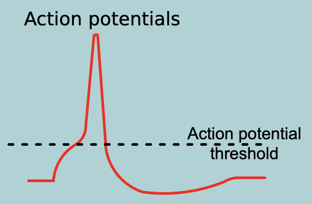
Action potentials:
WHAT event triggered when membrane potential is raised above a certain WHAT
Spread WHAT (self-regenerating) in non-decremented fashion
Travel over WHAT
Action potentials:
ALL-OR-NOTHING event triggered when membrane potential is raised above a certain THRESHOLD
Spread ACTIVELY (self-regenerating) in non-decremented fashion (Spread actively (self-regenerating)" = each region of the membrane actively generates the action potential anew. "Non-decremental" = the amplitude stays constant as the action potential moves down the axon, unlike graded potentials.)
Travel over LONG DISTANCES
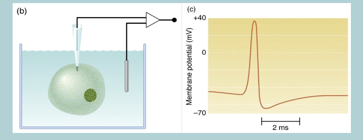
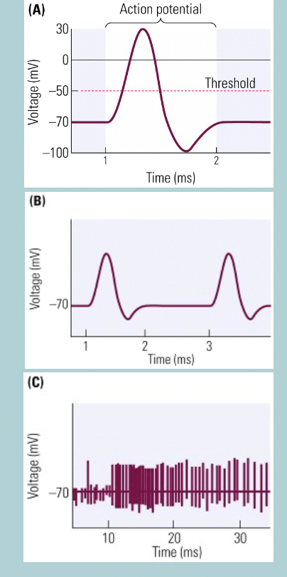
Action potentials in neurons:
Signals are conveyed along the neuron’s axon by an WHAT
Brief but large change in WHAT of the WHAT membrane
Shapes and firing patterns differ widely among WHAT
Vary in WHAT (1ms to 15ms)
Voltage potential WHAT, so the inside becomes WHAT relative to the outside
Reverts back to WHAT just as quickly
So rapid taht some neurons can have HOW MANY of action potentials per second
Action potentials in neurons:
Signals are conveyed along the neuron’s axon by an ACTION POTENTIAL
Brief but large change in POLARITY of the AXON’S membrane
Shapes and firing patterns differ widely among DIFFERENT types of neurons
Vary in DURATION (1ms to 15ms)
Voltage potential REVERSES, so the inside becomes POSITIVE relative to the outside
Reverts back to NEGATIVE INTERIOR just as quickly
So rapid taht some neurons can have 100 of action potentials per second
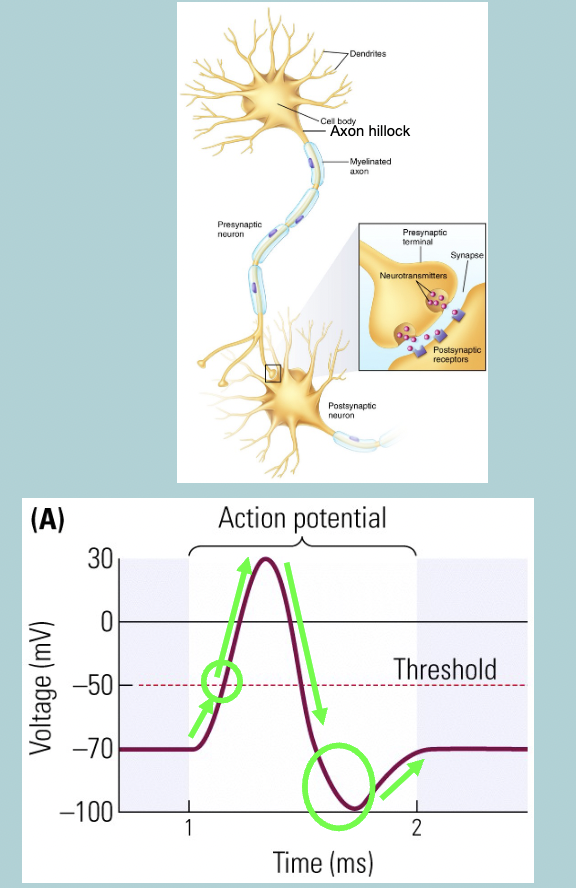
Generation of action potentials: Key steps
Depolarizing input(s) makes resting membrane potential WHAT
If this reaches the threshold potential:
Initiates start of WHAT in WHAT
Does not require further WHAT, will continue on its own
Membrane potential further WHAT and subsequently the interior becomes WHAT relative to the WHAT:
can reach WHAT
Total change can be more than WHAT
Membrane potential then WHAT
Overshoots WHAT and become WHAT
Returns to WHAT
Generation of action potentials: Key steps
Depolarizing input(s) makes resting membrane potential LESS NEGATIVE
If this reaches the threshold potential:
Initiates start of ACTION POTENTIAL in AXON HILLOCK
Does not require further STIMULATION, will continue on its own
Membrane potential further DEPOLARIZES and subsequently the interior becomes POSITIVE relative to the OUTSIDE:
can reach +40mV
Total change can be more than 100mV
Membrane potential then REPOLARIZES
Overshoots RESTING POTENTIAL and become HYPERPOLARIZED
Returns to RESTING POTENTIAL
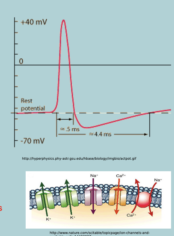
Action potentials: Key elements
Asymmetric concentration distribution of (mainly) WHAT and WHAT ions across the WHAT established by WHAT mechanisms
The presence of WHAT, WHAT K+ and Na+ channels in the plasmamembrane, these channels are normally WHAT when neuron is at rest
When a cell depolarizes, the WHAT change causes these channels to WHAT quickley:
Only found in WHAT and WHAT cells
WHAT after opening
Voltage gates WHAT channels open and close more slowly
Asymmetric concentration distribution of (mainly) K+ and Na+ ions across the PLASMAMEMBRANE established by ACTIVE TRANSPORT mechanisms
The presence of ION-SELECTIVE, VOLTAGE GATED K+ and Na+ channels in the plasmamembrane, these channels are normally CLOSED when neuron is at rest
When a cell depolarizes, the VOLTAGE change causes these channels to OPEN quickly:
Only found in NEURONS and MUSCLE cells
INACTIVE after opening
Voltage gates K+ channels open and close more slowly
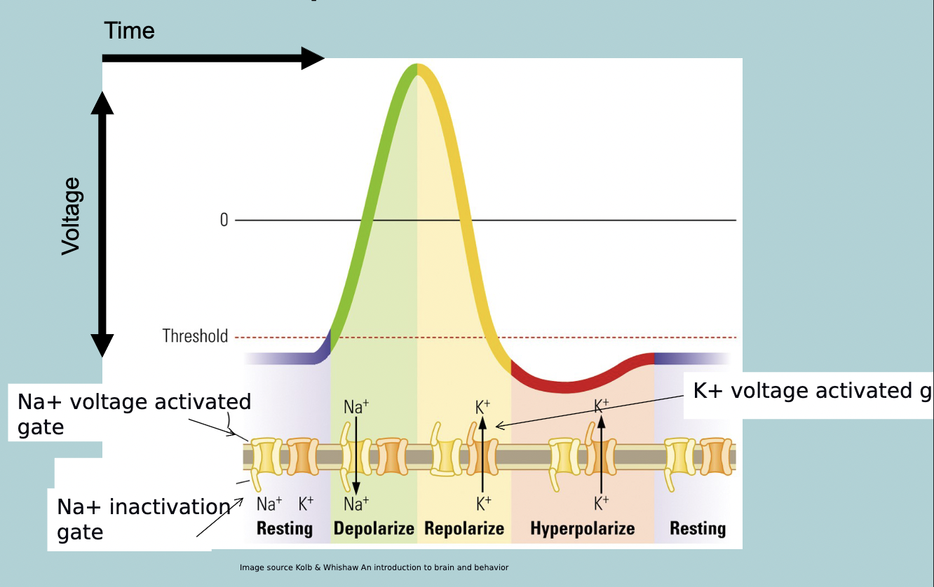
Ionic conductance events underlying the action potential summarized
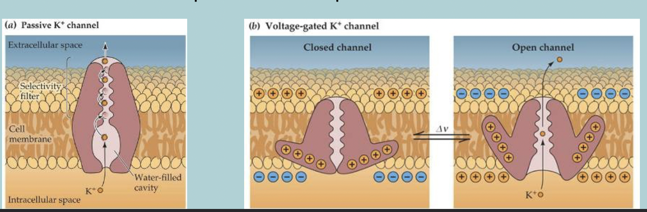
How do (voltage) gated channels work:
Passive (non-gated) WHAT (leakage channels) have one configuration → WHAT
All gated channels have HOW MANY (or WHAT) configurations:
WHAT (resting)
WHAT (inactive, mainly sodium channels)
WHAT
Part of the protein is WHAT (protein reshapes when cell WHAT)
How do (voltage) gated channels work:
Passive (non-gated) K+ CHANNELS (leakage channels) have one configuration → OPEN
All gated channels have 2 (or 3) configurations:
CLOSED (resting)
CLOSED (inactive, mainly sodium channels)
OPEN
Part of the protein is CHARGED (protein reshapes when cell DEPOLARIZES)
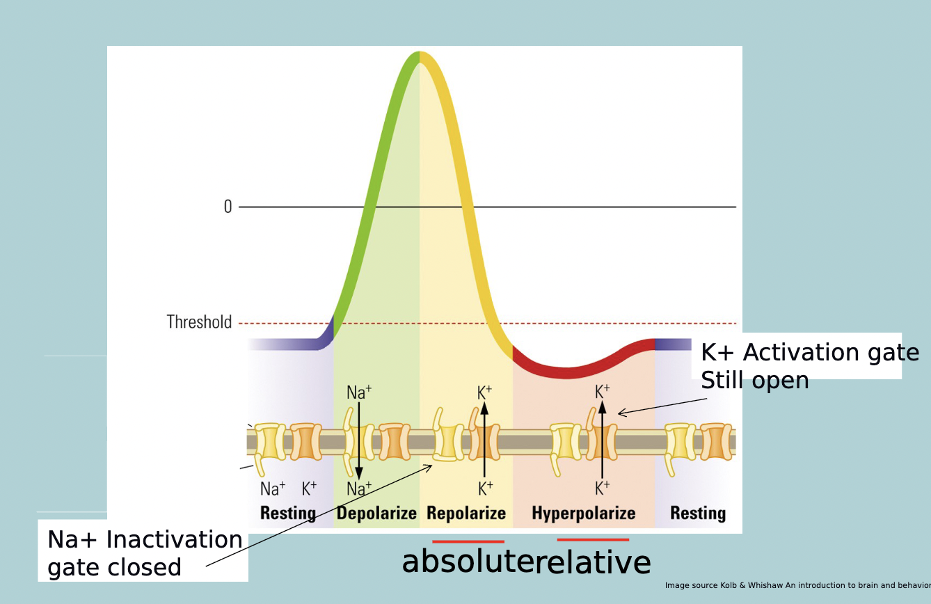
A refractory period is a period in which triggering another WHAT is much more difficult or even impossible
A refractory period is a period in which triggering another ACTION POTENTIAL is much more difficult or even impossible
The absolute refractory period is due to the WHAT. In this period NO new WHAT can be generated
The ABSOLUTE refractory period is due to the Na+ CHANNEL INACTIVATION GATE. In this period NO new ACTION POTENTIAL can be generated
The RELATIVE refractory period is due to the WHAT. The neuron needs a stronger WHAT input to reach threshold for an WHAT can be generated
The RELATIVE refractory period is due to the K+ CHANNEL ACTIVATION GATE. The neuron needs a stronger DEPOLARIZING input to reach threshold for an ACTION POTENTIAL can be generated
Refractory period diagram
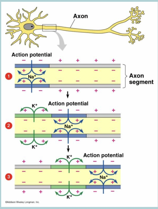
Propagation of impulse diagram
Nerve impulses are WHAT
Movement of action potential along the axon

When a segment of an axon generates an WHAT it depolarizes the WHAT section
When a segment of an axon generates an ACTION POTENTIAL it depolarizes the ADJACENT section
An action potential represents a WHAT change in the WHAT of the membrane
An action potential represents a -100mV change in the POTENTIAL of the membrane
You only need about HOW MUCH change to reach WHAT
You only need about 20mV change to reach THRESHOLD
Action potential brings the WHAT section to WHAT, activating the voltage sensitive WHAT channels in the adjacent section
Action potential brings the ADJACENT section to THRESHOLD, activating the voltage sensitive Na+ channels in the adjacent section
Each action potential WHAT the next WHAT
Each action potential PROPAGATES (moves) to the next neuron
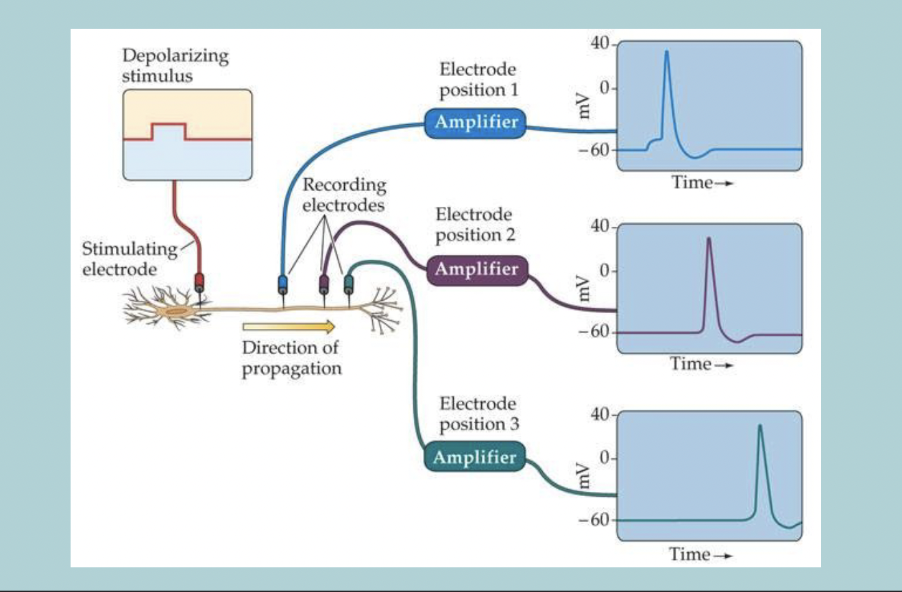
At each segment of the axon the action potential WHAT completely:
No loss in WHAT from the start of the axon to the end
It is the same WHAT at each stage
At each segment of the axon the action potential REGENERATES completely:
No loss in AMPLITUDE from the start of the axon to the end
It is the same SIZE at each stage
Stronger stimuli do not produce bigger WHAT, just more WHAT ones
Stronger stimuli do not produce bigger ACTION POTENTIALS, just more FREQUENT ones
Action potentials only move in WHAT
One direction
What are two solutions that speed up nerve impulses
Myelin
Saltatory conduction
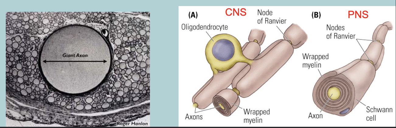
These solutions make your axon WHAT (larger WHAT, squids and other invertebrates)
These solutions make your axon HUGE (larger DIAMETER, squids and other invertebrates)

These solutions make it really hard for WHAT to escape by insulating the axon (WHAT, vertebrates)
These solutions make it really hard for IONS to escape by insulating the axon (MYELINATION, vertebrates)

What is multiple sclerosis
Loss of myelin around axons
Myelination increases WHAT
Nerve conduction
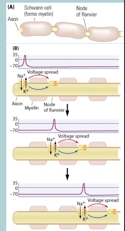
Saltatory conduction:
The action potential is WHAT fully at each WHAT
Action potential jumps from WHAT to WHAT
Moves WHAT (up to 150m/s) than opening millions of channels over a comparable portion of an WHAT neuron (0.5-10m/s)
Saltatory conduction:
The action potential is REGENERATE fully at each NODE of RANVIER
Action potential jumps from NODE to NODE
Moves FASTER (up to 150m/s) than opening millions of channels over a comparable portion of an UNMEYLINATED neuron (0.5-10m/s)
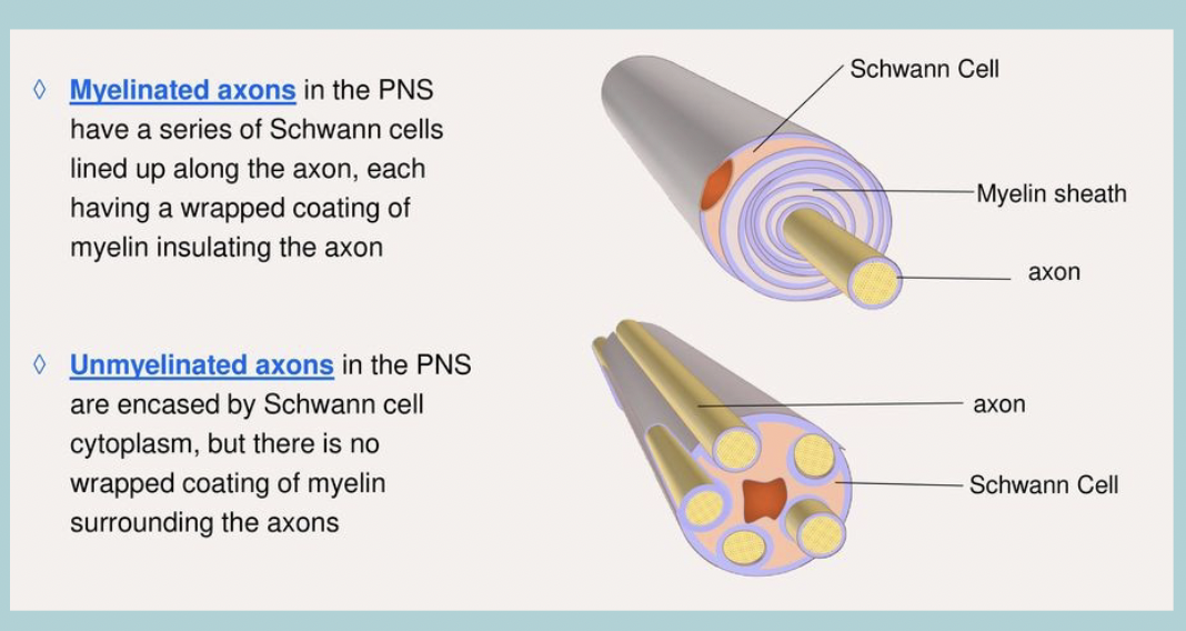
Not all axons are WHAT
Not all axons are MYELINATED
Unmyelinated axons: Group C fibers:
Group C fibres include WHAT in the Autonomic nervous system and nerve fibers at the WHAT roots (IV fiber)
Group C fibers carry WHAT information
Group C fibers, small in WHAT, WHAT conducting
Unmyelinated axons: Group C fibers:
Group C fibres include POST GANGLIONIC FIBERS in the Autonomic nervous system and nerve fibers at the DORSAL roots (IV fiber)
Group C fibers carry SENSORY information
Group C fibers, small in DIAMETER, SLOW conducting
(Voltage-gates) Ion channels shape electrical behaviour of neurons
WHAT
WHAT of action potential
WHAT
WHAT of action potentials
WHAT of action potential trains
(Voltage-gates) Ion channels shape electrical behaviour of neurons
EXCITABILITY (how eager are they to generate an action potential)
SHAPE of action potential
RESPONSE CHARACTERISTICS
FREQUENCY of action potentials
PATTERNING of action potential trains
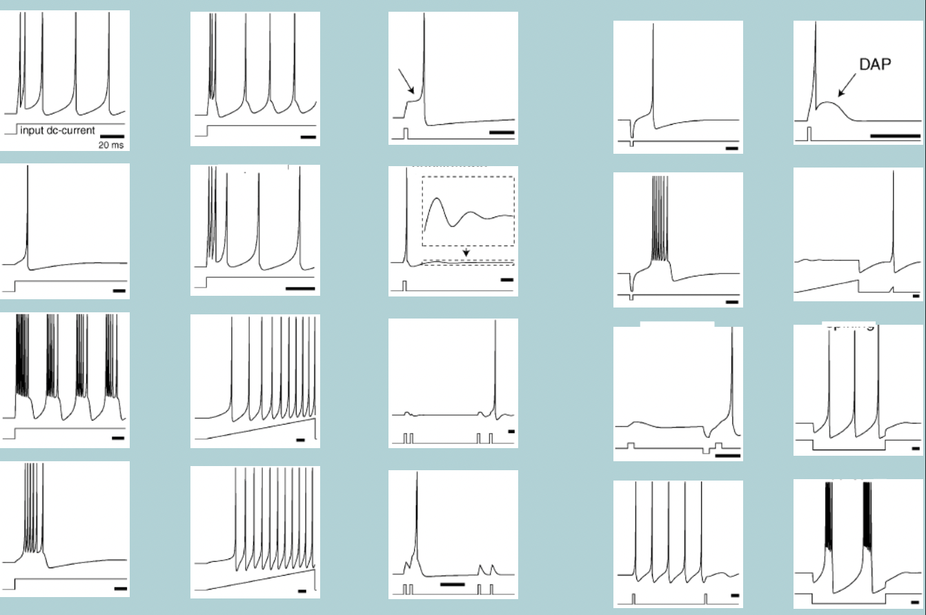
Each neuron has its own characteristic action potential (AP) WHAT and activity WHAT
Each neuron has its own characteristic action potential (AP) SHAPE and activity PATTERN
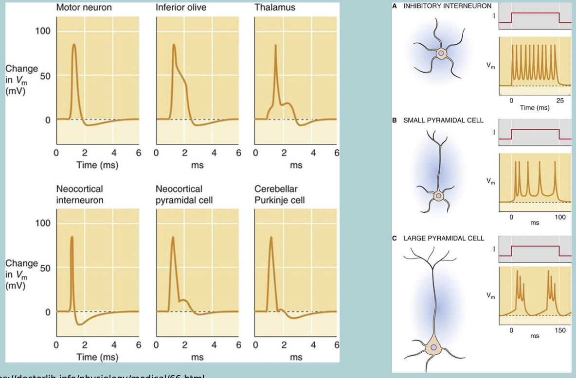
Electrical activity patterns diagrams
Since action potentials are a all-or-nothing response you can consider them as a WHAT (0 or 1) or WHAT signal
Since action potentials are a all-or-nothing response you can consider them as a BINARY (0 or 1) or DIGITAL signal
Sensory input is WHAT (continuous)
Sensory input is ANALOG (continuous)
The coding of these analog signals is in the WHAT (number of action potentials per second) and WHAT of action potentials
The coding of these analog signals is in the FREQUENCY (number of action potentials per second) and PATTERN of action potentials
Neurons can receive inputs from HOW MANY (sometimes HOW MANY) other neurons
Neurons can receive inputs from 1000s (sometimes 100000) other neurons
Neuron only has HOW MANY output, one WHAT
Neuron only has ONE output, one AXON
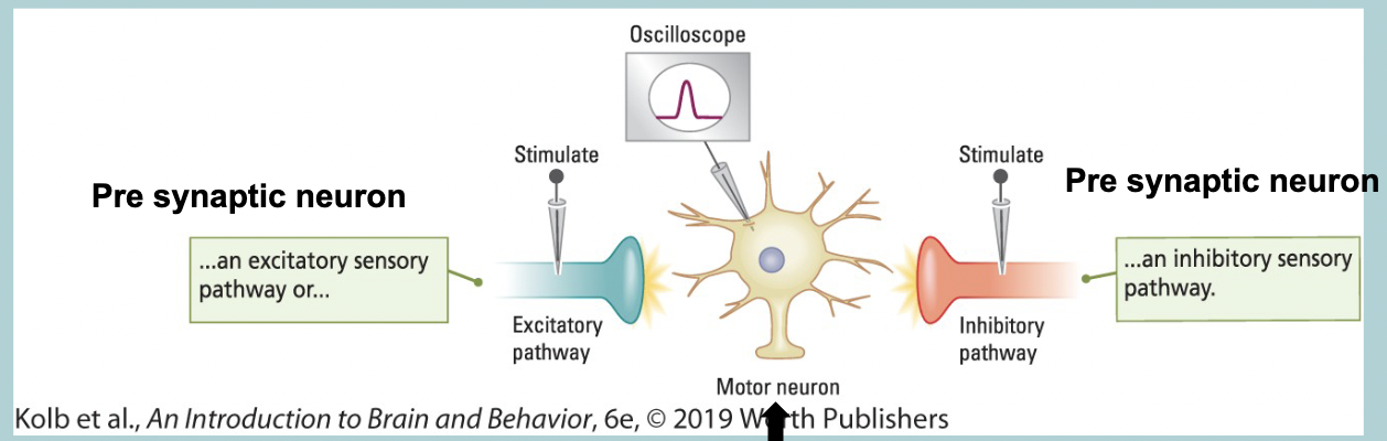
Post synaptic potentials (PSP) = WHAT
Put a recording electrode is WHAT (cell body) post synaptic neuron
Tease apart incoming WHAT
Stimulate individual WHAT
Some WHAT the post synaptic neuron (WHAT)
Some WHAT the post synaptic neuron (WHAT)
Post synaptic potentials (PSP) = GRADED POTENTIAL
Put a recording electrode is SOMA (cell body) post synaptic neuron
Tease apart incoming SENSORY FIBERS
Stimulate individual FIBERS
Some DEPOLARIZES the post synaptic neuron (EXCITATORY)
Some HYPERPOLARIZE the post synaptic neuron (INHIBITORY)
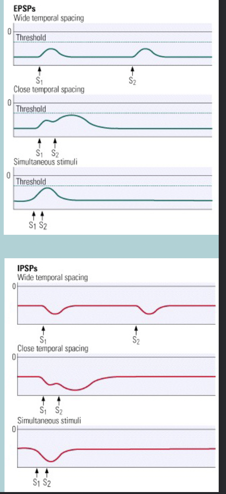
Excitatory postsynaptic potential (EPSP = WHAT graded potential)
Brings the WHAT closer to the threshold, thus increasing the probability that the cell will fire an WHAT
Excitatory postsynaptic potential (EPSP = DEPOLARIZING graded potential)
Brings the POTENTIAL MEMBRANE closer to the threshold, thus increasing the probability that the cell will fire an ACTION POTENTIAL
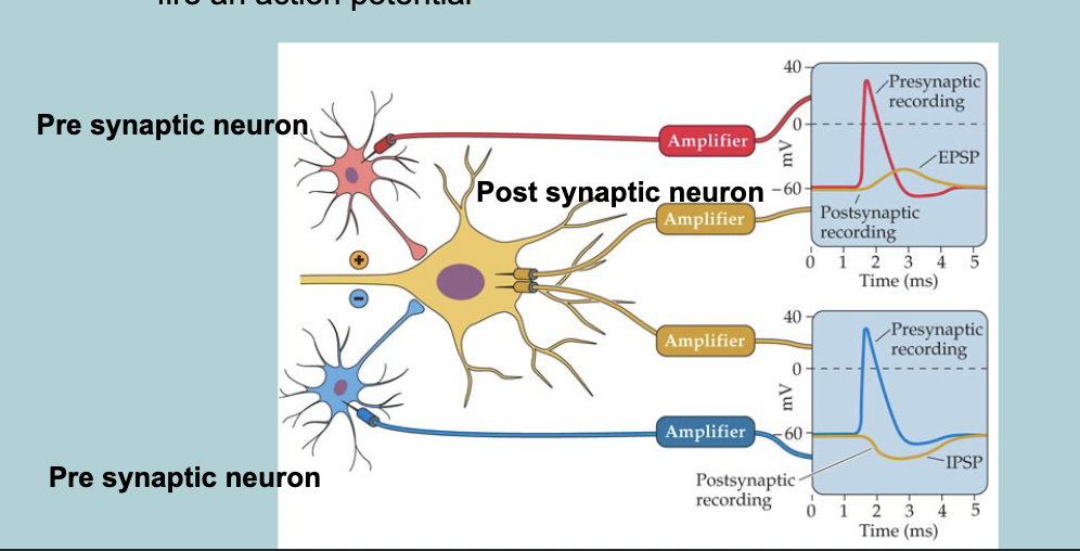
Inhibitory postsynaptic potential (IPSP = WHAT graded potential)
Move the WHAT away from the threshold, thus decreasing the probability that the cell will fire an WHAT
Inhibitory postsynaptic potential (IPSP = HYPERPOLARIZING graded potential)
Move the POTENTIAL away from the threshold, thus decreasing the probability that the cell will fire an ACTION POTENTIAL

Excitatory postsynaptic potential (EPSP)
Open WHAT channels
WHAT floes in the cell by its WHAT and WHAT
Excitatory postsynaptic potential (EPSP)
Open Na+ channels
WHAT floes in the cell by its VOLTAGE and CONCENTRATION GRADIENT
Inhibitory postsynaptic potential (IPSP)
Opens WHAT or WHAT channels
WHAT flows out and WHAT flows into the cell by its WHAT
Inhibitory postsynaptic potential (IPSP)
Opens K+ or Cl- channels
K+ flows out and Cl- flows into the cell by its CONCENTRATION GRADIENT
Inhibitory postsynaptic potential (IPSP) and Excitatory postsynaptic potential (EPSP) are proportional in WHAT to the strength of the stimulus
Inhibitory postsynaptic potential (IPSP) and Excitatory postsynaptic potential (EPSP) are proportional in SIZE to the strength of the stimulus
Neurotransmitters released by the WHAT neuron determines if the WHAT neuron will generate an WHAT or an WHAT
Neurotransmitters released by the PRE-SYNAPTIC neuron determines if the POST-SYNAPTIC neuron will generate an EPSP (eg, NE) or an IPSP (eg, GABA)
Neurons receive many WHAT and WHAT potentials
Neurons receive many SIMULTANEOUS and CONSECUTIVE potentials
These signals will have a WHAT effect and determine whether the electrical message is WHAT or if it WHAT at that point
These signals will have a SUMMATIVE effect and determine whether the electrical message is CONVEYED or if it STOPS at that point
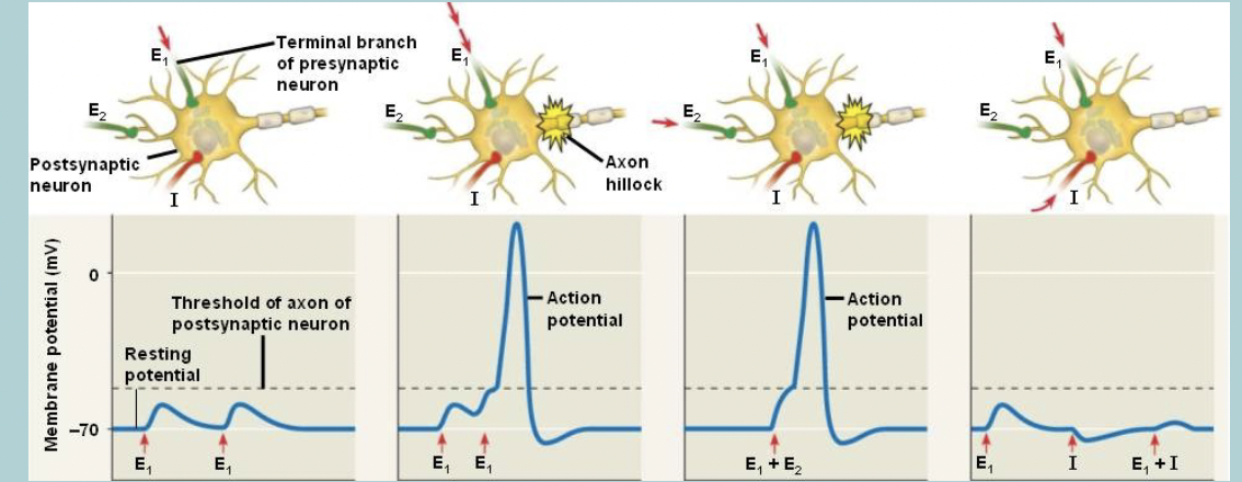
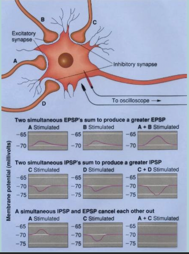
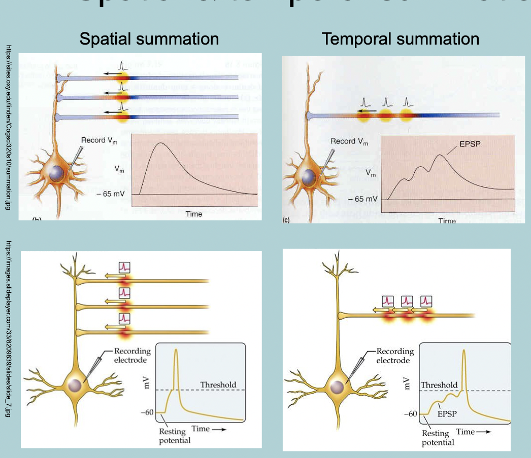
Spatial summation:
If PSPs occur simultaneously they will WHAT
This can occur cause either:
WHAT
WHAT
WHAT
Spatial (in space) summation:
If PSPs occur simultaneously they will SUMMATE
This can occur cause either:
Larger EPSP
Larger IPSP
Cancel each other out
Temporal (in time) summation:
If two signals occur close enough together in WHAT, the send one has its WHAT before the first has WHAT
Temporal (in time) summation:
If two signals occur close enough together in TIME, the send one has its EFFECT before the first has DEGRADED
In reality there is a combination of both WHAT
In reality there is a combination of both SPATIAL and TEMPORAL SUMMATION
Where does integration take place
The axon hillock
The cell body (soma) does not have WHAT channels, it cannot fire an WHAT
The cell body (soma) does not have VOLTAGE GATED channels, it cannot fire an ACTION POTENTIAL
Only an axon can generate and conduct an WHAT
Action potential
The action potential is generated at the WHAT:
Rich in WHAT channels
The WHAT of all the EPSPs and IPSPs at the axon hillock will determine if the cell will fire an WHAT
The action potential is generated at the AXON HILLOCK:
Rich in VOLTAGE GATED channels
The SUM of all the EPSPs and IPSPs at the axon hillock will determine if the cell will fire an ACTION POTENTIAL
PSP (post synaptic potential) spreads though WHAT/WHAT:
Loses WHAT as it spreads
PSP (post synaptic potential) spreads though DENDRITES / CELL BODY
Loses INTENSITY as it spreads