Echo Board Pt. 1
 Call Kai
 Learn
Learn Practice Test
 Spaced Repetition
 Match
Match Flashcards
 Knowt Play
1/286

There's no tags or description
Looks like no tags are added yet.
Name | Mastery | Learn | Test | Matching | Spaced | Call with Kai | Chat |
|---|
No analytics yet
Send a link to your students to track their progress
287 Terms
When are all four chambers of the heart fully developed?
8 weeks GA
What is the sino-atrium?
Fetal anatomy that forms atria
What is the primitive ventricle?
Fetal anatomy that forms LV
What are bulbous cords?
Fetal anatomy that forms RV and truncus arteriosus
What is the truncus arteriosus?
Fetal anatomy that forms aorta and pulmonary artery
What is the sinus venosus?
Fetal anatomy that forms SVC, IVC, and atrial floor
(T/F) Right heart pressures are higher than left heart pressures in the fetus.
True; Right heart pressures are higher than left heart pressures in the fetus because lungs are not in use until birth
What composes the fetal circulation system?
Umbilical vein carries OXYGENATED blood into IVC
Umbilical arteries carry DEOXYGENATED blood to placenta
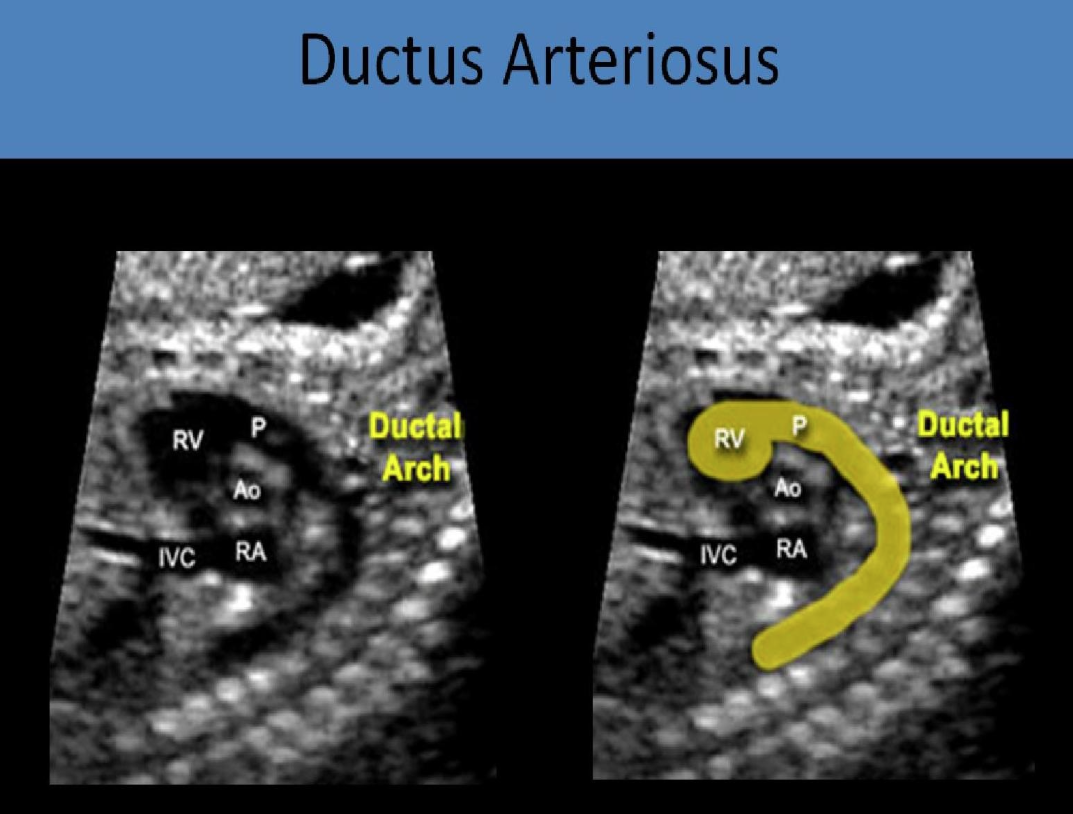
What is the ductus arteriosus?
Part of fetal circulation system that connects pulmonary artery to aorta and closes within 72 hours after birth to become ligamentum arteriosum
What is the ductus venosus?
Part of fetal circulation system that connects umbilical vein to IVC and closes when umbilical cord is cut to become ligamentum venosum
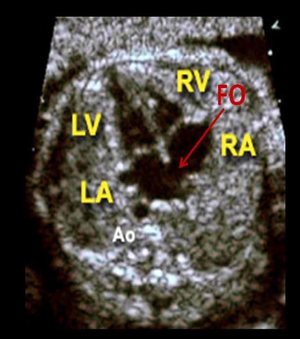
What is the foramen ovale?
Part of fetal circulation system that connects RA to LA and leaves small indentation after birth called fossa ovalis
Where is the heart located?
Posterior to sternum within middle mediastinum
What angle does the heart lie at?
45 degrees towards left side between third and fifth intercostal space
What is levocardia?
Normal heart axis
What is dextrocardia?
Abnormal heart axis in which heart points towards right side of chest
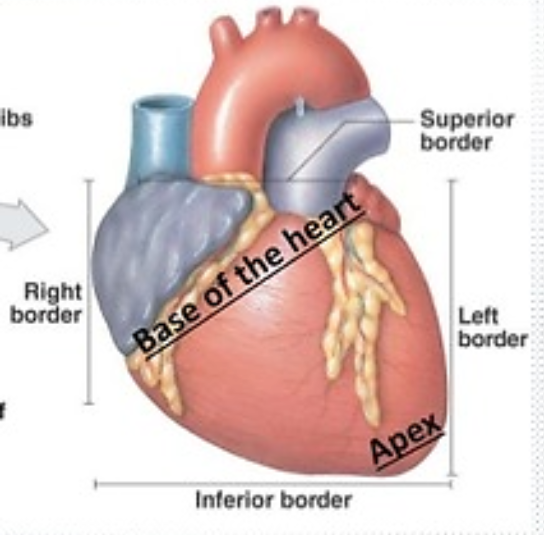
What is the apex of the heart?
Cone or bottom of heart that consists of tip of left ventricle and rests on diaphragm below seventh rib
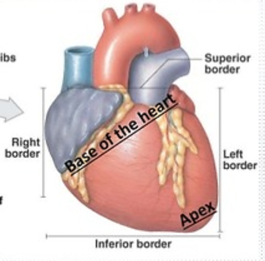
What is the base of the heart?
Top of heart that consists of atria and great vessels
What is the most anterior part of the heart?
RV
What is the crux of the heart?
Posterior portion of heart that is junction of atrial and ventricular septums or all four parts of heart
What is the pericardium?
Thin sac that surrounds heart and roots of great vessels
What is located within the pericardial cavity?
10-20 mL or 50 cc of serous fluid used to lubricate for smooth contractions
What are the two layers of the pericardium?
Serous: Innermost layer composed of visceral and parietal layers
Fibrous: Outermost layer
What is the epicardium or visceral layer of the heart?
Outermost layer composed of connective tissue
What is the myocardial layer of the heart?
Middle layer composed of muscle fibers
What is the endocardial layer of the heart?
Innermost layer composed of epithelial cells
What is the right atrium?
Chamber composed of two portions separated by crista terminalis
Anterior: Connected to right atrial appendage
Posterior: Where IVC and SVC enter heart
What is a Eustachian valve?
Valve that protects opening of IVC into RA
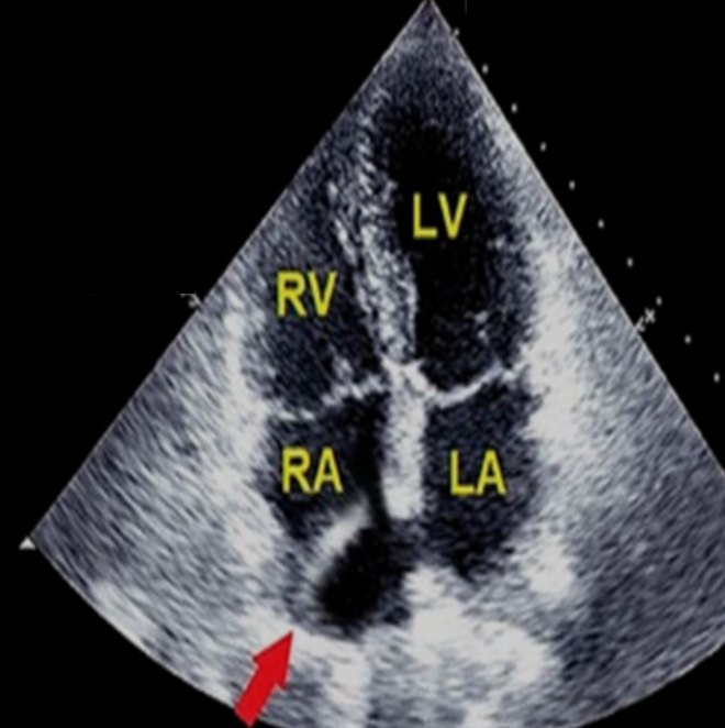
Identify this image.
Eustachian valve
What is the Chiari Network?
Net-like remnant of fetal development in RA
What is the Thebesian valve?
Valve that protects coronary sinus opening into RA
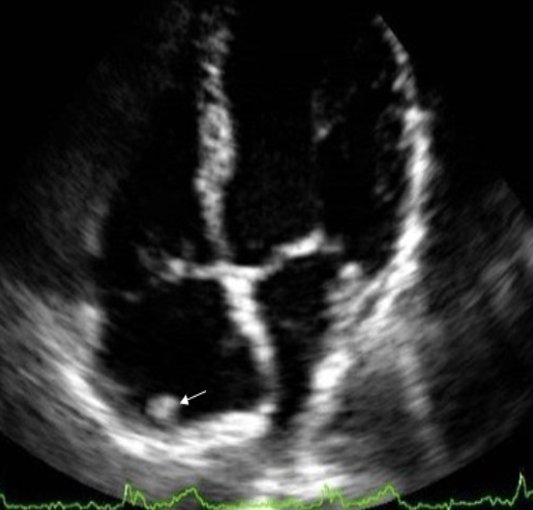
Identify this image.
Crista terminalis resembling cardiac mass
What is the normal size of RA?
VOLUME USED TO ASSESS RA SIZE
Women: < 27ml/m²
Men: < 32 ml/m²
What is the right ventricle?
Chamber composed of three portions
Posterior inflow: Trabeculated portion containing papillary muscles and TV
Apex: Heavily trabeculated portion
Anterior outflow: Smooth walled infundibulum
What is the moderator band?
Muscular tissue that connects IVS to anterior RV free wall to create a quick path for conduction system
What is the normal size of RV?
WIDTH USED TO ASSESS RV SIZE
Basal < 4.1 cm
Mid < 3.5 cm
Apex < 3.5 cm
Where is the moderator band best visualized?
A4C
What is the left atrium?
Chamber composed of two portions
True left atrium: Where pulmonary veins enter heart
Left atrial appendage (LAA): Posterior portion where blood is stored
(T/F) The left atrial appendage is typically seen transthoracically.
False; LAA is seen with TEE
Where is the left atrial appendage best visualized?
A2C
What is the normal size of the LA?
AREA AND AP USED TO ASSESS LA SIZE
Area < 34 ml/m²
PLAX AP < 4 cm
What is the left ventricle?
Largest chamber composed of three portions
Inflow: Contains MV
Apex
Outflow: Portion below AV
How much cardiac mass does the left ventricle compose?
75%
What is the normal size of the LV?
WALL THICKNESS AND CHAMBER SIZE USED TO ASSESS LV SIZE
Wall thickness in women: 0.6-0.9 cm
Wall thickness in men: 0.6-1.0 cm
End diastolic chamber size in women: 3.8-5.2 cm
End diastolic chamber size in men: 4.2-5.8 cm
What are the differences between the LV and RV?
LV
No moderator band
Smooth walled
2 papillary muscles
Bi-leaflet valve
RV
Moderator band present
Trabeculations present
3 papillary muscles
Tri-leaflet valve
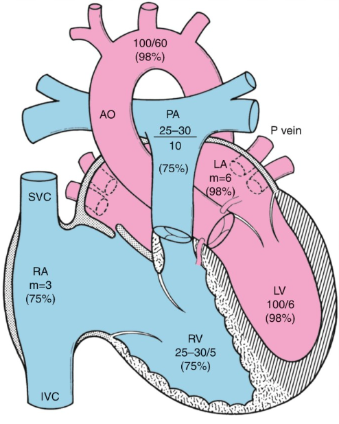
What oxygen percentage does “deoxygenated” blood have?
75%
RA
RV
MPA
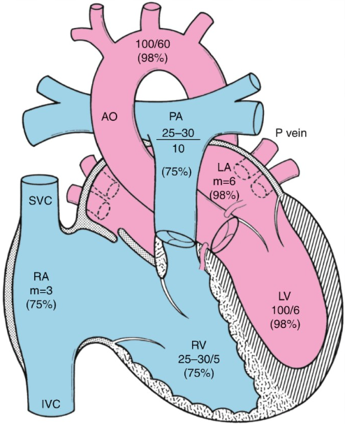
What oxygen percentage does “oxygenated” blood have?
98%
Pulmonary veins
LA
LV
Aorta
What is the interatrial septum (IAS)?
Septum located between right and left atrium that is best evaluated SUBCOSTALLY
What is the interventricular septum (IVS)?
Septum between right ventricle and left ventricle that is best evaluated in SHORT AXIS OR SUBCOSTAL
What are papillary muscles?
Muscles attached to atrioventricular valves by chordae tendineae
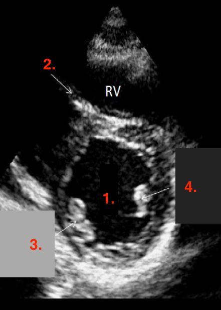
Identify this image.
LV
Moderator band
Posterior medial papillary muscle
Anterior lateral papillary muscle
What are chordae tendineae?
Fibrous strands connecting papillary muscles to leaflet
What are atrioventricular (AV) valves?
Valves located between atria and ventricles
TV
MV
What is the tricuspid valve (TV)?
Valve located between RA and RV that contains three leaflets
Anterior
Posterior
Septal or medial
What is the normal valve area for the TV?
5-8 cm²
What is the mitral or bicuspid valve (MV)?
Valve located between LA and LV that contains two leaflets
Anterior
Posterior
What is the normal valve area for the MV?
4-6 cm²
(T/F) TV is located higher in the heart than MV.
False; TV is located downset or further down in heart than MV
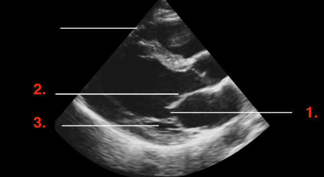
Identify this image.
PLAX MV
MV
Anterior leaflet
Posterior leaflet
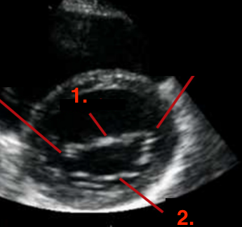
Identify this image.
PLAX MV
Anterior leaflet
Posterior leaflet
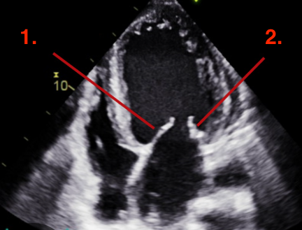
Identify this image.
A4C MV
Anterior leaflet
Posterior leaflet
What are semilunar valves?
Valves located between ventricles and great vessels or outflow tracts
PV
AV
What is the aortic valve (AV)?
Valve between LV and aortic root that contains three leaflets
Right coronary
Left coronary
Non-coronary cusp
What is the normal valve area for the AV?
3-4 cm²
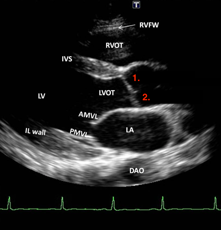
Identify this image.
PLAX AV
Right coronary cusp
Non-coronary cusp
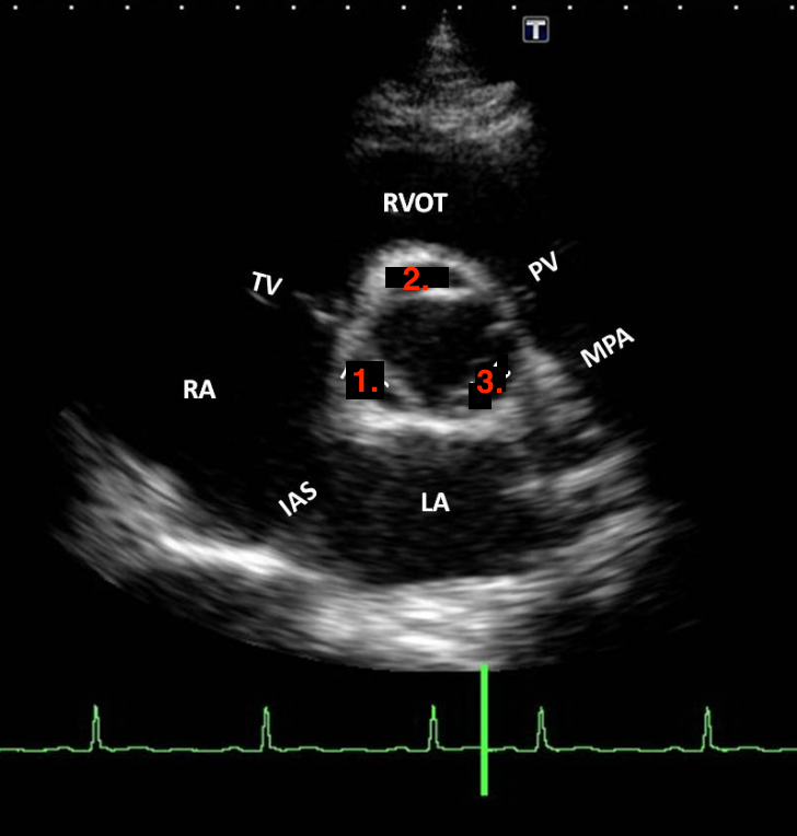
Identify this image.
Non-coronary cusp
Right coronary cusp
Left coronary cusp
What is the pulmonary valve (PV)?
Valve located between RV and MPA or outflow tract and contains three leaflets
Right leaflet
Left leaflet
Anterior leaflet
Which valve is the most anterior?
PV
What is the superior vena cava (SVC)?
Vessel that drains deoxygenated blood from superior portions of body and empties into posterior RA
What is the inferior vena cava (IVC)?
Vessel that drains deoxygenated blood from inferior portions of body and empties into posterior RA
What is the coronary sinus?
Vessel that drains deoxygenated blood from myocardium and empties into RA
What is the main pulmonary artery (MPA)?
Vessel that carries deoxygenated blood from RV to lungs
What is the normal value for the main pulmonary artery (MPA)?
Mean pulmonary artery pressure (MPAP): 9-18 mmHg
What are the pulmonary veins?
Four vessels that bring oxygenated blood from lungs to heart
What is the right coronary artery (RCA)?
Vessel that arises from right coronary cusp at sinuses of Valsalva and supplies blood to
Atria
IAS
RV free wall
RV apex
Inferoseptal LV wall
Inferior LV wall
Posterior medial papillary muscle
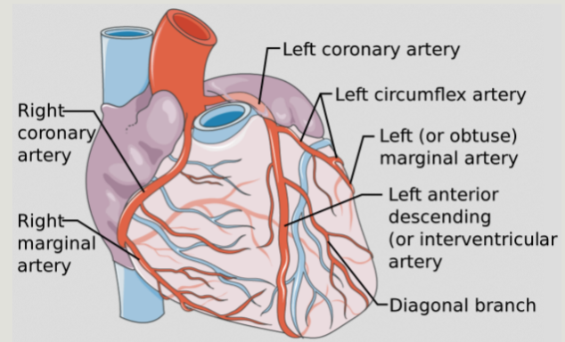
What is the distal portion of the right coronary artery (RCA)?
Continuation of RCA that gives rise to posterior descending artery (PDA) and supplies blood to
IVC
Inferior LV wall
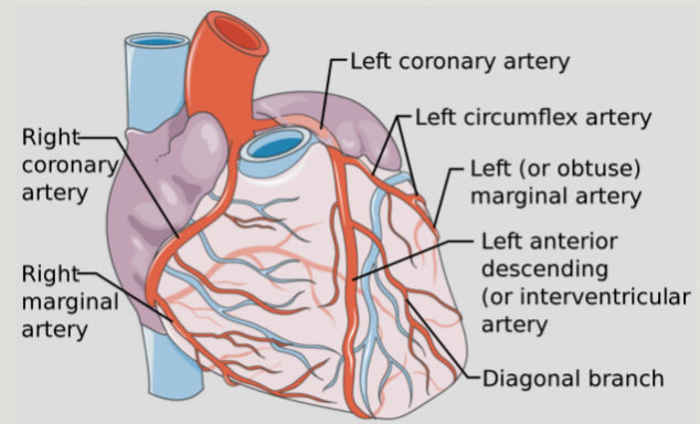
What is the left coronary artery (LCA)?
Vessel that arises from left coronary cusp at sinuses of Valsalva
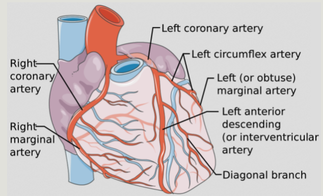
What is the left anterior descending artery (LAD)?
Branch of LCA that supplies blood to
Anterior septal wall
Anterior LV wall
LV apex
Anterior lateral papillary muscle
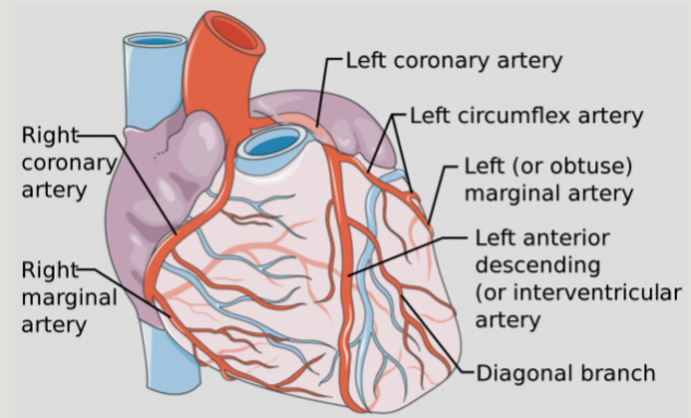
What is the left circumflex (LCX)?
Branch of LCA that supplies blood to
Inferior lateral LV wall
Anterior lateral LV wall
Anterior lateral papillary muscle
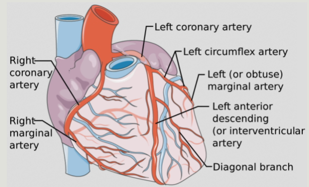
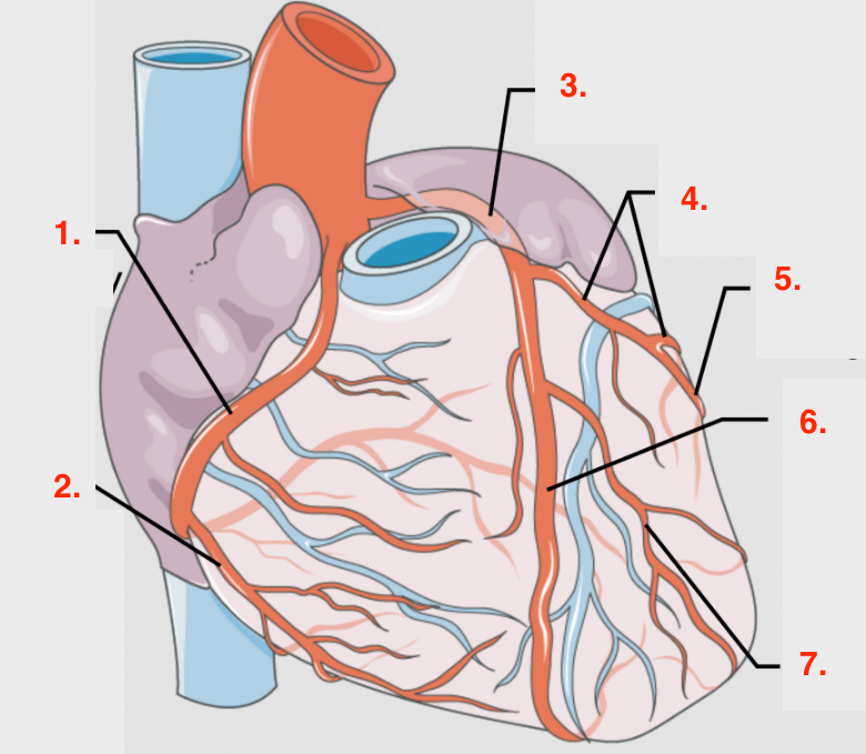
Identify this image.
RCA
Right marginal artery
LCA
LCX
Left marginal artery
LAD
Diagonal branch
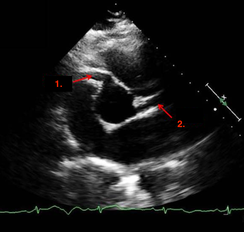
Identify this image.
RCA
LCA
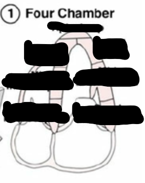
Identify this image.
A4C
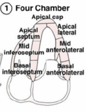
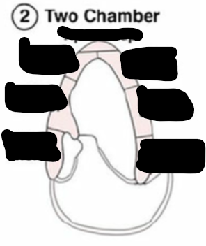
Identify this image.
A2C

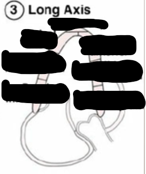
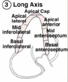
Identify this image.
A3C or LONG AXIS
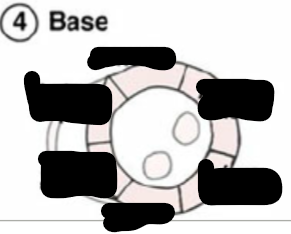
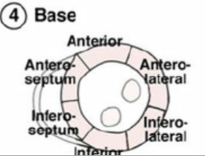
Identify this image.
Base or top
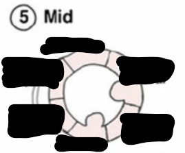
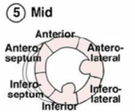
Identify this image.
Mid
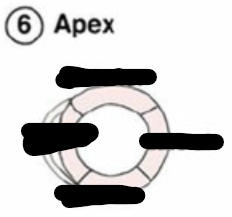
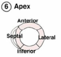
Identify this image.
Apex or bottom
What is the “scout view”?
First image taken with an increased depth to visualize effusion
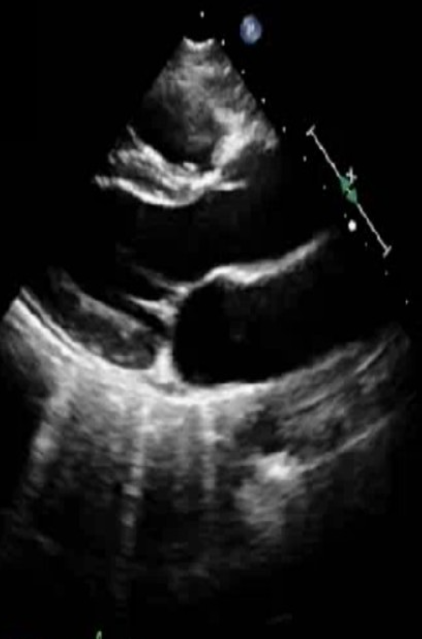
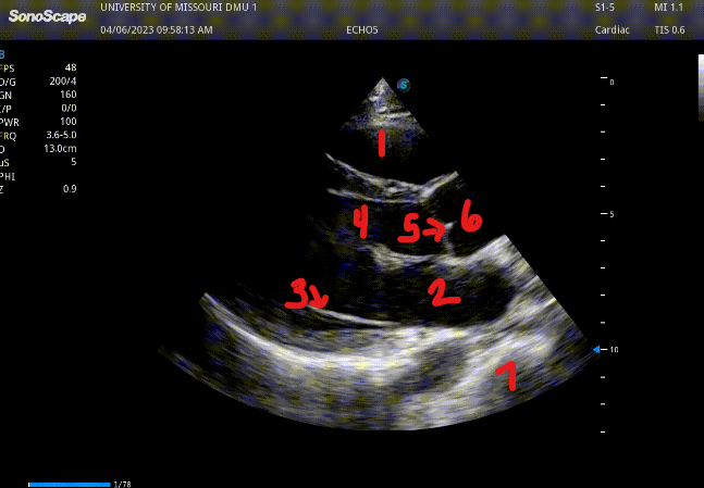
Identify this image.
PLAX
RV
LA
MV
LVOT
AV
Aortic root
Descending AO
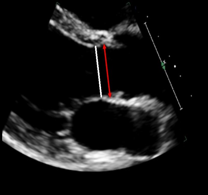
Identify this image.
PLAX AV measurements during MID SYSTOLE
White: LVOT
Red: Aortic annulus
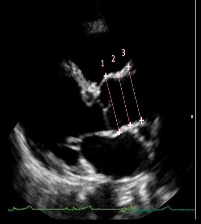
Identify this image.
PLAX AV measurements during END DIASTOLE
Sinus of Valsalva or aortic root
Sinotubular junction (STJ)
Ascending aorta
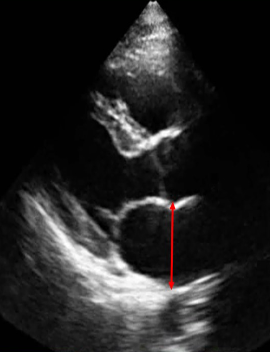
Identify this image.
PLAX LA diameter measured during END SYSTOLE
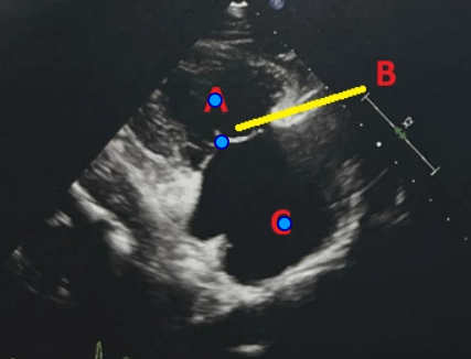
Identify this image.
PLAX RVIT
A. RV
B. TV
C. RA
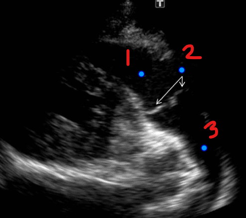
Identify this image.
PLAX RVOT
RVOT
PV
Main pulmonary artery
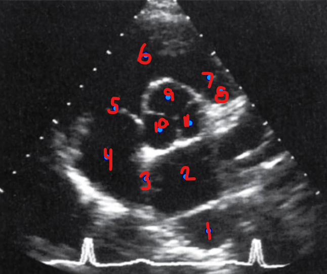
Identify this image.
PSAX AV Basal
Descending aorta
LA
IAS
RA
TV
RVOT
PV
Main pulmonary artery
Right coronary cusp (adjacent to RVOT)
Noncoronary cusp
Left coronary cusp (adjacent to LA)
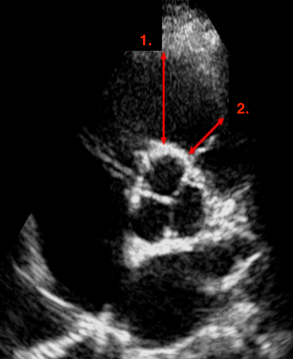
Identify this image.
Proximal RVOT measured at END DIASTOLE
Distal RVOT measured at END DIASTOLE
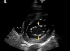
Identify this image.
PSAX MV level
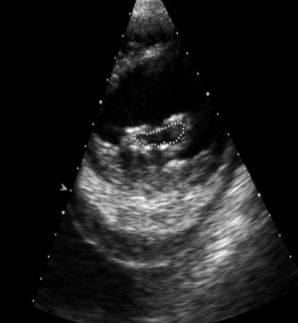
Identify this image.
Planimetry or most accurate way to measure MVA
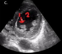
Identify this image.
PSAX LV
Papillary muscle
LV
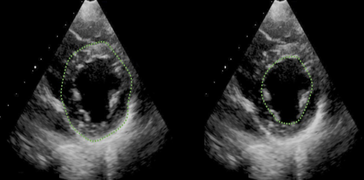
Identify this image.
LV mass measured at END DIASTOLE
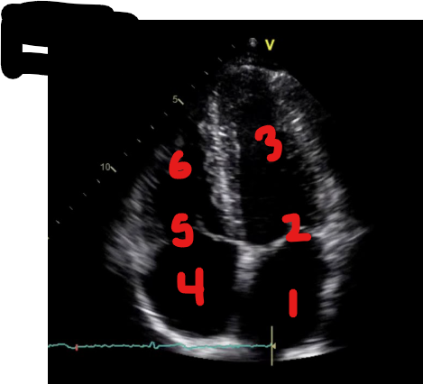
Identify this image.
A4C
LA
MV
LV
RA
TV
RV