bruh
1/16
There's no tags or description
Looks like no tags are added yet.
Name | Mastery | Learn | Test | Matching | Spaced | Call with Kai |
|---|
No analytics yet
Send a link to your students to track their progress
17 Terms
definitions
Phagocytes
Cells that protect the body by engulfing and digesting pathogens, dead cells, and foreign particles through phagocytosis. Examples include neutrophils, macrophages, and dendritic cells.
Leucocytes
Also called white blood cells. They are all the immune cells in the blood and tissues that help defend the body against infection and disease. Includes lymphocytes, monocytes, neutrophils, eosinophils, basophils, and others.
Monocytes
A type of leucocyte produced in the bone marrow that circulates in the blood. They can leave the bloodstream and develop into macrophages or dendritic cells in tissues.
Macrophages
Large phagocytic cells formed from monocytes after entering tissues. They engulf pathogens, remove dead cells, and help activate other immune cells by presenting antigens.
Neutrophils
The most common type of leucocyte and a fast-acting phagocyte. They are usually the first cells to arrive at an infection site and mainly destroy bacteria and fungi.
Dendritic cells
Immune cells that act as phagocytes and antigen-presenting cells. They capture pathogens and display antigens to T cells, helping activate the adaptive immune response.
process of phagocytosis
Internal Non-Specific Defences - Phagocytosis
• Phagocytes – specialised white blood cells (leucocytes) that engulf and digest/destroy micro-organisms & cell debris (foreign or unwanted material)
• Most common; monocytes/macrophages, Neutrophils & Dendritic Cells.
Process of Phagocytosis:
Phagocyte engulfs foreign material forming phagosome
Lysosome fuses and forms phagolysosome
Enzymes break down foreign material into smaller pieces
Small waste fragments expelled from phagocyte via exocytosis – vesicle fuses with plasma membrane and contents are released.
inflammation
• Purpose:
i. Reduce the spread of any pathogens to destroy them and prevent entry of more pathogens
ii. Remove damaged tissue and cell debris
iii. Begin the repair of damaged tissue
• Signs of inflammation;
i. redness
ii. swelling
iii. heat
iv. Pain
v. Loss of function in affected area
steps to inflammation
Complement proteins activate specialised leucocytes: mast cells.
Mast cells stimulate and coordinate inflammation by releasing chemicals
• Histamine, heparin and other substances into tissue fluid
Histamine increase blood flow through the area, causing the walls of the blood capillaries to become more permeable so fluid is filtered from blood
• Increased blood flow causes heat and redness
• Escape of fluid from blood causes swelling
Heparin prevents clotting so the release of heparin from the mast cells prevent clotting in immediate area of injury
• Clot of fluid around damaged area forms which slows spread of pathogen to healthy tissues
Complement proteins and chemicals released by mast cells attract phagocytes
• Macrophages and neutrophils actively consume micro-organisms and debris by phagocytosis
Abnormal conditions in tissue stimulate pain receptors; person feels pain in inflamed area
Phagocytes, filled with bacteria, debris and dead cells begin to die
• Dead phagocytes and tissue fluid form a yellow liquid called pus
New cells are produced by mitosis and repair of damaged tissue takes place
fever
· Increase in body temperature is due to the hypothalamus resetting the body’s thermostat to a higher level
· When the body’s thermostat is changed to a higher level, the person feels cold,
· Vasoconstriction of blood vessels in the skin and shivering occurs
· Both these responses are conserving heat and increasing heat production
· Body temperature increases rapidly
· When a fever ‘breaks’, this point is referred to as crisis point
· Bodies thermostat has been reset to normal
· Person feels hot and appears flushed
· Blood vessels in skin vasodilate
antigen presenting cells
Antigen-presenting cells work by:
1. Detecting the presence of non-self-antigens
2. Engulfing the pathogen (source of non-self-antigen)
3. Digest the pathogen, producing small fragments that move to the surface of the cell
4. Present the antigen to certain lymphocytes (usually Helper T-cells)
specific line of defence (adaptive)
The specific immune system is made up of 2 different systems;
1. Antibody-mediated response (humoral response)
2. Cell-mediated response
· There are 2 types of lymphocytes involved that are both produced in the bone marrow but mature in different locations:
- B cells: mature in the bone marrow (BB) and are involved in the antibody mediated response
- T-cells: mature in the thymus (TT) and are involved in the cell-mediated response
· After maturation both cells will migrate to lymphoid tissue or circulate in the blood.
Strategies which antibodies combat pathogens
Neutralisation
- Deactivating a pathogen or toxin by blocking its active site
Precipitation
- Antibodies bind to soluble antigens causing them to form insoluble clumps
Agglutination
- Antibodies bind to antigens on the surface of cells to form clumps of cells
Activating complement system
- To help disarm pathogens, enhancing phagocytosis, inflammation and pathogen removal by cell lysis
Antigen presenting cells
· These are specific cells that recognise when a non-self antigen enters the body.
· Includes dendritic cells, macrophages and undifferentiated B-cells
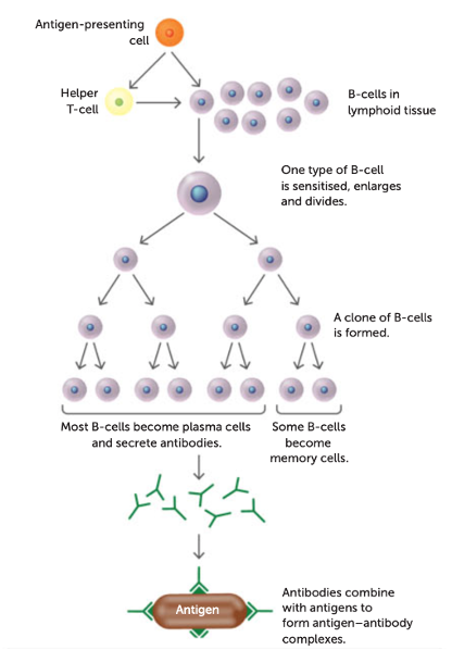
Antibody mediated (humoral) response
• Provides resistance against viruses, bacteria and bacterial toxins before they enter the body’s cells (extracellular defence)
• Involves B-cells that produce antibodies (proteins) that inactivate antigens.
• 1000s of different B-cells, each specific to a particular non-self antigen.
• For a B-cell to initiate an antibody-mediated response it must be activated by an antigen-presenting cell or Helper T-cell.
• When antigen is presented to Helper T-cells, cytokines are released
• This causes T-cells to clone themselves, releasing other cytokines which activate B-cells
how it works
· When activated by a Helper T-cell or antigen the B-cell enlarges.
· It then divides into a group of cells forming a clone.
· Some of these cells develop into memory B-cells.
· The rest develop into plasma cells.
· The plasma cells secrete antibodies into the blood, lymph or tissue fluid.
·
The antibodies then attach to the active site on a specific antigen to form an antigen-antibody complex.
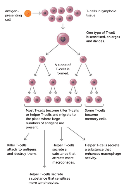
Cell mediated response
· Provides resistance to intracellular (host cell) viruses and bacteria.
· Provides resistance to fungi & parasites (’whole cells’)
· Involved in the rejection of transplants of foreign tissues and also appears to help fight cancer cells
· Involves T-cells that attack ‘foreign’ organisms directly
- 1000s of different T-cels, each specific to one particular antigen.
· For a T-cell to initiate a cell-mediated response it must be presented with an antigen by antigen-presenting cells.
how it works
Each T-cell responds to a specific antigen
When a foreign antigen enters the body the following occurs;
1. Macrophages phagocyte the pathogen, then present the antigen on their cell surface (antigen presenting cell)
2. They then travel to the nearest lymph node and present the antigen to helper T-cells
3. The specifically programmed T-cell for that antigen becomes activated or sensitised
4. Sensitised T-cells enlarge and divide, each giving a rise to a clone of identical T-cells
5. Most clone cells develop into other T-cell types, migrate to infection site and fight infection.
6. A small number develop into memory T-cells and remain in lymphoid tissue for future infections by the same antigen.
table
Types of Vaccines
1. Live-attenuated vaccines: they use a weakened form (reduced virulence) of the living pathogen. It’s antigen is intact but its ability to cause disease has been reduced
- E.g. tuberculosis, yellow fever & MMR (measles, mumps & rubella) vaccines
2. Inactivated vaccines: Vaccine containing dead form of the pathogen with an intact surface antigen, immunity from this type of vaccine is not as prolonged.
- E.g. whooping cough, typhoid
3. Toxoid vaccines: They use an inactivated form of a toxin produced by the bacteria or pathogen (For pathogens that cause disease by producing toxins)
- Eg: diptheria and tetanus.
4. Sub-unit vaccine: a fragment of the pathogen containing the antigen is used
- E.g. hepatitis B, HPV
methods of vaccine manufacturing
1. Modifying the pathogen’s characteristics by slightly changing the pathogen’s DNA to make it less virulent
2. The use of recombinant DNA technology to produce recombinant vaccines (Eg: Hepatitis B & HPV vaccines).
3. DNA vaccines: DNA for the antigen is introduced into the vaccine and then the human cells receiving this DNA will produce the antigen themselves.
immunity
Passive Immunity | Active Immunity |
|
|
Immunity can be either:
Natural Active immunity to a disease can develop from having the disease and recovering
Artificial injection of the antigens associated with the disease
Natural and artificial can be either passive or active
Two types of antibiotics;
Bactericidal antibiotics: kill bacteria by changing the structure of the cell wall/membrane or disrupting the action of essential enzymes
Bacteriostatic antibiotics: stop bacteria from reproducing by disrupting protein synthesis
• Different antibiotics offer range of different effects:
- Broad-spectrum antibiotics affect a wide range of different bacteria types
- Narrow-spectrum antibiotics only affect specific types of bacteria