Genetics Part 1 (Review of basic genetics)
1/104
There's no tags or description
Looks like no tags are added yet.
Name | Mastery | Learn | Test | Matching | Spaced | Call with Kai |
|---|
No analytics yet
Send a link to your students to track their progress
105 Terms
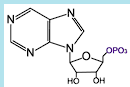
Define a Nucleotide
BUILDING block of DNA or RNA (nucleic acids)
Purine or pyrimidine base attached to a ribose phosphate
EX: Adenine, Guanine, Thymine, Cytosine, Uracil
DON’T have the phosphate
Define a Codon
Set of three nucleotides that codes for a particular amino acid
Define an amino acid
BUILDING blocks of PROTEIN
How many amino acids are in human proteins?
20
How many amino acids are essential in human proteins?
9
Cannot be synthesized in humans
Must be provided by dietary sources
How many amino acids are non-essential in human proteins?
11
Can be synthesized in the body
Not required in the diet
Define a Protein
Polymers of amino acids
Enzymes
Structural
Carrier
Receptor
Regulatory
Enzymes….
carry out biochemical reactions
Structural…
holds things together (ex: collagen, keratin)
Carrier…
transfer small molecules (ex: hemoglobin, ferritin)
Receptors…
bind circulating molecules (ex: insulin, cholesterol)
Regulatory….
turn genes on and off (ex: growth factors)
Explain what DNA is?
Double stranded
Deoxyribose nucleotides (A, T, G, C)
Repository of genetic information
Replicated in cell divisions
Stored as chromosomes
Explain what RNA is?
SINGLE stranded
Ribose nucleotides (A,U,G, C)
Short half life
mRNA, tRNA, rRNA
Regulatory functions (snRNA)
Each chromosome is _____ molecule of ____ and associated protein
one
DNA
What are associated with chromosomes?
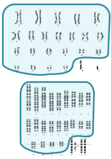
Karyotype
Autosomes
Sex Chromosomes
Homologs
Sister Chromatids
Alleles
How many pairs of chromosomes are there in normal content?
23
What explains how Nuclear chromosomes come in pairs?
Humans are diploid
ONE from MOTHER (maternal) and ONE from FATHER (paternal)
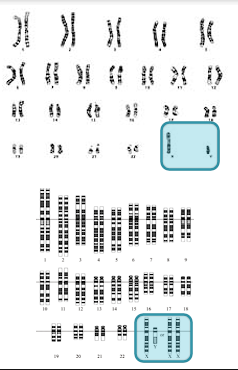
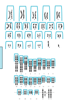
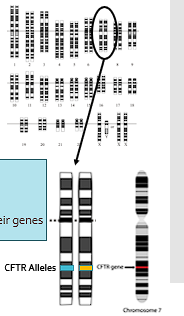
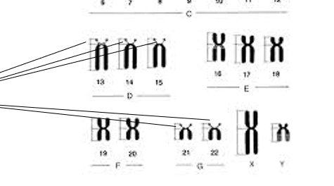
Define a Karyotype
Graphic arrangement of chromosomes in a cell
Arranged by size, position of centromere
Numbered 1-22, X and Y
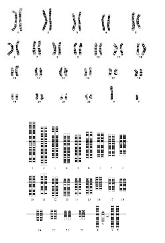
Define autosomes
Chromosomes that are the same in men and women
Numbered in order of size (sort of)
Chromosome 1-22
Pairs
Define sex chromosomes
chromosomes that are different in men and women
X and Y
Y contains genes that determine male sex and little else
X contains A LOT of genes that have nothing to do with SEX DETERMINATION
color blindness, hemophilia, muscular dystrophy
Define Homologs
Pairs of the same chromosome
One maternal, one paternal
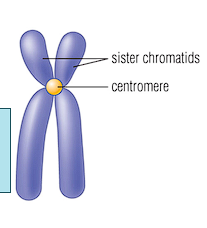
Define Sister chromatids
Duplicated copies of chromosomes
After DNA replication, before cell division
Define Alleles
Variants of individual genes
Variants at a single genetic locus
An individual has two alleles for each of their gene
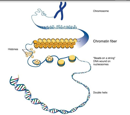
Explain chromatin structure
Each chromosome is a molecule of DNA→ Coiled into chromatin→ Coiled around central cores called nucleosomes→ Associated with histone proteins →TIGHTNESS of coiling controls GENE EXPRESSION
Each Chromosome is a molecule of____
DNA
Chromosomes are numbered in order of _____
size
Chromosomes are grouped by position of ______
centromere
Metacentric
Submetacentric
Acrocentric
What is a centromere
(Chromosome structure)
Divides chromosomes into TWO parts (arms)
Attachment point for SPINDLE FIBERS during cell division
Define what “Arms” are in the structure of chromosome
Human chromosomes that have “arms”
Short arm (p)
Long arm (q)
Short arms of acrocentric chromosomes contains REPETITIVE sequences for rRNA
Advanced staining techniques reveal dark and light _____ in _______
Bands
Chromosomes (Giemsa banding)
Each Chromosome has a unique and consistent banding pattern. What are they?
Bands numbered from centromere (band 0) to end
Subbands given second and third number
15q11.2
Dark bands contains heterochromatin
Light bands contain euchromatin
Dark bands are…
More condensed
Fewer expressed genes
Light bands are…
Less condensed
More expressed genes
Explain the components of Chromosome nomenclature
First part is the number of chromosomes
46 normal
Second part is the sex chromosome constitution
XX, XY, XXY etc
Third part is any abnormal chromones
46, XX and 46, XY is normal
Is 46 chromosomes normal or abnormal?
Normal
Is 45 or 47 chromosomes normal or abnormal?
Abnormal
45, X is a _________ and associated with what disease?
Monosomy, Turner syndrome
“47, XY +21 is a male with trisomy 21” is correlated with what disease
Down syndrome
Define Trisomy
extra chromosome
21,13 and 18 are only seen at birth
Define Monosomy
missing chromosome
Monosomy X (Turner syndrome) is only viable monosomy
Explain Translocations
Piece of one chromosome attached to another
RECIPROCAL, balanced (pieces of chromosomes interchanged)

Individuals with balanced translocation at risk for having children with _________
Unbalanced rearrangements
Missing part of one chromosome, trisomic for a piece of another chromosome
Explain Deletions and the types of chromosomal deletions
Part of a chromosome is missing, Involve many genes, Cause of syndromes (Prader-Willi, Angelman, Williams, DiGeorge)
Terminal
Interstitial
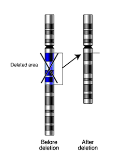
Terminal deletion
at end of chromosome
Interstitial deletion
in middle of chromosome
Define insertions
COUNTERPART of deletion
Part of chromosome is inserted of duplicated
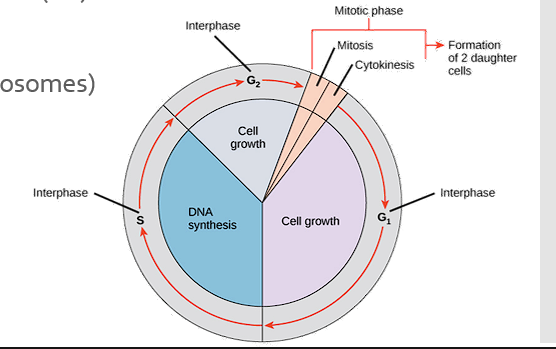
What is the interphase in the cell cycle?
“Resting” part of the cycle
Cell metabolism and growth (G1)
DNA synthesis (S)- chromosome duplication
Cell metabolism and growth (G2)
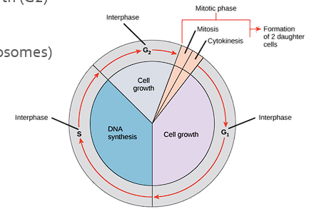
Describe the Mitotic phase (M) in the cell cycle
Mitosis (division of chromosomes)
Cytokinesis (cell division)
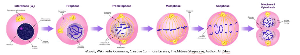
Describe the process of Mitosis in Cell Division
Binary division of a cell
DNA replication → sister chromatids of each chromosome, 92 sister chromatids
Chromosomes condense (prophase)
Chromosomes align along cell midline (metaphase)
Chromosomes split at centromere→ One chromatid drawn to each side of cell →Spindle fibers →Anaphase
•Cell divides →Telophase→ Cytokinesis →46 chromosomes in each cell
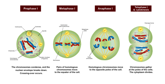
Describe the process of Meosis in Cell Division
REDUCTION division of a cell
Occurs in gonadal tissues
In testes continuously after puberty
In Ovaries before birth →Ova held in stasis in meiosis I → Meiosis completed after fertilization
Produces haploid gametes-1 chromosome of each pair
DNA replication produces 92 sister chromatids
TWO DIVISONS
Describe Meiosis I in Cell Division
DNA replication
92 sister chromatids
Chromosome condensation
Chromosomes associate in homologous pairs (tetrads)
Crossing over between arms of homologous chromosomes
One homologous chromosome (2 chromatids) goes into EACH daughter cell
Cell now haploid
2 sister chromatids of one chromosome
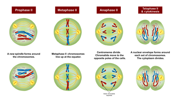
Describe Meiosis II in Cell Division
Daughter cell now has 46 chromatids, 23 chromosomes
NO further DNA replication
Meiosis II is like mitosis
Sister chromatids divide at centromere
One chromatid into each daughter cell
Daughter cells have 23 chromosomes, one of each homologous pair
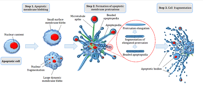
Define Apoptosis and the type of pathways
Programmed cell death
Important process in embryogenesis
Culls “sick” or stressed cells
Produces apoptotic bodies → Cleared by phagocytic cells
Intrinsic pathway
Extrinsic pathway
Intrinsic pathway
More like cell suicide
Internal signals-SMACs and MACs
Extrinsic pathway
External activation
TNF
Describe the relationship between apoptosis and cancer
Acts as a natural defense mechanism to eliminate potentially malignant cells.
Its evasion is a hallmark of cancer development and progression.
Normally, apoptosis removes damaged or dysfunctional cells to maintain tissue health →Cancer cells subvert this process to survive, proliferate, and resist treatment.
Dysregulated BCL-2 proteins
TP53 (Tumor Protein 53) Tumor suppressor gene inactivation
BCL- 2 inhibitors and P53 reactivators are new drugs used in treatment of cancers
What are Dysregulated BCL-2 proteins and how do they affect apoptosis?
Cancer cells often overexpress anti-apoptotic proteins like BCL-2 and BCL-xL, which INHIBIT a critical step in the intrinsic apoptotic pathway.
Over 50% of cancers exhibit elevated BCL-2 levels, rendering cells resistant to DNA damage or chemotherapy.
What are TP53 (Tumor Protein 53) Tumor Suppressor Gene Inactivation and how do they affect apoptosis?
Mutations in the p53 gene DISABLE its ability to trigger apoptosis in response to DNA damage. This allows cells with genetic errors to survive and accumulate oncogenic mutations. Over 50% of tumor cells have p53 mutations.
What is the function of the Mitochondria
Main generator of ATP
Kerbs cycle
Urea cycle
Outer membrane = permeable to small molecules
Inner membrane = impermeable except by specific transporters, inner contains bound enzymes
Matrix= soluble enzymes
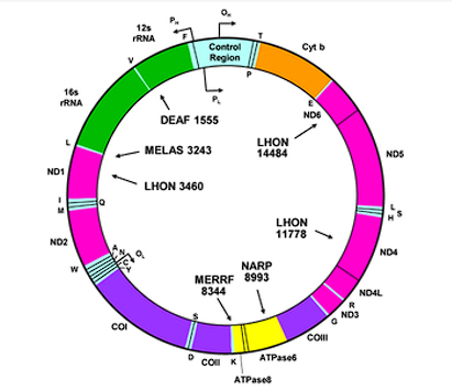
Describe the Mitochondrial genome
Mitochondria contain DNA
Mitochondrial chromosome is circular, bacterial-like →No introns→ Replication start site
Encodes some proteins →DNA replication enzymes→ Some respiratory chain components →Set of t-RNAs for protein synthesis
Several copies in each mitochondrion
Come from ovum (maternal)
Most mitochondrial proteins and enzymes are encoded by nuclear genes and are imported into the mitochondria.
Describe the Mitochondrial disorders/ dysfunction
Defects in respiratory chain activity
DECREASED ATP generation from oxidation (O2)
Wide range of symptoms: Weakness, myopathy, Neurologic symptoms, Diabetes, Blindness, Hearing loss
Some due to mutations in mitochondrial genome
Variable due to heteroplasmy (number of mutant copies in each mitochondrion)
Show maternal pattern of inheritance
Many due to nuclear genome mutations
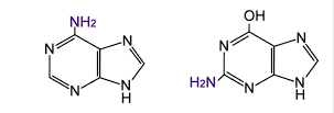
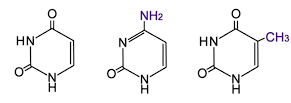
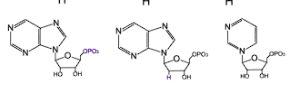
What are the components of DNA?
Purine bases
Pyrimidine bases
Nucleotides
Purine bases
Adenine
Guanine
Aminopurines
Pyrimidine bases
Uracil
Cytosine
Thymine
Nucleotides
Ribose
Deoxyribose
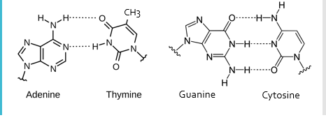
Amino groups of_____ form hydrogen bonds with keto groups of ________
Purines
Pyrimidines
Adenine-Thymine
Guanine-Cytosine
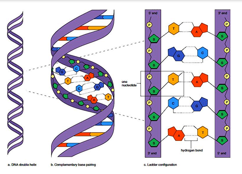
Describe the structure of DNA
Bases are connected through ribose links
5’ carbon of one nucleotide attached to 3’ carbon of the next by phosphate bridge
Gives DNA strand directionality – 5’ to 3’
Describe the process of DNA Replication
Semiconservative replication
One strand is the template, unchanged
Second strand is newly synthesized
DNA unwinds
A’s in DNA strand bind with free dTTP
T’s in DNA strand bind with free dATP
G’s in DNA strand bind with free dCTP
C’s in DNA strand bind with free dGTP
DNA polymerase creates new strand
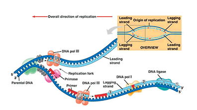
What enzymes are involved in DNA replication
DNA polymerase
Helicase
Primase
Topoisomerase
Telomerase
Ligase
Describe the function of Helicase in DNA replication
Unwinds the double strand
Describe the function of DNA polymerase in DNA replication
Creates the new DNA strand in the 5’ to 3’ direction
Describe the function of Primase in DNA replication
Synthesizes the RNA Primer needed to start sythesis
Describe the function of DNA Ligase in DNA replication
Connects Okazaki fragments on the lagging strand
Describe the function of Topoisomerase in DNA replication
Prevents tangling as the DNA unwinds
Describe the function of Telomerase in DNA replication
Extends telomeres at the end of chromosomes
A gene is a……
linear stretch of DNA that codes for something
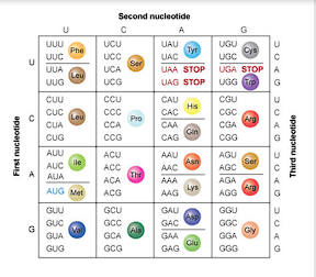
Describe the process of transcription
RNA polymerase binds to a promoter sequence (like the TATA box) and synthesizes RNA in a 5’ to 3’ direction using uracil instead of thymine. This binding is regulated by enhancers and repressors.
5-methylguanin = beginning of mRNA strand (capping)
PolyA tail= end of mRNA
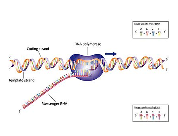
Describe the purpose of transcription
To copy a DNA sequence into mRNA for PROTEIN SYTHESIS
What is the Function of mRNA?
Provides templates for protein synthesis
synthesized by RNA polymerase II
What is the Function of tRNA?
Binds to specific amino acids and has an anticodon to align them correctly during protein synthesis
Synthesized by RNA polymerase III
What is the Function of rRNA
Provides RNA for ribosomes. Particles containing mRNA binding and protein synthesis machinery
synthesized by RNA polymerase I
Describe post-transcriptional modifications
Primary mRNA transcripts undergo capping (adding 5-methylguanine), addition of a PolyA tail, and splicing, where introns are removed by the spliceosome
Alternative splicing can produce multiple proteins from one gene
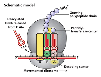
Describe the purpose of translation
To synthesize a polypeptide chain based on the mRNA template
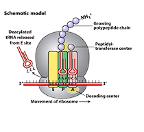
Describe the process of translation
mRNA binds to a ribosome (two subunits, rRNA and Proteins-elongation factors)→ translation starts at an AUG (methionine) codon→ tRNAs bring amino acids to the ribosome’s A site → they are added to the chain at the P site → empty tRNA leaves via the E site
Polypeptides fold based on _____and _______ interactions, often aided by ______proteins (protein folding)
Ionic
Hydrophobic
Chaperone
Post-translational modifications include cleavage of _______, association of _______, adding cofactors (heme), or _______ in the Golgi apparatus
pro-proteins
Multiple subunits
glycosylation
Describe the mechanisms of gene regulation
Regulation occurs via DNA methylation (inhibits unwinding), RNA polymerase binding
Histone acetylation (activates chromatin)
Use of repressors or enhancers
mRNA splicing
miRNA & RISC binding
mRNA Stability
Protein folding, Post translational modifications
Explain the various causes of mutations
Include single base changes, nucleotide deletions/insertions (causing frameshifts)
Trinucleotide repeat expansions (leading to anticipation)
Genome/chromosome level (Copy number variants, Recurrent deletions )
Cell division: Frequency 10-8 bases/division, Most are not in coding sequences, Most are benign
Describe Nucleotide level mutations
Single base changes
May change a single amino acid
May not change anything (synonymous mutation)
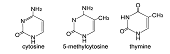
Most common is C-T change at a GC doublet
The C in a CG doublet may be methylated
Deamination changes the C to a T
During DNA replication the T would pair with an A instead of a G
Describe Nucleotide deletions or insertions
Single nucleotide additions or losses
Within coding sequence will cause a frameshift
Codons after the mutation will all be changed
Leads to severely defective protein after mutation
Early stop codon
Small deletions or insertions
In multiples of 3 can be in frame
Delete single or a few amino acids
Protein may still function
e.g. delF508 in cystic fibrosis deletes a single phenylalanine
Other changes will cause a frameshift
Describe trinucleotide repeat expansion
Some genes contain repetitive sequences of trinucleotides
Huntington disease - CAG – glutamine repeat in coding region
Fragile-X – CGG near promotor
Myotonic dystrophy – CTG in 3’ untranslated region
In meiosis, the repeat region may expand
If expansion is greater than some threshold, the gene will not function properly causing a genetic disorder
Huntington disease: <26 CAG nl, >36 causes disorder
Fragile-X: 5-44 CGG nl, >200 causes disorder
Myotonic dystrophy: 5-27 CTG nl, >50 causes disorder
Expansion occurs preferentially in maternal meiosis (FraX, MD) or paternal meiosis (HD)
Causes “anticipation” in pedigrees
Earlier onset in later generations
Describe copy number variant mutations
Genome contains many duplicated genes
Beta-globin
Alpha-globin
Color vision locus
Genes may misalign during meiosis
Crossover will delete gene on one chromosome, duplicate gene on the other
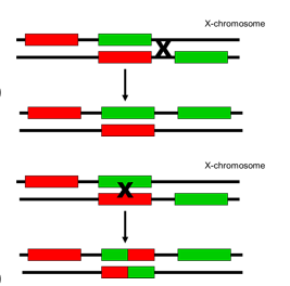
Describe X-linked color blindness mutation
X-chromosome contains one red-sensitive pigment gene and several green sensitive ones
Only first green sensitive gene is expressed
Color pigment genes have very similar DNA sequences and may misalign during Meiosis I
Crossover between genes causes deletion of green sensitive visual pigment
Crossover within a gene causes fusion gene with differing color sensitivity
Describe recurrent deletion mutation
Several genetic disorders are caused by deletions which occur in the same place in affected individuals
Severe Hemophilia A – X-chromosome, Factor VIII gene
DiGeorge syndrome – chromosome 22q11.2
Prader-Willi syndrome – chromosome 15q11.2-13
Williams syndrome – chromosome 7q11.23
Regions flanked by repetitive or inverted sequences
Misalignment and crossover causes deletion in the same places
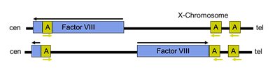
Describe severe hemophilia A mutation
Duplicated, inverted small gene (A) in Factor VIII gene
Pairs with homologous gene telomeric to Factor VIII
Crossover causes inverted deletion of Factor VIII
Responsible for 40% of severe Hemophilia A
Mutation occurs only in male meiosis
What are the types of DNA repair?
Nucleotide excision repair
Base excision repair
Mismatch repair
Nucleotide excision repair
Damaged bases cause DNA helix distortion, e.g. thymidine dimers caused by UV light
Damaged segment cut out by endonucleases
Segment removed by helicase
New segment synthesized by DNA polymerase
Reconnected by ligase
Base excision repair
Single chemically damaged base cut out by glycosylase
New base inserted by ligase
Mismatch repair
Mismatched bases introduced during errors in DNA replication
Mismatched segment recognized in newly replicated strand by repair system
Segment cut out by endonuclease
New strand made by DNA polymerase
Reconnected by ligase