OMFPR 2 - Principles of Tomography and Panoramic Imaging (Dr. Ramesh)
 Call Kai
 Learn
Learn Practice Test
 Spaced Repetition
 Match
Match Flashcards
 Knowt Play
1/85

There's no tags or description
Looks like no tags are added yet.
Name | Mastery | Learn | Test | Matching | Spaced | Call with Kai | Chat |
|---|
No analytics yet
Send a link to your students to track their progress
86 Terms
Panoramic
_____________ radiography is based on body-section radiography and it blurs out the shadows of superimposed structures
- Broad coverage of facial bones and structures
- Low patient dose
- Simple and convenient
- Can be used on patients with trismus (inability to open mouth)
- Minimal time
What are the five advantages of a panoramic x-rays?
- Critical positioning
- Inherent magnification
- Poor resolution (compared to intraoral radiographs)
- Incorrect interpretation of ghost image
What are the four disadvantages of panoramic x-rays?
All are indications!
Which of the following are an indication for panoramic x-ray?
A) Evaluation of trauma
B) Evaluation of third molars
C) Extensive disease
D) Suspected large lesions
E) Mixed dentition
F) Developmental abnormalities
horizontal
Panoramic radiographs are obtained by rotating a narrow beam of radiation in the __________ plane
narrow
Panoramic radiographs are obtained by rotating a __________ beam of radiation in the horizontal plane
Opposite direction
What direction is the film rotated in while the object (jaw) is stationary?
tomography
Define the following:
Body section radiography
body section radiography
Define the following:
- Technique that blurs out shadows of superimposed structures
- Object of interest less blurred
- Doesn't improve sharpness
focal trough (image layer)
when taking a tomographic image the part of the body that you want to appear the most clearly should be positioned within the _________
focal trough (image layer)
Define the following:
A three-dimensional curved zone or image layer in which structures are reasonably well defined
false (often inaccurate)
t/f: Measurement on a panoramic is often very accurate
true
t/f: Magnification of radiographic structures varies with error in patient positioning
midline
_______ objects may project as single or double images
3
how many centers of rotation are used to take a pano?
double image formation
What has the following characteristics:
- Objects are imaged (intercepted) twice by beam
- One image is mirror image of the other
midline objects
double image formation often occurs with what type of objects?
real image
What type of image is formed when the object is located between the rotation center and film?
ghost image
What type of image is formed when the object is located between the x-ray source and the rotational center?
film
A real image is formed when the object is located between the rotation center and the _____________
x-ray source
A ghost image is formed when the object is located between the rotational center and the _____________
real and double image
the shaded regions will produce what type of images?

ghost image/shadows
the shaded regions will produce what type of images?

3
the shaded region within the blue diamond will produce a total of __ images

2 real
1 ghost
the shaded region within the blue diamond will produce a total of 3 images. What types of images are they?

real image
the following patient has a surgical plate. What type of image is the purple arrow pointing to?
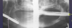
ghost shadows
______________ arise from structures located on the posterior aspect of the head of opposite side
true
t/f: ghost shadows have same general shape and appear on the opposite side
higher
ghost shadows are positioned __________ than real structures
more
ghost shadows are __________ blurred than real structures
more, highly
vertical component of ghost shadows are __________ blurry and __________ magnified
- Antero-posteriorly (too far back or front)
- Supero-inferiorly (chin up or down)
- Mid-sagittal plane (face turned towards right or left)
- Tongue positioning
- Spinal Column positioning
What are five common types of errors with panoramic radiographs?
None - ideal
ID the positional error from the radiograph:

right rotation (pts right side is larger)
(And too far back)
ID the positional error from the radiograph:
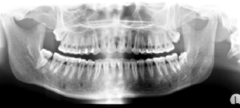
left rotation (pts left side is wider)
ID the positional error from the radiograph:

too far front (narrow/elongated incisors)
(And improper tongue positioning)
ID the positional error from the radiograph:
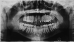
too far forward
when you're looking on a radiograph and the mandibular anteriors look narrow and tall, the pt is ...
too far back
when you're looking on a radiograph and the mandibular anteriors look wider and short, the pt is...
too far back (short/wider incisors)
(and right rotation)
ID the positional error from the radiograph:
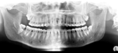
chin down
ID the positional error from the radiograph:

chin up
ID the positional error from the radiograph:
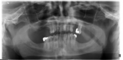
none - ideal
ID the positional error from the radiograph:
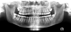
Improper tongue placement
ID the positional error from the radiograph:
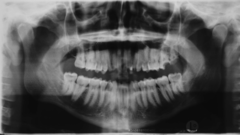
Improper tongue placement
(and too far front?)
ID the positional error from the radiograph:
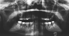
minification
if a patient is positioned where the incisors are anterior to the center of the focal trough, the image of the incisors will experience ________
magnification
if a patient is positioned where the incisors are posterior to the center of the focal trough, the image of the incisors will experience ________
downward
in order to prevent the cervical vertebrae from appearing on a pano, you can make sure that the patient's chin is tilted slightly _____
ghost image
the blue arrow is pointing to what type of image of the cervical vertebrae?

floor of max. sinus
ID the hard tissue anatomy indicated by the arrow pointing from the red box:

infra orbital canal and foramen
ID the hard tissue anatomy indicated by the arrow pointing from the orange box:
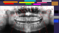
hard palate/floor of nasal fossa
ID the hard tissue anatomy indicated by the arrow pointing from the yellow box:
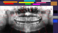
nasal fossa
ID the hard tissue anatomy indicated by the arrow pointing from the green box:
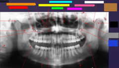
ant. wall of max. sinus
ID the hard tissue anatomy indicated by the arrow pointing from the light blue box:

coronoid process
ID the hard tissue anatomy indicated by the arrow pointing from the dark blue box:
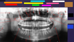
infra orbital rim
ID the hard tissue anatomy indicated by the arrow pointing from the hot pink box:

pterygomaxillary fissure
ID the hard tissue anatomy indicated by the arrow pointing from the pink/salmon box:

external auditory meatus/ear canal
ID the hard tissue anatomy indicated by the arrow pointing from the brown box:

articular eminence
ID the hard tissue anatomy indicated by the arrow pointing from the white box:
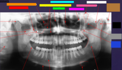
mandibular condyle
ID the hard tissue anatomy indicated by the arrow pointing from the black box:
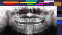
zygomatic bone
ID the hard tissue anatomy indicated by the arrow pointing from the gray box:

mandibular fossa
ID the hard tissue anatomy indicated by the arrow pointing from the red box:
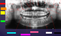
lateral pterygoid plate
ID the hard tissue anatomy indicated by the arrow pointing from the orange box:

zygomatic process of maxilla
ID the hard tissue anatomy indicated by the arrow pointing from the yellow box:

cervical spine
ID the hard tissue anatomy indicated by the arrow pointing from the green box:

external oblique ridge
ID the hard tissue anatomy indicated by the arrow pointing from the light blue box:
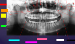
inferior boarder of the mandible
ID the hard tissue anatomy indicated by the arrow pointing from the hot pink box:
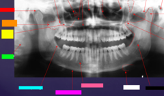
mental foramen
ID the hard tissue anatomy indicated by the arrow pointing from the pink/salmon box:

inferior alveolar canal
ID the hard tissue anatomy indicated by the arrow pointing from the black box:

hyoid bone
ID the hard tissue anatomy indicated by the arrow pointing from the white box:
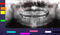
posterior pharyngeal wall
ID the soft tissue anatomy indicated by the arrow pointing from the red box:

soft palate
ID the soft tissue anatomy indicated by the arrow pointing from the orange box:
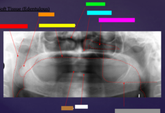
dorsal surface of tongue
ID the soft tissue anatomy indicated by the arrow pointing from the yellow box:

middle nasal meatus
ID the soft tissue anatomy indicated by the arrow pointing from the green box:

inferior nasal meatus
ID the soft tissue anatomy indicated by the arrow pointing from the light blue box:
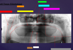
inferior nasal concha (turbinate)
ID the soft tissue anatomy indicated by the arrow pointing from the hot pink box:

upper lip
ID the soft tissue anatomy indicated by the arrow pointing from the brown box:

lower lip
ID the soft tissue anatomy indicated by the arrow pointing from the white box:

ghost image of opposite mandible
ID the soft tissue anatomy indicated by the arrow pointing from the grey box:

ala of nose
ID the soft tissue anatomy indicated by the arrow pointing from the red box:

nasal septum
ID the soft tissue anatomy indicated by the arrow pointing from the orange box:

columna (the septum separating the nostrils)
ID the soft tissue anatomy indicated by the arrow pointing from the yellow box:

tragus
ID the soft tissue anatomy indicated by the arrow pointing from the green box:

ear lobe
ID the soft tissue anatomy indicated by the arrow pointing from the light blue box:

- Hard palate
- Epiglottis
-Hyoid bone
- Cervical vertabrae
What midline objects have the ability to project as single/double images? (4)
More (ex: hands on a desk with light above)
The farther the sensor is from anatomy, the ____________ magnification
Buccinator (attaches to the external oblique ridge of the mandible)
What muscle attaches here?
