L3: Liver Path
 Call Kai
 Learn
Learn Practice Test
 Spaced Repetition
 Match
Match Flashcards
 Knowt Play
1/41

There's no tags or description
Looks like no tags are added yet.
Name | Mastery | Learn | Test | Matching | Spaced | Call with Kai | Chat |
|---|
No analytics yet
Send a link to your students to track their progress
42 Terms
fx (5)
-processing dietary carbohydrates, lipids, amino acids, and vitamins
-metabolic balance
lipid and glucose turnover, synthesis and storage
-synthesis and turnover of plasma proteins
clotting factors, albumin, vitamin K, etc
-detoxification and biliary excretion of endogenous wastes and xenobiotics
-innate immunity
Kupffer cell filtration, cytokine synthesis, and Pit cells
what is the functional unit of the liver?
-wedge shaped acinus or polyhedral lobule
summary:
zone 1 hepatocytes
-surrounded by blood with the highest concentration of O2, insulin, glucose, and AAs
-principal site of gluconeogenesis, protein synthesis, aerobic metabolism, urea, lipid and cholesterol metabolism
summary:
zone 3 hepatocytes
-furthest from blood inflow
-site of glycolysis, lipogenesis, and biotransformation due to ^ cytochrome p450 levels
term:
limiting plate
-area that separates the portal tract CT
note:
pig lobules
-delineated by fibrous tissues
-easy to visualize
what happens to defect hepatocytes in zone 2?
-can dissociate from cords if the adhesions are lost
blood flow:
portal v
-from forestomach/glandular stomach, intestines, spleen, pancreas
-rapid clearance of nutrients, xenobiotics, microorganisms
-blood flow to sinusoids
blood flow:
hepatic a
-oxygenated blood
-disperse into the peribiliary capillary plexus > sinusoids
summary:
hepatocytes
-arranged in single-cell plates/cords bordered with sinusoids
-sinusoids are lined with endothelial cells
-gap between the endothelial cells and hepatocytes – Space of Disse - usually not visible by light microscopy
-Kupffer cells – within the sinusoid
-Stellate cells – within space of Disse
summary:
sinusoids
-special fenestrated endothelium facilitates passage of fluids and particulates into the perisinusoidal space (Space of Disse)
-with damage, 'capillarization'/ loss of fenestrations interferes with uptake and secretion.
summary;
Kupffer cells
-fixed tissue macrophages within the sinusoidal lumens
-remove particulates, dead cells and bacteria-filtering organ
-cytokine and pro-inflammatory mediator secretion
summary:
pit cells
-large granular lymphocytes with NK cell activity in sinusoidal lumens
-innate immunity
summary:
ito cells/hepatic stellate cells
-within the perisinusoidal space
-secretes extracellular matrix of sinusoids
-inflammation causes extra secretion = capillarization + contractile element production restricting blood flow
-regulate microvascular tone and sinusoidal flow
-store lipid droplets rich in vitamin A
-notable aged carnivores
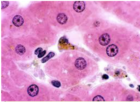
Kupffer cell
-visible from accumulated intracytoplasmic pigment (age or injury)
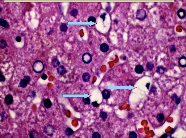
Ito cells
-perisinusoidal space
-ntracytoplasmic lipids (why they look like fat droplets)
blood flow:
central vv
sinusoidal blood flow → central vv → CaVC
flow:
lymphatic drainage
-perisinusoidal space → lymphatics (capsule & portal tracts) → hepatic nodes (hilus)
summary:
canaliculi
-intercellular space between 2 hepatocytes isolated by tight junctions
-secretes bile.
-difficult to recognise unless distended
summary:
biles ductules
-canaliculi connect to bile ductules via the canal of Hering (w/ oval cells)
-multiply with severe hepatic injury
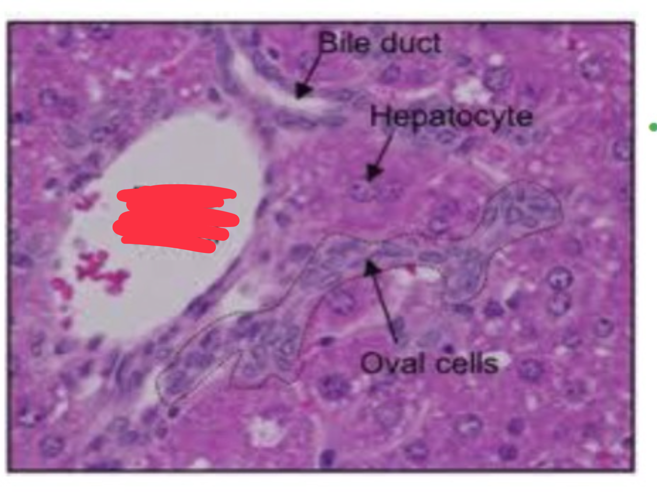
-blood vessels
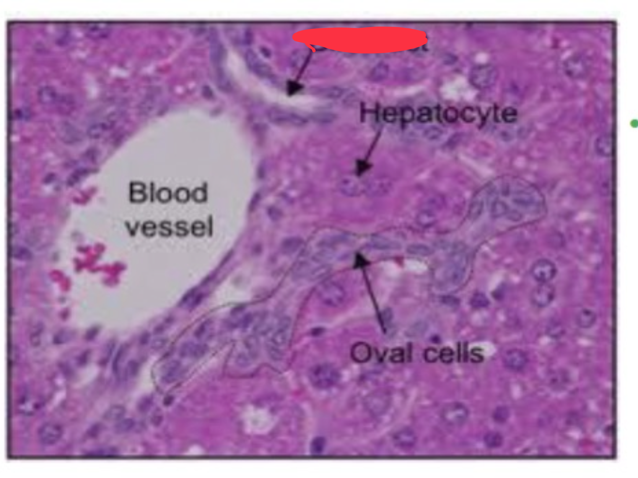
-bile duct
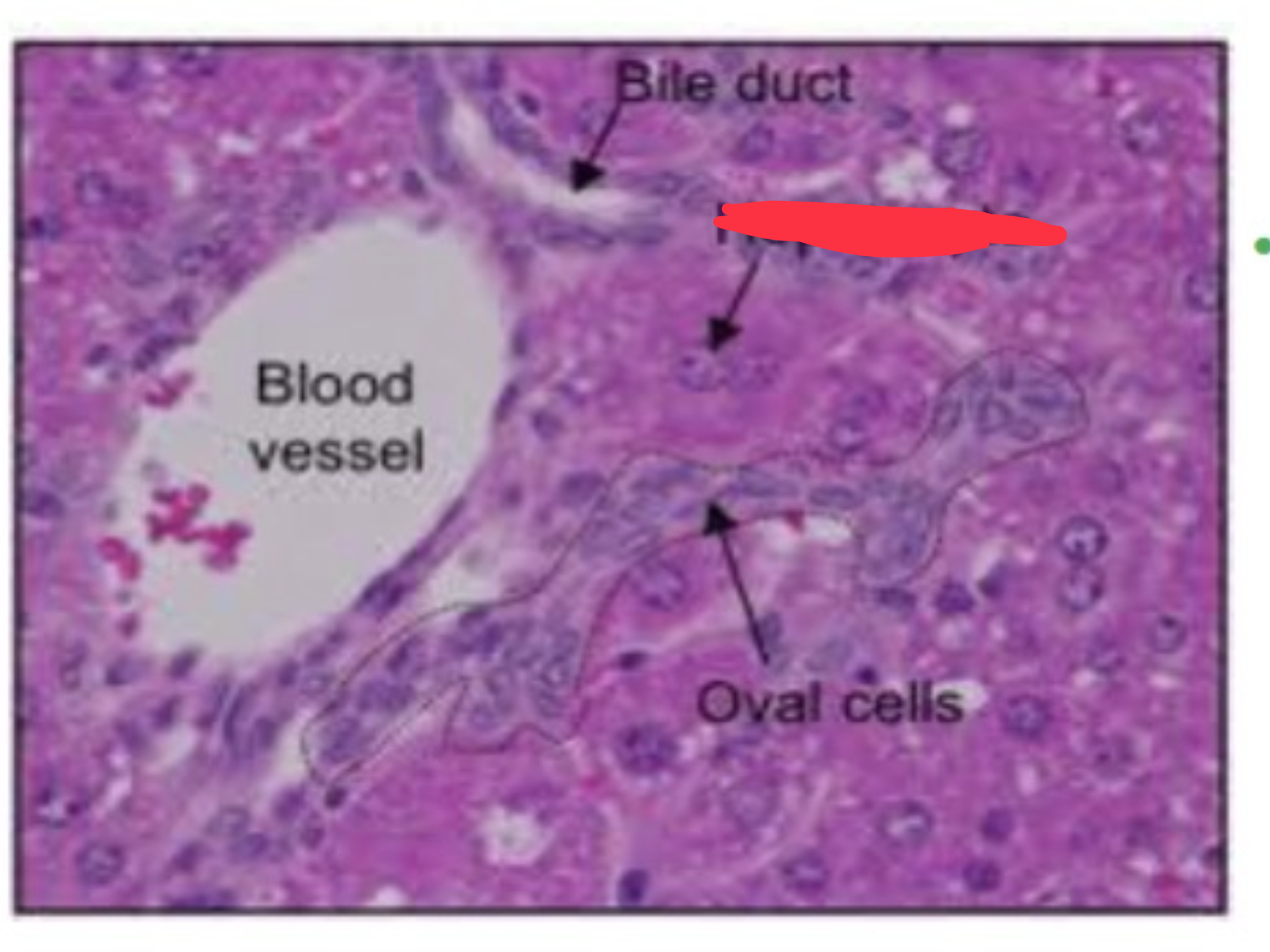
-hepatocyte
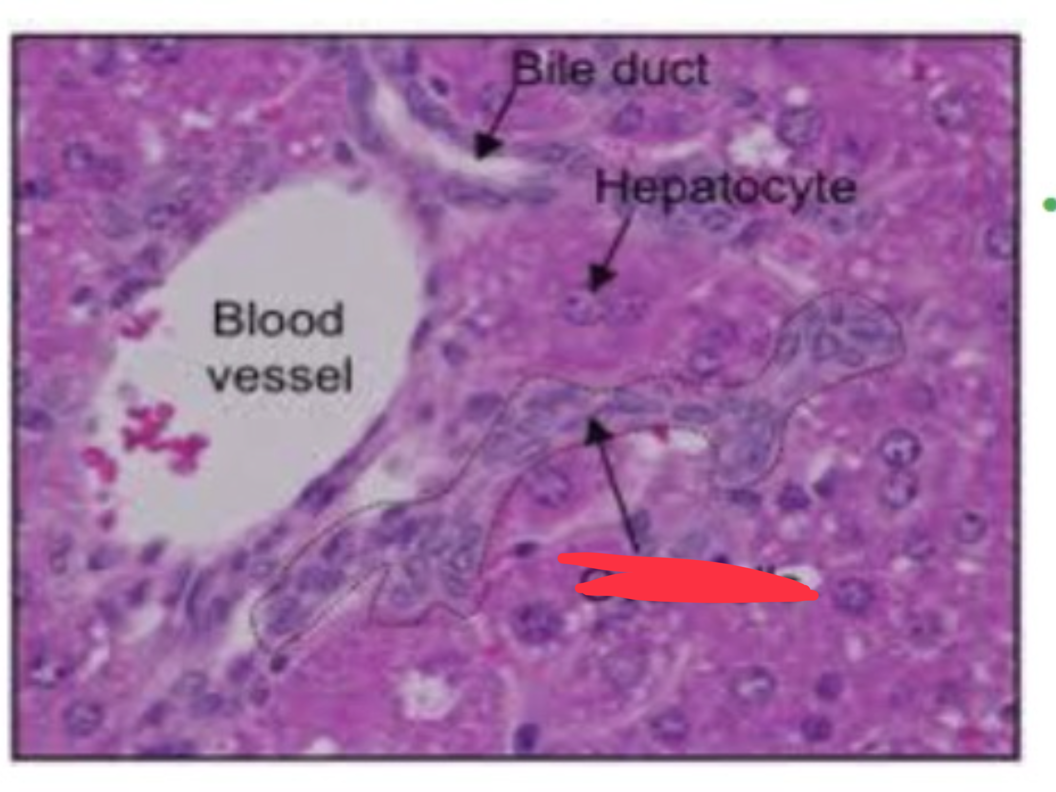
-oval cells
summary:
bile ducts
-cuboidal or low columnar epithelium
-modify bile composition and secrete substances
-recognisable as part of the portal triads
hepatic artery, hepatic portal venule, and bile duct
fx:
gallbladder
-bile storage before release into the GIT
liver:
postmortem
-breaks down fast
-core site within body → kept at a higher temperature
-bacteria rapidly spread from the gut
-gas bubbles + bile imbibition in surrounding tissues
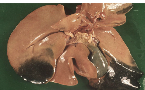
-bile imbibition
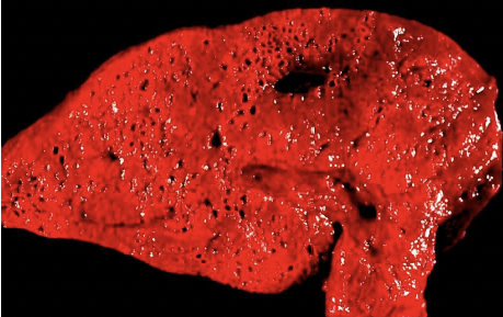
-gas bubbles
Which hepatocytes are most susceptible to direct-acting toxicants?
A. zone 1- Periportal
B. zone 2- Midzonal
C. zone 3- Periacinar/ centrilobular
C.
-receive the least amt of blood so can not handle more stress
-lots of P450 enzymes that metabolize toxins
Which hepatocytes are most susceptible to hypoxia and/ or indirect or metabolically activated toxicants?
A. zone 1- periportal
B. zone 2- midzonal
C. zone 3- periacinar/ centrolobular
C.
-least amt of oxygen so can will experience hypoxia first
developmental anomalies (2)
-cysts
-biliary atresia
summary:
cysts
-serous cysts: young animals, capsular, incidental
-biliary cysts
associated with polycystic kidney disease in cats, Westies, and Swiss Freiberger horses
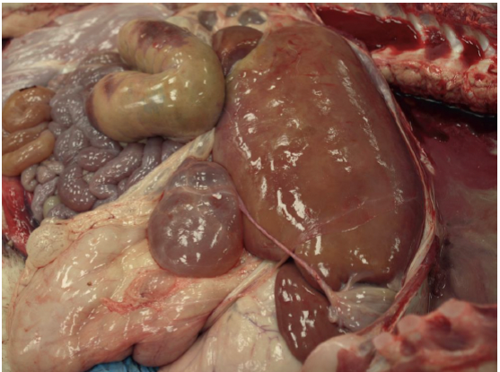
-serous cyst attached by a stalk to the hepatic capsule
(calf)
types:
congenital vascular anomalies
-congenital arteriovenous fistulae- dogs & cats
-portal vein hypoplasia- dogs
-portal microvascular dysplasia- dogs
-sequelae:
portal hypertension & acquired extrahepatic shunts
congenital vascular anomalies
how?
-arteriolar proliferation and parenchymal fibrosis
-hepatic parenchyma atrophy (serum bile acids)
types:
portosystemic shunts
-congenital
-acquired
portosystemic shunts types:
congenital
-intrahepatic = patent foetal ductus venosu
large breed dogs; Irish wolfhounds
-extrahepatic = portal vein/ tributory> vena cava or azygos vein
small breed dogs (Yorkies, Maltese), cats
portosystemic shunts types:
acquired
-secondary to portal hypertension: post necrotic scarring
-thin wall, tortuous portal v/tributary → VC
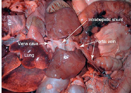
K9
Congenital intrahepatic shunt, persistent patent ductus venosus
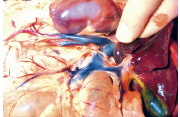
Congenital extrahepatic portocaval shunt in a dog
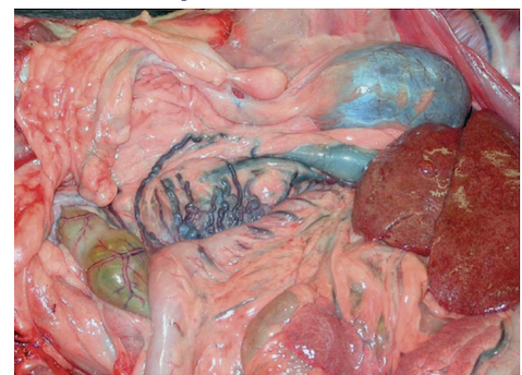
Multiple acquired portosystemic vascular shunts in a dog with chronic liver disease.