neurophysiology 2 and 3
1/60
There's no tags or description
Looks like no tags are added yet.
Name | Mastery | Learn | Test | Matching | Spaced |
|---|
No study sessions yet.
61 Terms
Olfaction
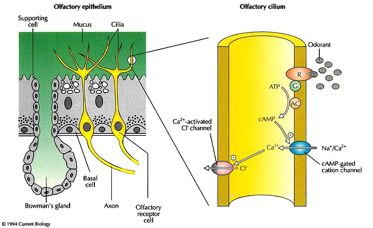
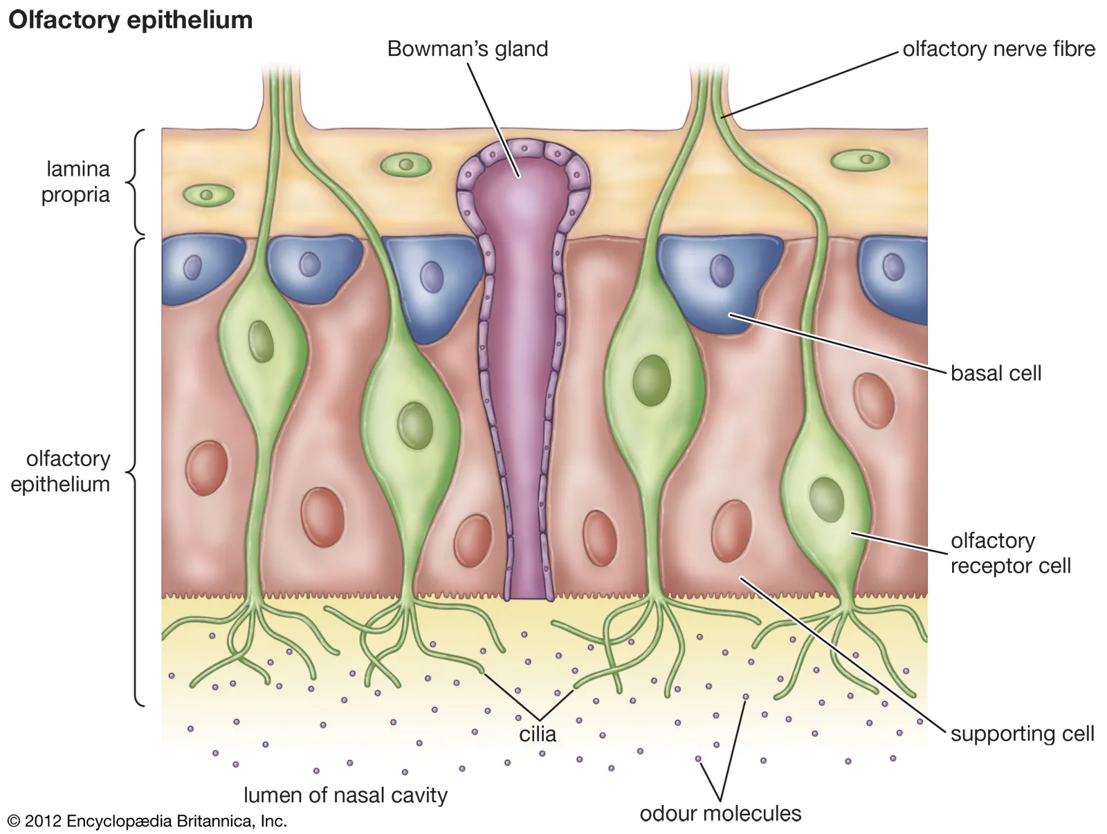
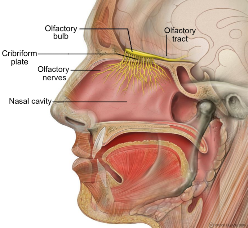
The sense of smell, which involves the detection of odorants in the air. This process begins with olfactory receptors in the nasal cavity and culminates in the perception of scent in the brain.
Olfactory Transduction
The process by which odorant molecules interact with smell receptors in the nasal cavity, leading to the generation of action potentials. These signals travel along the olfactory pathways to the brain, allowing us to perceive smells.
Olfactory Sensory Neurons
Specialized neurons located in the olfactory epithelium that possess cilia projections extending from their dendrites. These cilia are crucial for signal transduction, converting chemical signals (odorants) into electrical signals that the brain can interpret.
Olfactory Receptors
Proteins located on the olfactory cilia that bind to specific odorant molecules. This binding initiates the olfactory transduction process, leading to the perception of different smells.
Olfactory Signal Pathway
Unlike other sensory pathways, olfactory signals bypass the thalamus before reaching the cerebral cortex. This direct route may contribute to the strong emotional connections associated with smells.
Olfactory Axon Paths
Incoming olfactory axons follow two main paths: 1. Temporal lobe → Olfactory cortex (for conscious smell perception). 2. Limbic system (responsible for emotional responses to smells).
Gustation
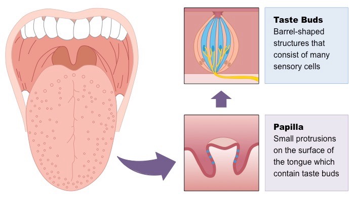
The sense of taste, which involves the detection of taste molecules in the mouth. Taste perception is a complex process involving taste receptors, taste buds, and neural pathways to the brain.
Taste Transduction
The process by which tastant molecules dissolve in saliva and are detected by taste buds, leading to the generation of action potentials. These signals travel along the gustatory nerve pathway to the brain, allowing us to perceive tastes.
Taste Receptor Cells
Specialized cells contained within taste buds that detect tastant molecules. These cells play a crucial role in initiating the taste transduction process.
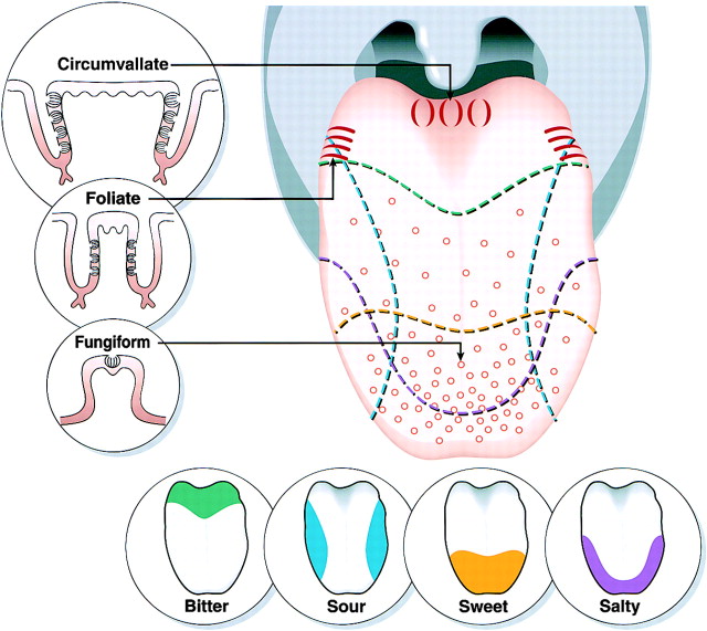
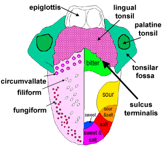
Lingual Papillae
Bumps on the tongue that increase surface area and contain taste buds. There are four types: filiform (provide texture), fungiform (contain taste buds), foliate (contain taste buds), and vallate (contain taste buds).
Taste Signal Pathway
Tastant molecules stimulate depolarization in gustatory epithelial cells, causing the release of neurotransmitters. These neurotransmitters trigger action potentials in first-order taste neurons.
Facial Nerve (VII)
This cranial nerve conducts taste signals from taste buds located in the anterior 2/3 of the tongue.
Glossopharyngeal Nerve (IX)
This cranial nerve conducts taste signals from taste buds located in the posterior 1/3 of the tongue.
Destinations of Taste Signals
Taste signals travel through nerves to the medulla oblongata, passing through the gustatory nucleus. From there, they go to: 1. Limbic system (emotional response to taste). 2. Hypothalamus (regulating appetite). 3. Thalamus to the insula (gustatory cortex, for conscious taste perception).
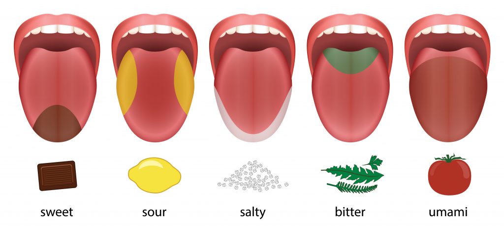
Five Primary Taste Categories
The five basic taste qualities that humans can perceive: sour, sweet, salty, bitter, and umami (savory).
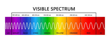
Visible Light Spectrum
The range of electromagnetic wavelengths (approximately 400-700 nanometers) that our eyes are capable of detecting. These wavelengths are perceived as different colors.
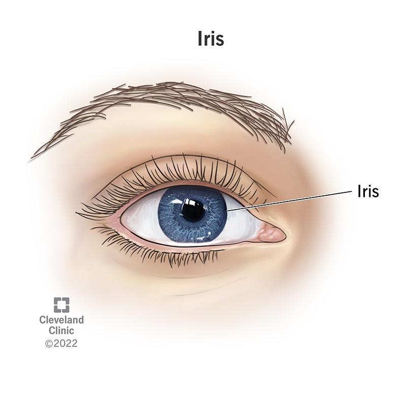
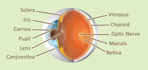
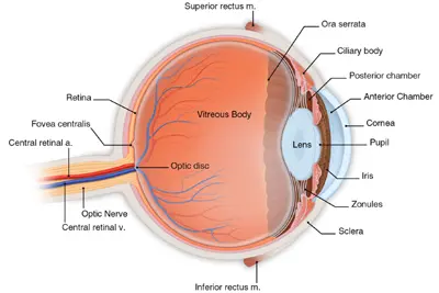
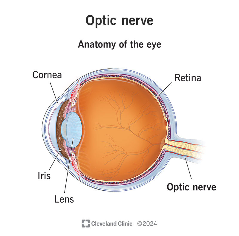
Iris
The colored, circular part of the eye, located behind the cornea. It controls the size of the pupil and, therefore, the amount of light that enters the eye.
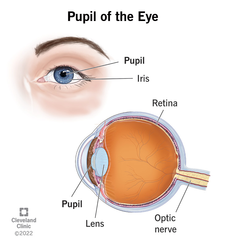
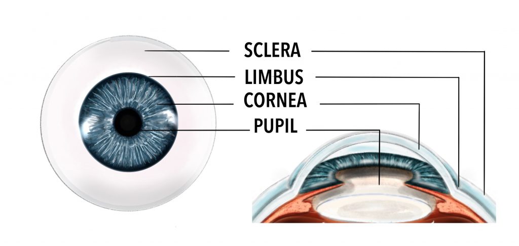
Pupil
The black circular opening in the center of the iris through which light enters the eye. Its size is adjusted by the iris to regulate light flow.
Cornea
The transparent, dome-shaped membrane that covers the iris and pupil. It is the eye's primary refractive surface, helping to focus light onto the retina.
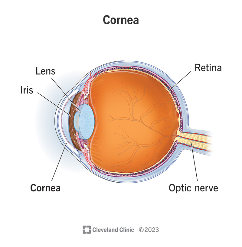
Sclera
The white, outer layer of the eyeball. It provides protection and structure to the eye.
Retina
The inner, light-sensitive layer of the eyeball. It contains photoreceptor cells (rods and cones) that convert light into electrical signals.
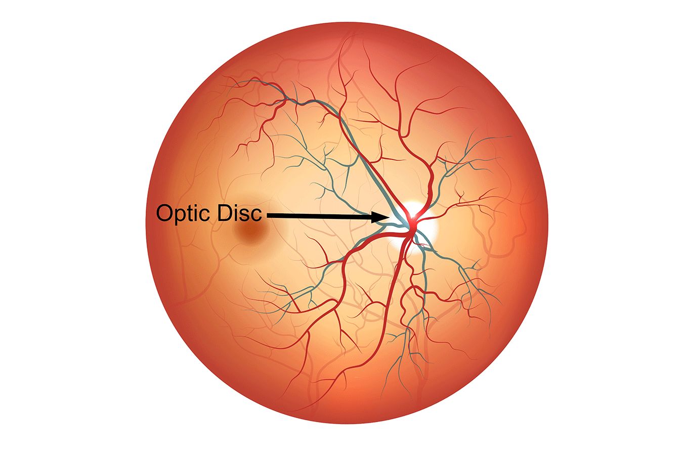
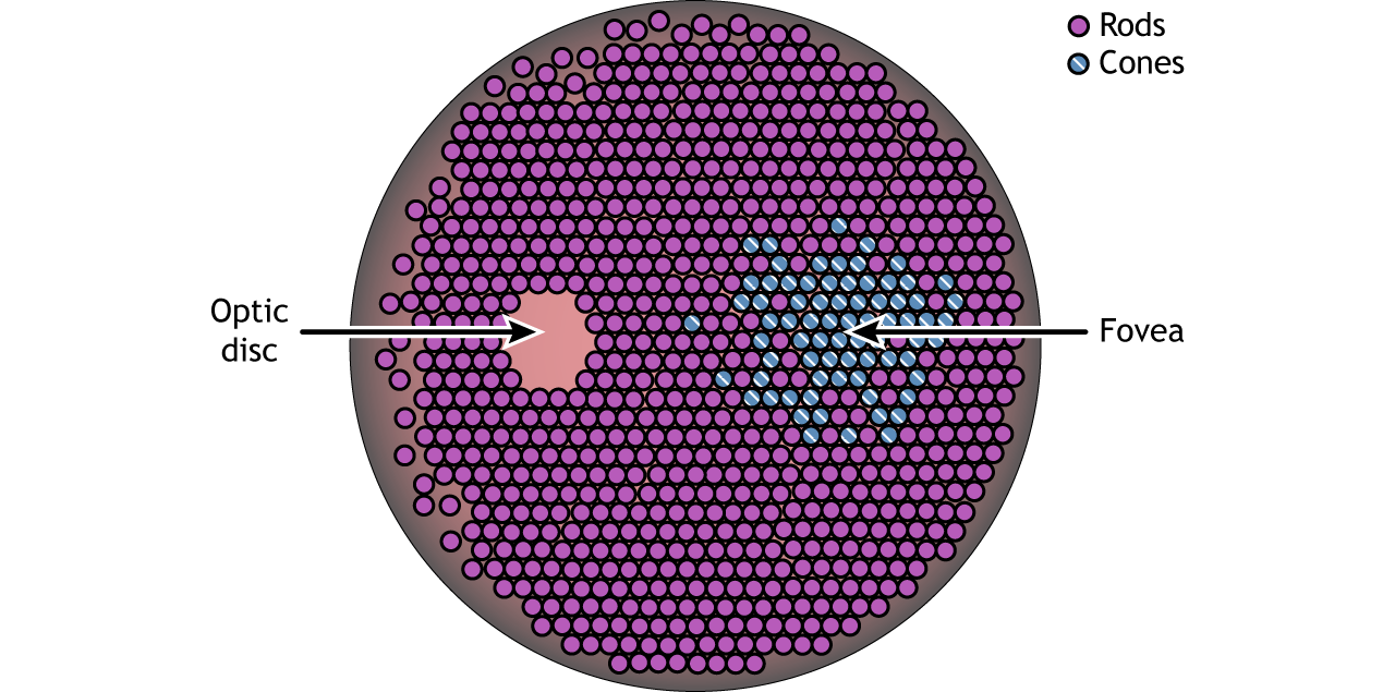
Optic Disc
The area on the retina where the optic nerve exits the eye. Because it lacks photoreceptors, it creates a blind spot in our visual field.
Macula
The central area of the retina, responsible for sharp, central vision. It is critical for activities such as reading and driving.
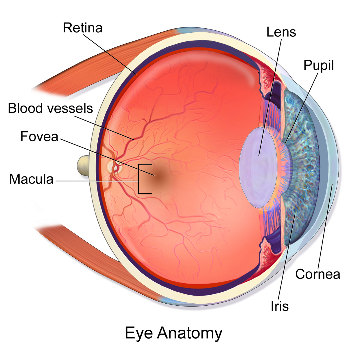
Fovea Centralis
A small depression in the center of the macula, containing the highest concentration of cones. It is the area of the retina with the greatest visual acuity (sharpness).
Pupil Function
The pupil's primary function is to regulate the amount of light entering the eye, allowing for optimal vision under varying light conditions. It constricts in bright light and dilates in dim light.
Layers of the Retina
The retina consists of three main layers of neurons: 1. Ganglion cell layer (outermost). 2. Bipolar cell layer (middle). 3. Photoreceptor cell layer (innermost).
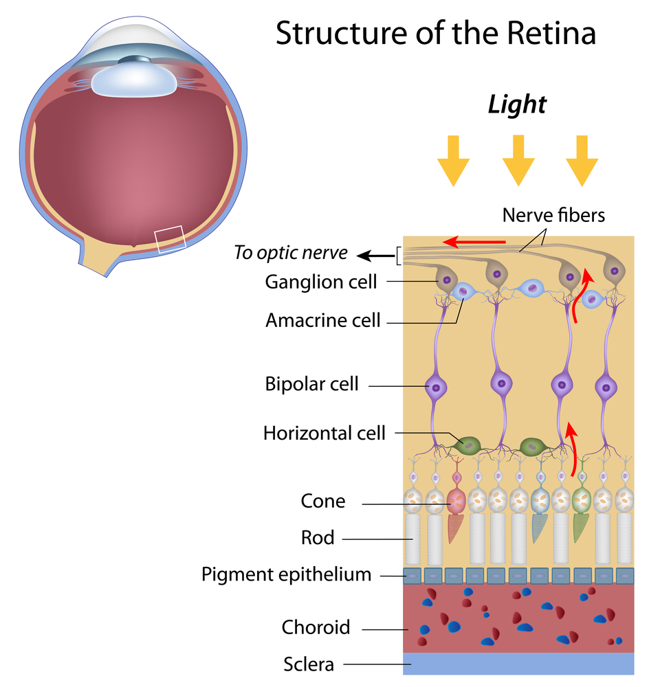
Ganglion Cell Layer
The outermost layer of retinal neurons, containing ganglion cells. Axons of these cells form the optic nerve, which carries visual information to the brain.
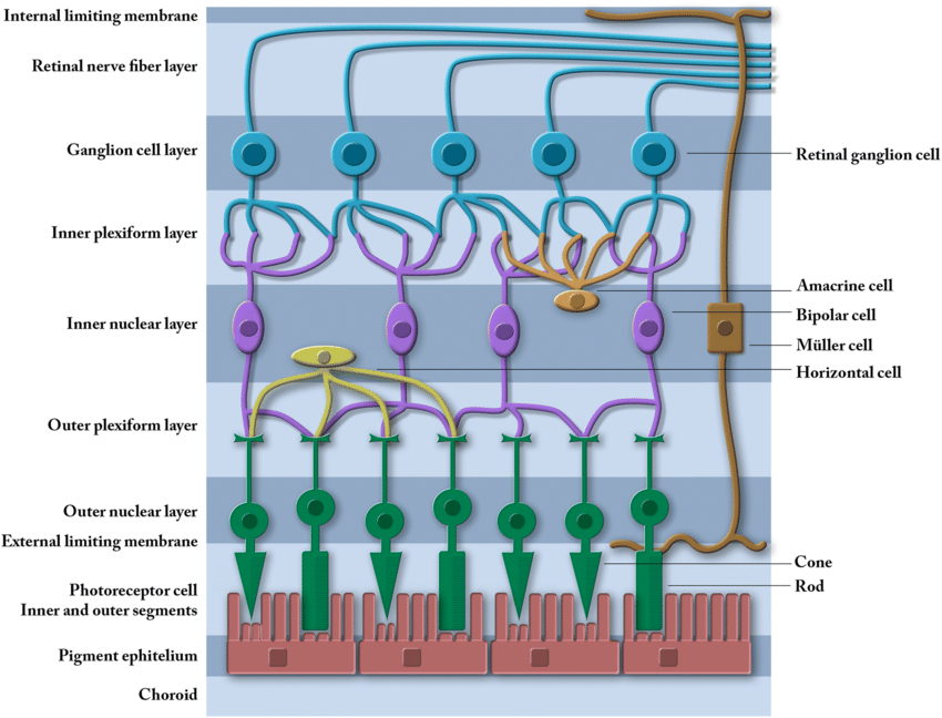
Bipolar Cell Layer
The middle layer of retinal neurons, containing bipolar cells, horizontal cells, and amacrine cells. These cells modify visual signals before they are passed on to ganglion cells.

Photoreceptor Cell Layer
The innermost layer of the retina, containing rods and cones. These photoreceptors transduce light into electrical signals that are processed by other retinal neurons.
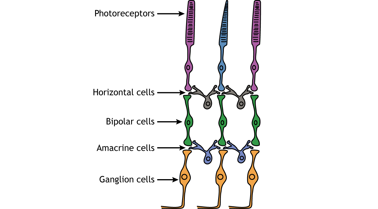
Photoreceptors
Specialized cells in the retina that transduce light rays into nerve signals. The two main types are rods (for dim light vision) and cones (for color vision).
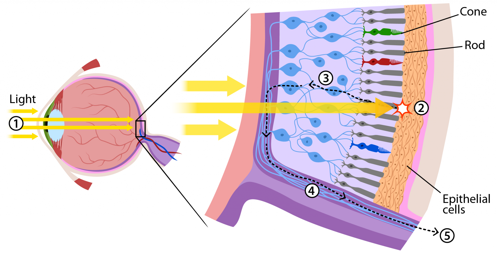
Rods
Photoreceptor cells in the retina that are highly sensitive to dim light. They are responsible for night vision and do not detect color.
Cones
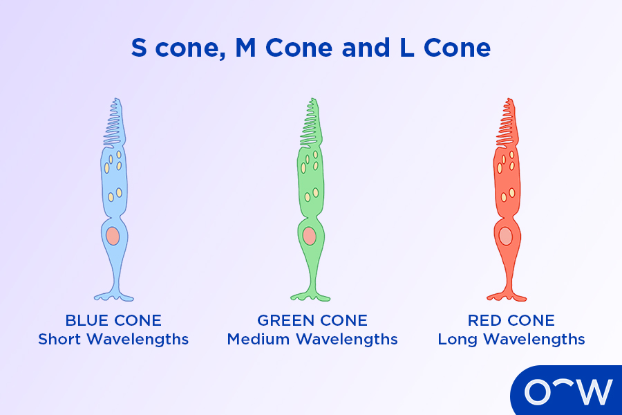
Photoreceptor cells in the retina that are sensitive to bright light and responsible for color vision. There are three types of cones, each sensitive to different wavelengths of light.
Types of Cones
There are three types of cones: blue cones (sensitive to short wavelengths), green cones (sensitive to medium wavelengths), and red cones (sensitive to long wavelengths). The combination of signals from these cones allows us to perceive a wide range of colors.
Fovea Centralis Composition
The fovea centralis is solely comprised of cones, which allows for high-acuity color vision in the center of our gaze.
Isomerization
The process where light interacts with photoreceptor molecules such as rhodopsin. Isomerization triggers an intracellular cascade within the photoreceptors, ultimately leading to a receptor potential and nerve signal.
Rhodopsin
A light-sensitive pigment found in the rods of the retina. It absorbs light and initiates the phototransduction process, enabling vision in low-light conditions.
Phototransduction
The process of converting light into electrical nerve signals in the photoreceptor cells of the retina. This process involves a cascade of biochemical reactions that ultimately lead to a change in the membrane potential of the photoreceptor.
Optic Nerve (II)
The cranial nerve that carries visual signals from the retina to the brain. It is formed by the axons of ganglion cells in the retina.
Optic Chiasm
The point in the brain where the optic nerves from each eye cross. This crossover allows information from the visual field to be processed by the opposite hemisphere of the brain.
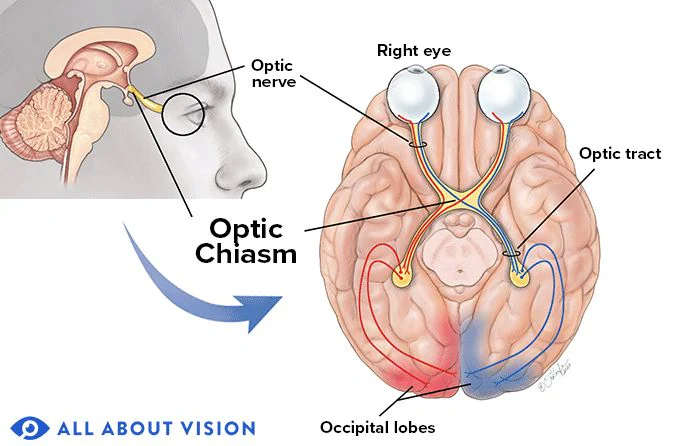
Lateral Geniculate Nucleus (LGN)
A part of the thalamus that relays visual signals from the optic nerve to the primary visual cortex in the occipital lobe.
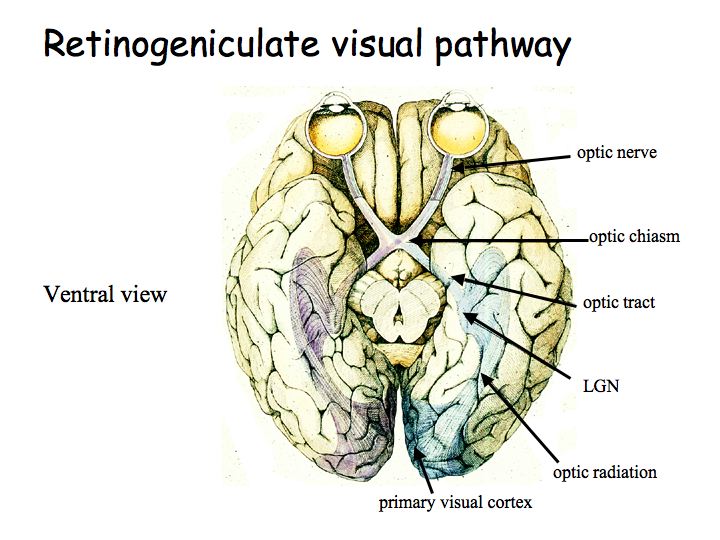
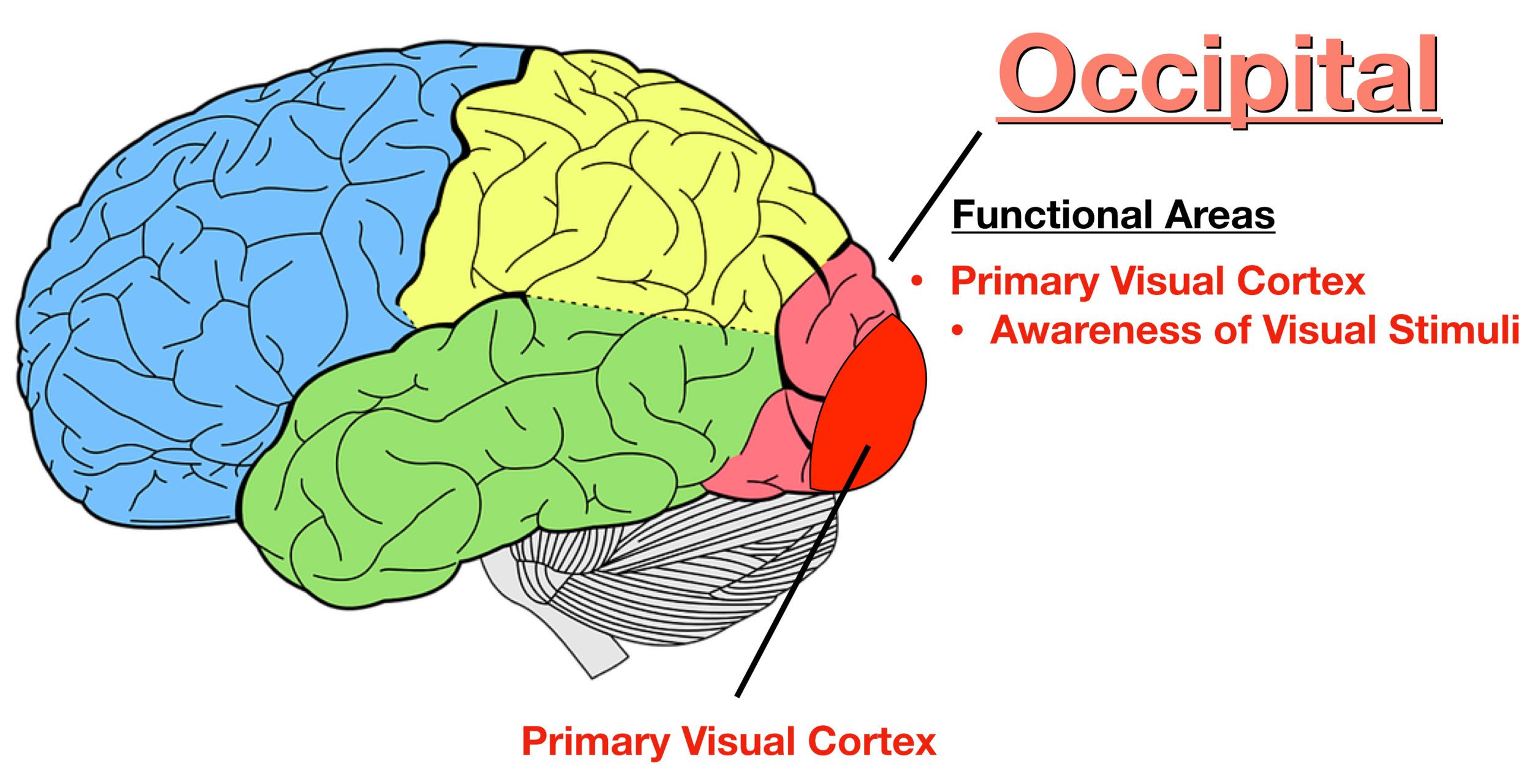
Primary Visual Cortex (V1)
Located in the occipital lobe, the primary visual cortex is where we consciously perceive vision. It processes basic visual features such as lines, edges, and colors.
Special Senses
Senses that have specialized organs devoted to them, such as hearing and equilibrium (balance). Unlike general senses (e.g., touch, temperature), special senses are localized to specific parts of the body.
Ear
The organ responsible for hearing and equilibrium. It converts sound/mechanical vibrations into electrical nerve signals that the brain can interpret as auditory information.
Equilibrium
Our sense of balance, which allows us to maintain our posture and orientation in space. It is mediated by the vestibular system in the inner ear.
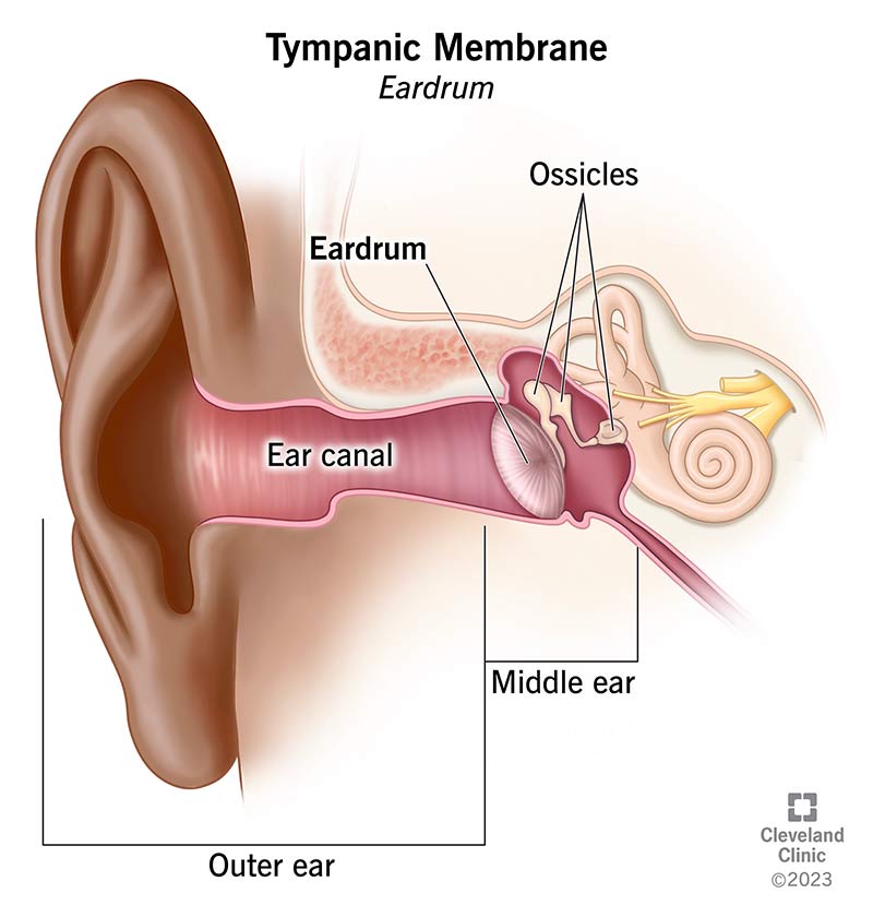
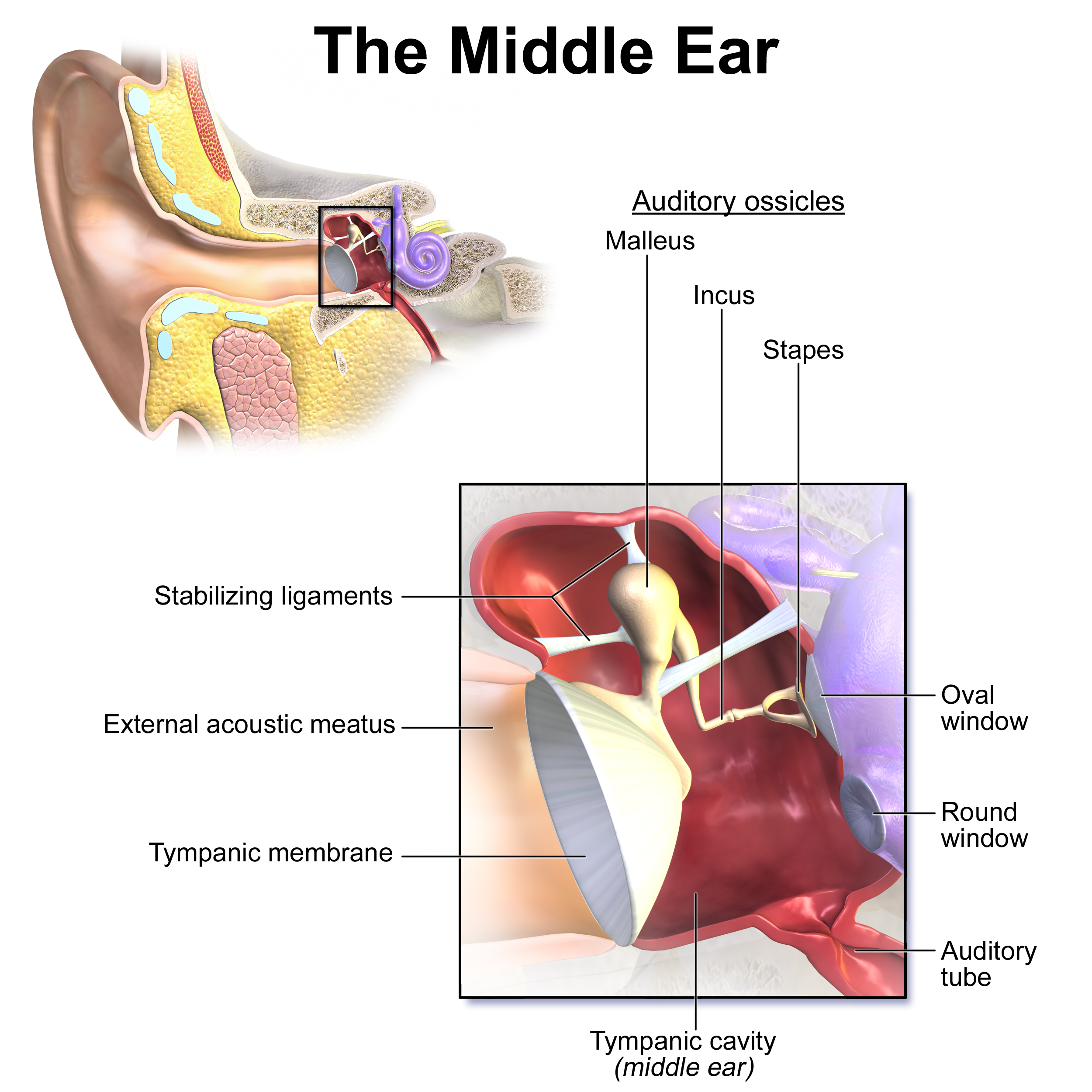
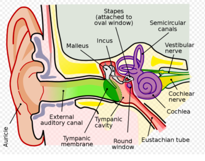
Ear Drum
Also known as the tympanic membrane, it separates the external ear from the middle ear. It vibrates in response to sound waves, transmitting these vibrations to the auditory ossicles.
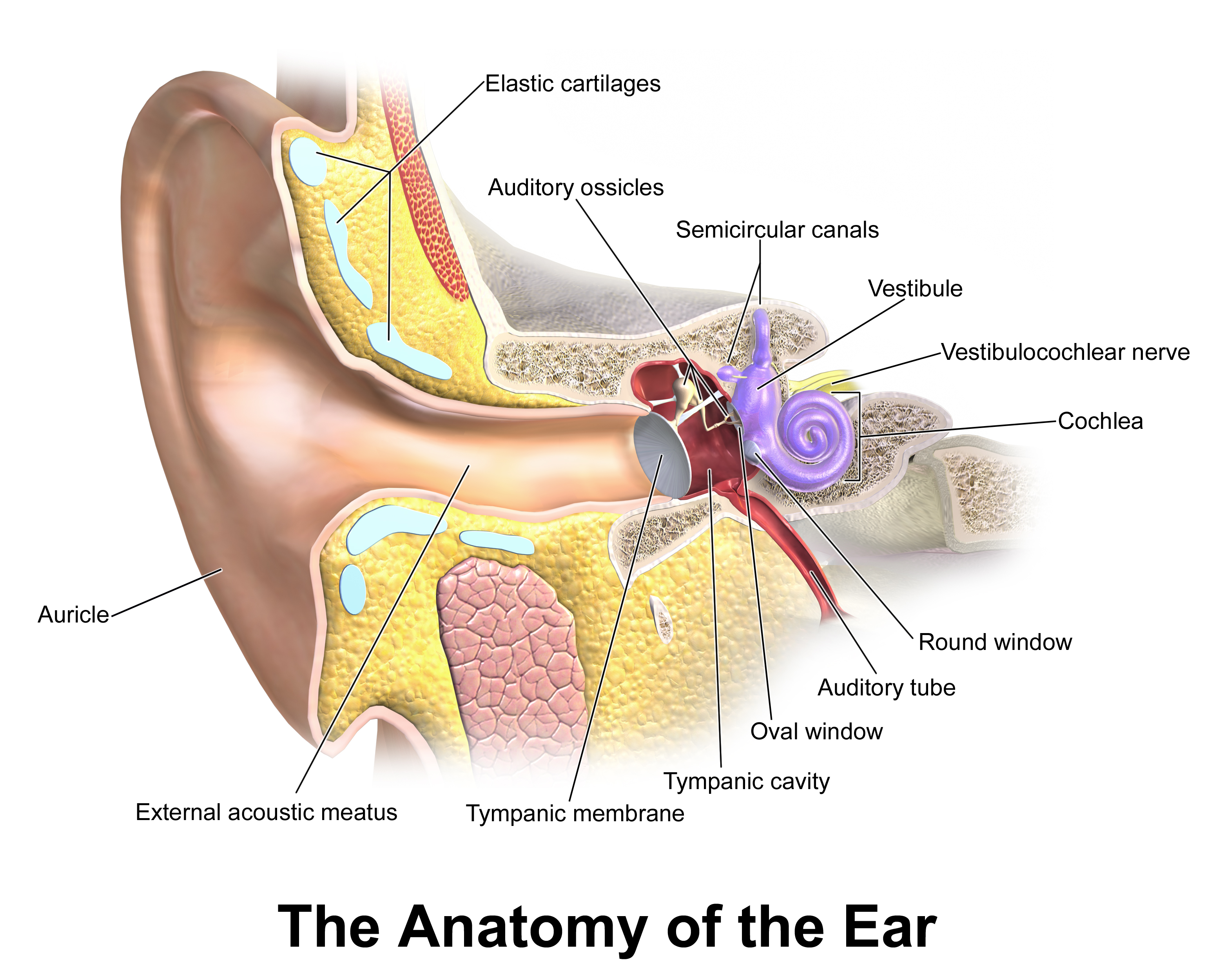
Middle Ear
An air-filled cavity within the temporal bone that contains the three auditory ossicles: malleus (hammer), incus (anvil), and stapes (stirrup). These bones amplify and transmit vibrations from the eardrum to the inner ear.
Vestibular and Cochlear Windows
Two openings where the middle ear meets the inner ear: the vestibular window (oval window) and the cochlear window (round window). The stapes transmits vibrations to the inner ear via the vestibular window.
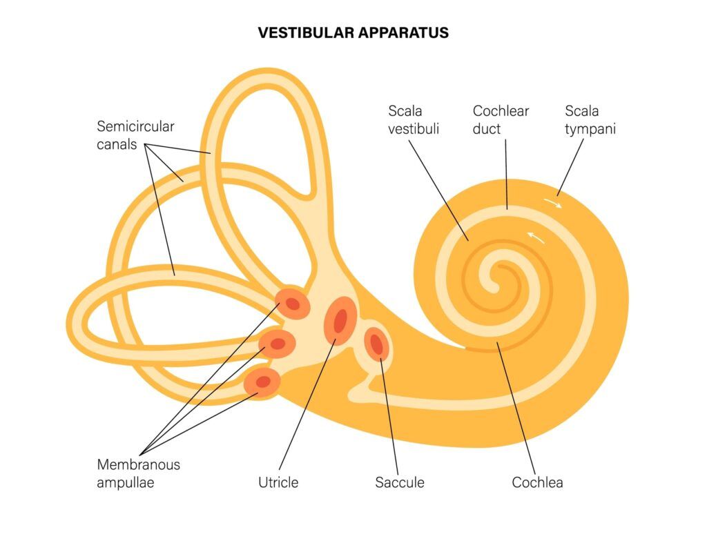
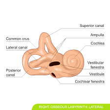
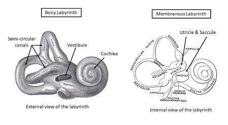
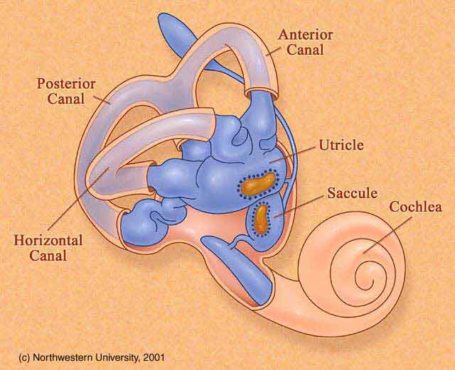
Bony Labyrinth
A series of connected cavities within the temporal bone that house the inner ear structures, including the cochlea, vestibule, and semicircular canals.
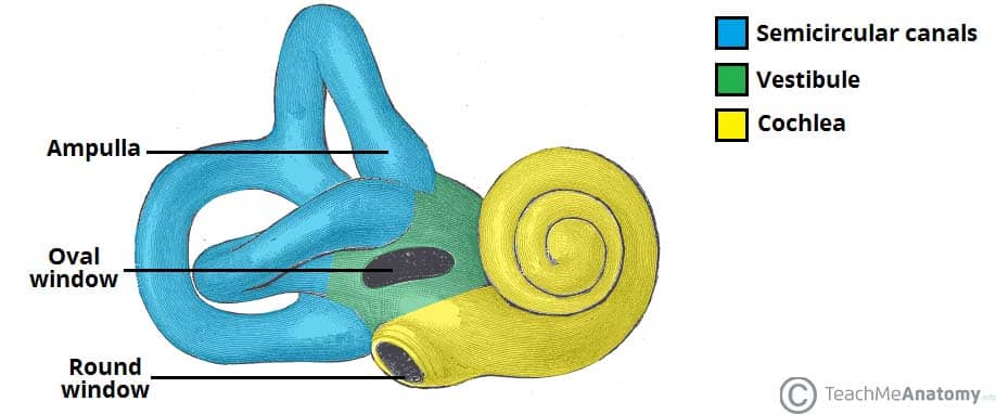
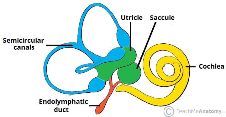
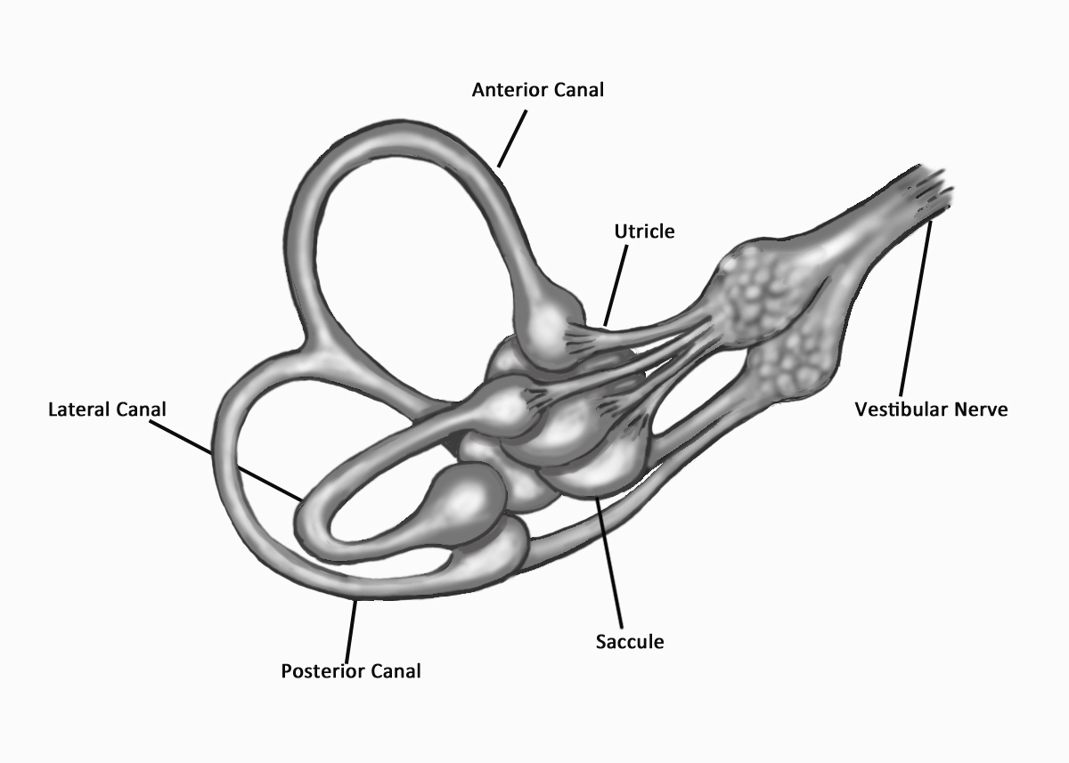
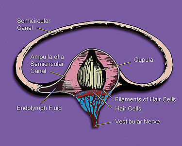
Semicircular Canals
Three fluid-filled canals in the inner ear that are responsible for detecting rotational movements of the head, contributing to our sense of equilibrium.

Vestibule (Inner Ear)
A central chamber of the bony labyrinth in the inner ear, which contains the utricle and saccule. These structures are responsible for detecting linear acceleration and head tilt, contributing to our sense of equilibrium.
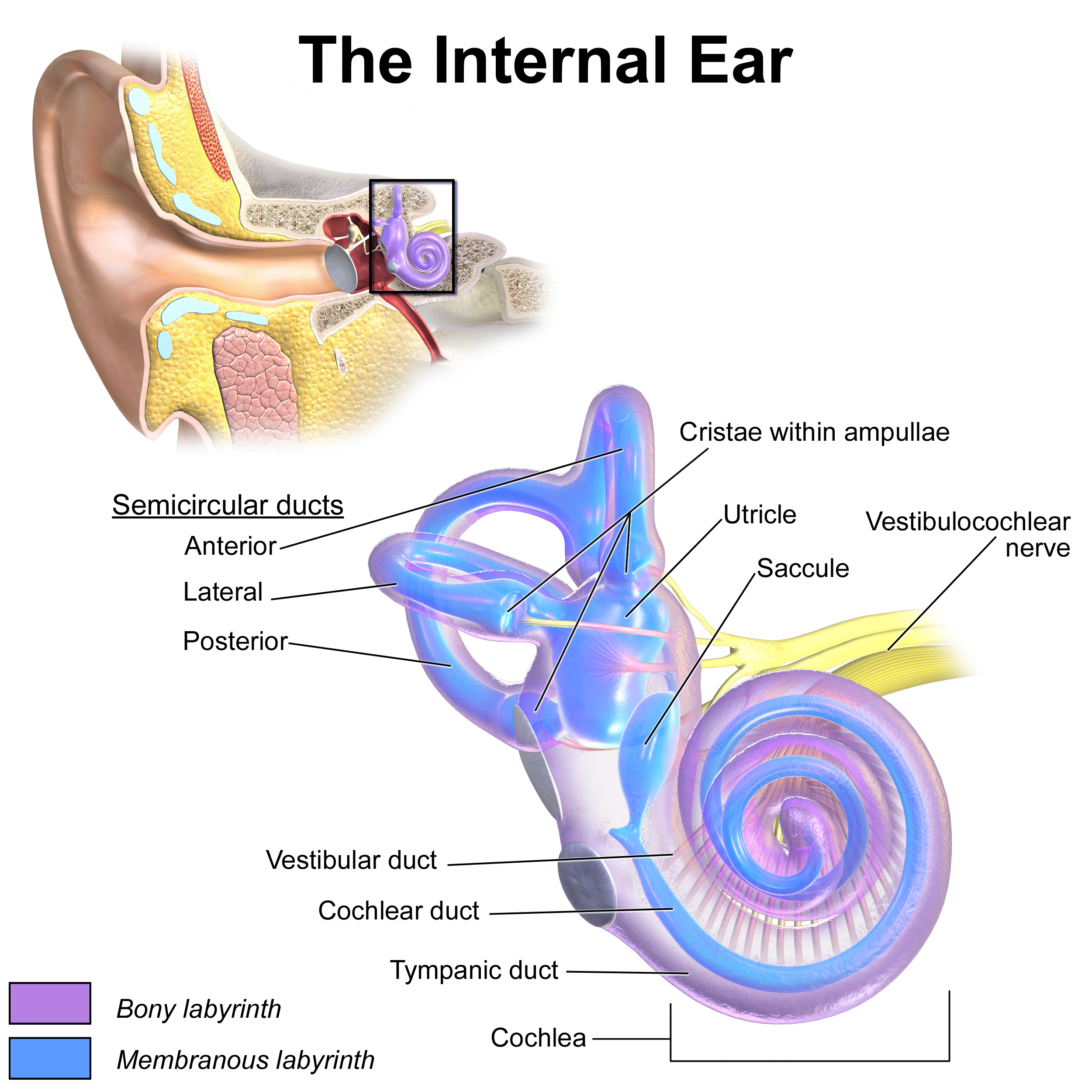
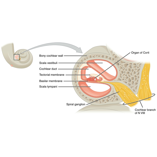
Cochlea
A spiral-shaped structure in the inner ear that contains the sensory receptors for hearing (hair cells). It converts mechanical vibrations into electrical signals that are sent to the brain.
Membranous Labyrinth
A system of interconnected sacs and ducts within the bony labyrinth, filled with endolymph fluid. It contains the sensory receptors for hearing and equilibrium.
Auditory and Equilibrium Receptors
Sensory receptor cells located within the membranous labyrinth that are responsible for detecting sound and changes in head position and movement.
Vestibular Branch (VIII)
The branch of the vestibulocochlear nerve (CN VIII) that carries signals related to equilibrium from the inner ear to the brain. It includes the ampullary, utricular, and saccular nerves.
Spiral Organ (Organ of Corti)
The sensory epithelium located within the cochlea that contains hair cells, which are the auditory receptors responsible for transducing sound vibrations into electrical signals.
Cochlear Branch (VIII)
The branch of the vestibulocochlear nerve (CN VIII) that carries signals related to hearing from the cochlea to the brain. It is formed by axons originating from the cochlea.
Stereocilia
Small, hair-like projections on the surface of hair cells in the inner ear. Bending of the stereocilia opens mechanically gated ion channels, initiating the process of auditory or equilibrium transduction.
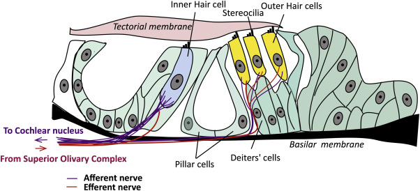
Potassium Ions (K+)
In the inner ear, the influx of K+ ions into hair cells (when stereocilia are bent) results in a depolarizing receptor potential. This depolarization triggers the release of neurotransmitters, leading to the generation of nerve signals.
Utricle and Saccule
Two otolith organs located in the vestibule of the inner ear. They contain crystals (otoliths) and a gel layer that sense linear acceleration and deceleration, as well as head tilt.
Gel Layer (Cupula)
A gelatinous structure located in the ampulla of the semicircular canals. Displacement of the cupula, due to fluid movement, is involved in sensing rotational acceleration and deceleration.
Equilibrium Signals Pathway
Equilibrium signals travel from the inner ear to the brainstem and cerebellum, which then relay the information to other brain areas. This allows us to consciously perceive body position and movements.