Help
 Call Kai
 Learn
Learn Practice Test
 Spaced Repetition
 Match
Match Flashcards
 Knowt Play
1/31

There's no tags or description
Looks like no tags are added yet.
Name | Mastery | Learn | Test | Matching | Spaced | Call with Kai | Chat |
|---|
No analytics yet
Send a link to your students to track their progress
32 Terms
Fibrous Pericardium
Tough outer CT layer, anchors the heart
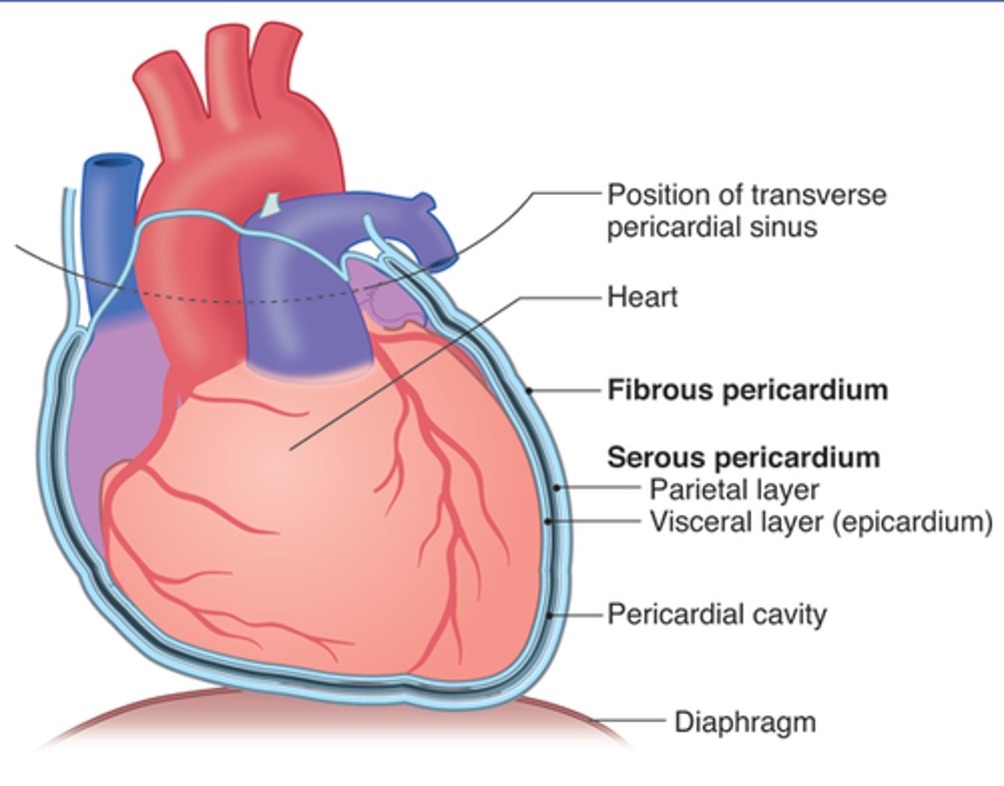
Parietal Serous Pericardium
Lines fibrous pericardium, contracts fibers (outer)
Pericardial Cavity
Space between visceral and parietal. Contains serous fluid
Visceral Serous Pericardium
Outer layer of the heart wall, contracts epicardium (inner)
Heart Layers (Outer -> Inner)
Epicardium
Outer layer of the heart wall. Comprised of meseothelial cells + CT.
Function: protection
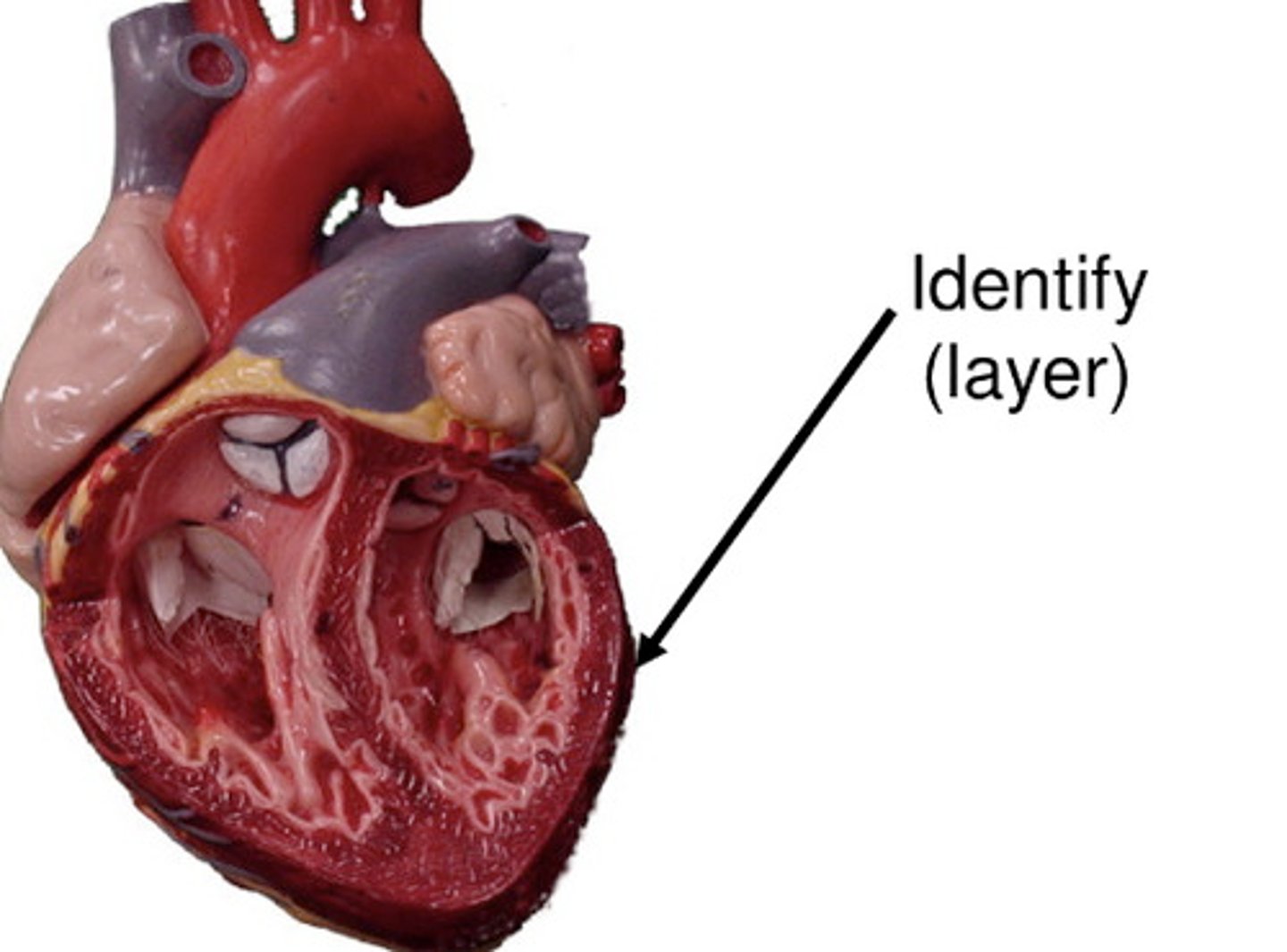
Myocardium
Thick cardiac muscle layer
Function: contraction
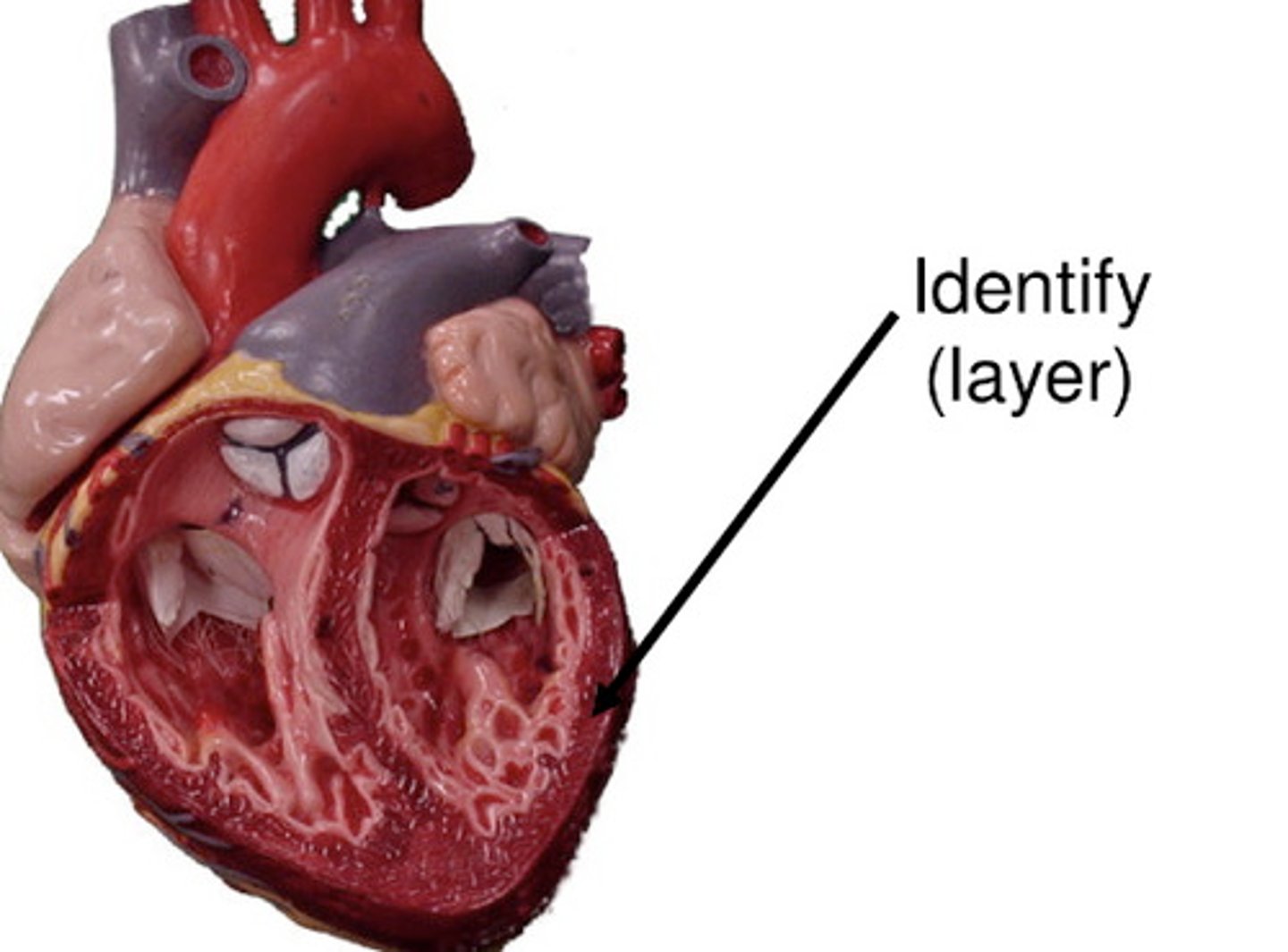
Endocardium
Inner endothelial lining. Comprised of Endothelium + CT
Function: Smooth blood flow
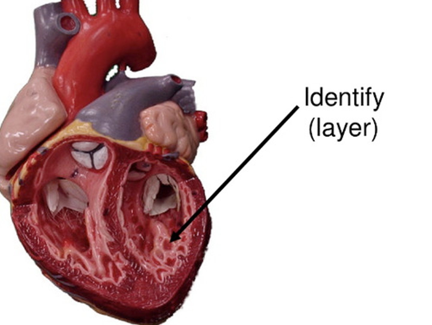
Subendocardium
Purkinje fibers; most ischemia-prone
Function: Electrical conduction
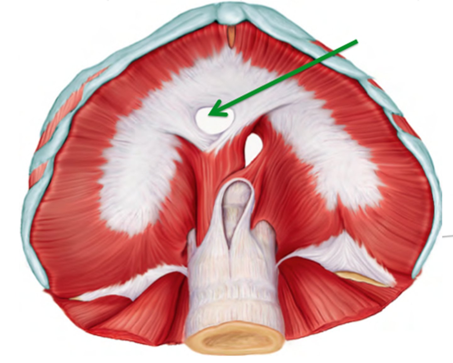
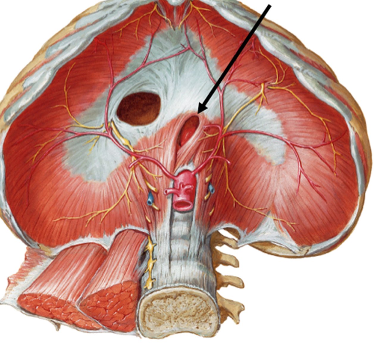
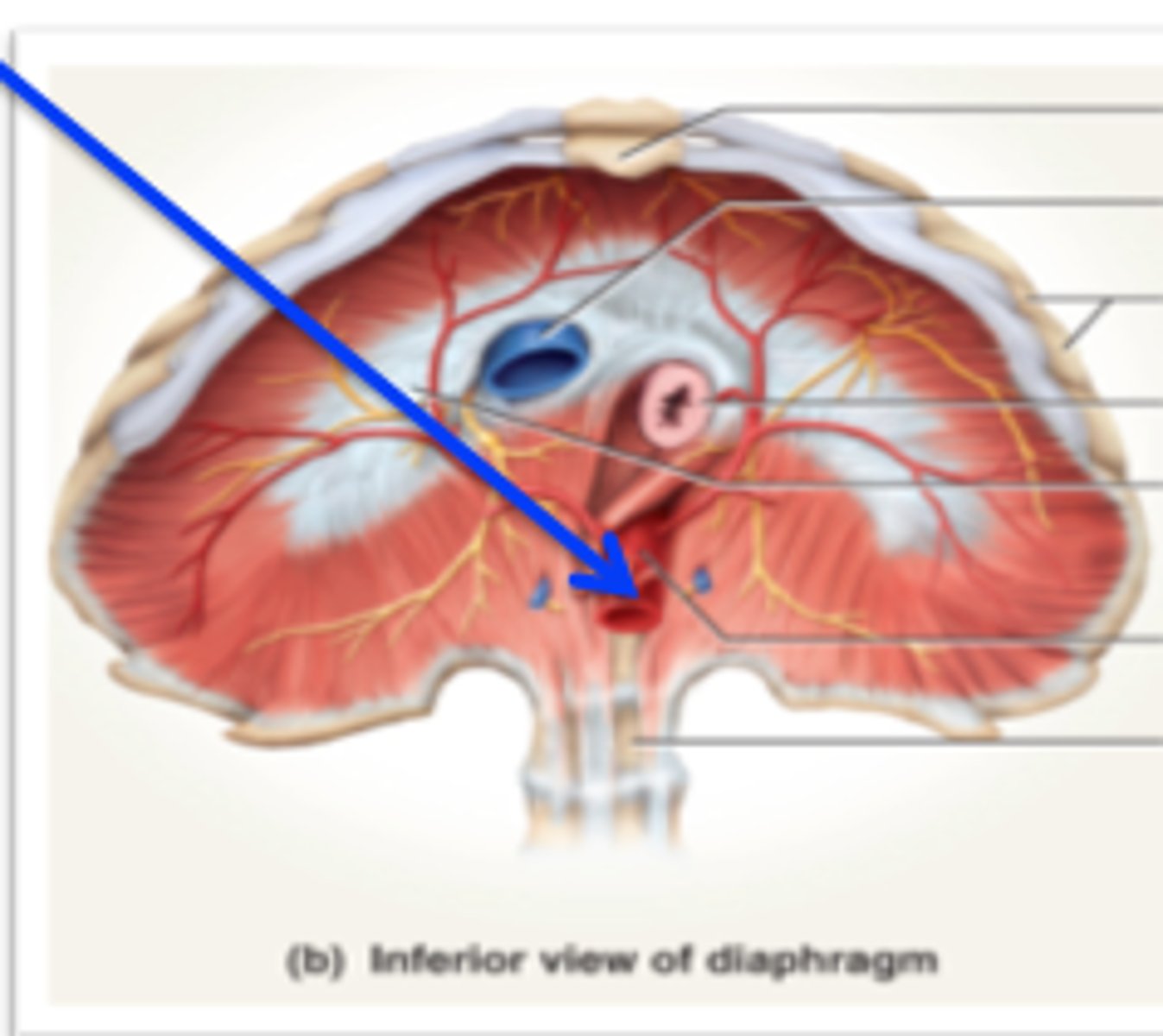
Diaphragm - openings, levels, structures
Caval Opening
Level : T8
Structures passing through: IVC, right phrenic nerve branches
Esophageal hiatus
Level : T10
Structures passing through: Esophagus, vagal trunks
Aortic hiatus
Level : T12
Structures passing through: Aorta, thoracic duct, azygos vein
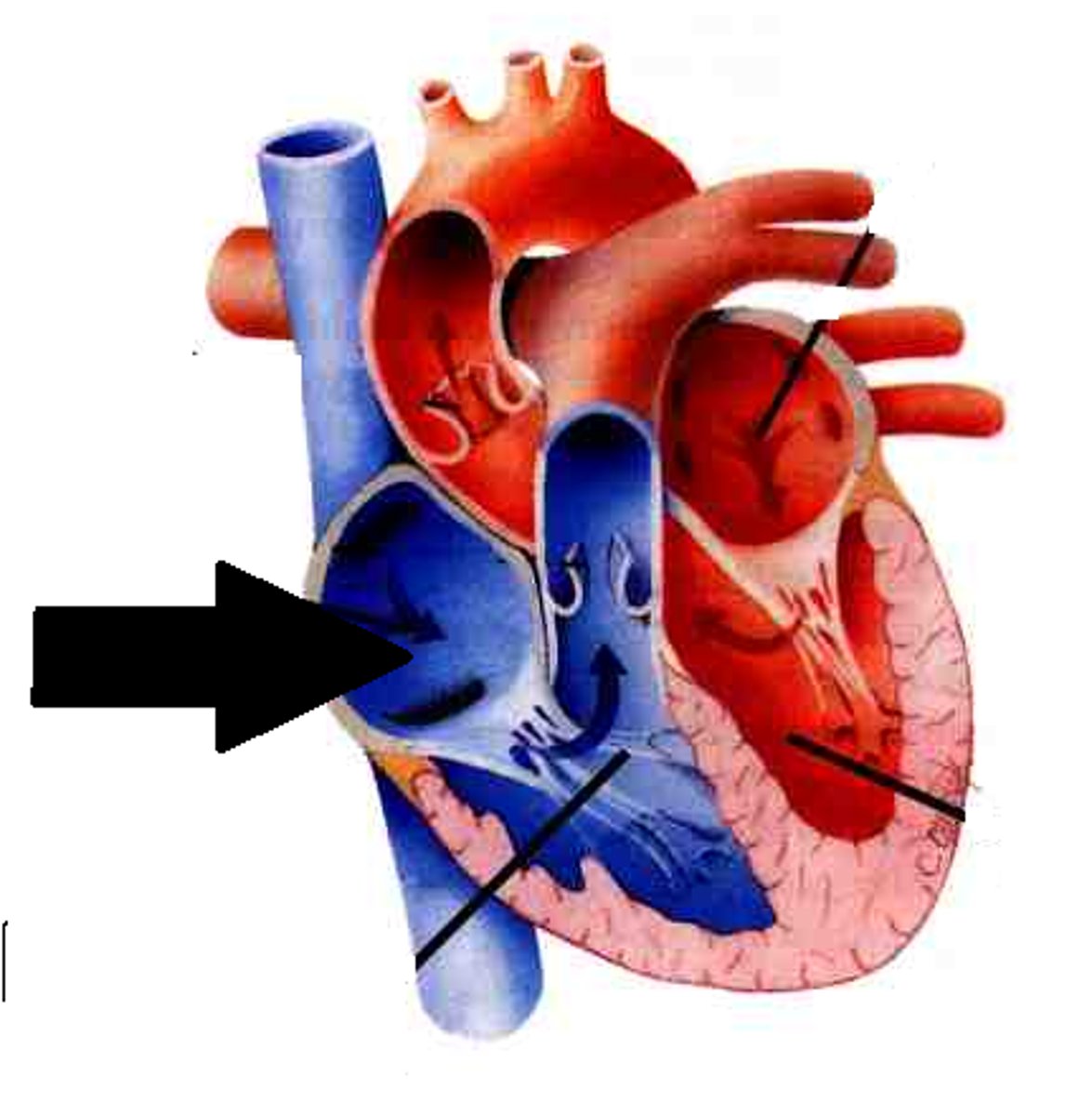
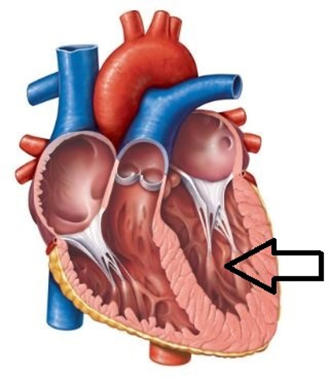
Heart Chambers and Their Structures
Right Atrium
SVC
IVC
Coronary sinus
Fossa ovalis
SA node
AV node
Pectinate muscles
Crista terminalis
Right Ventricle
Tricuspid valve
Chordae tendineae
Papillary muscles
Moderator band
Trabeculae carneae
Pulmonary valve
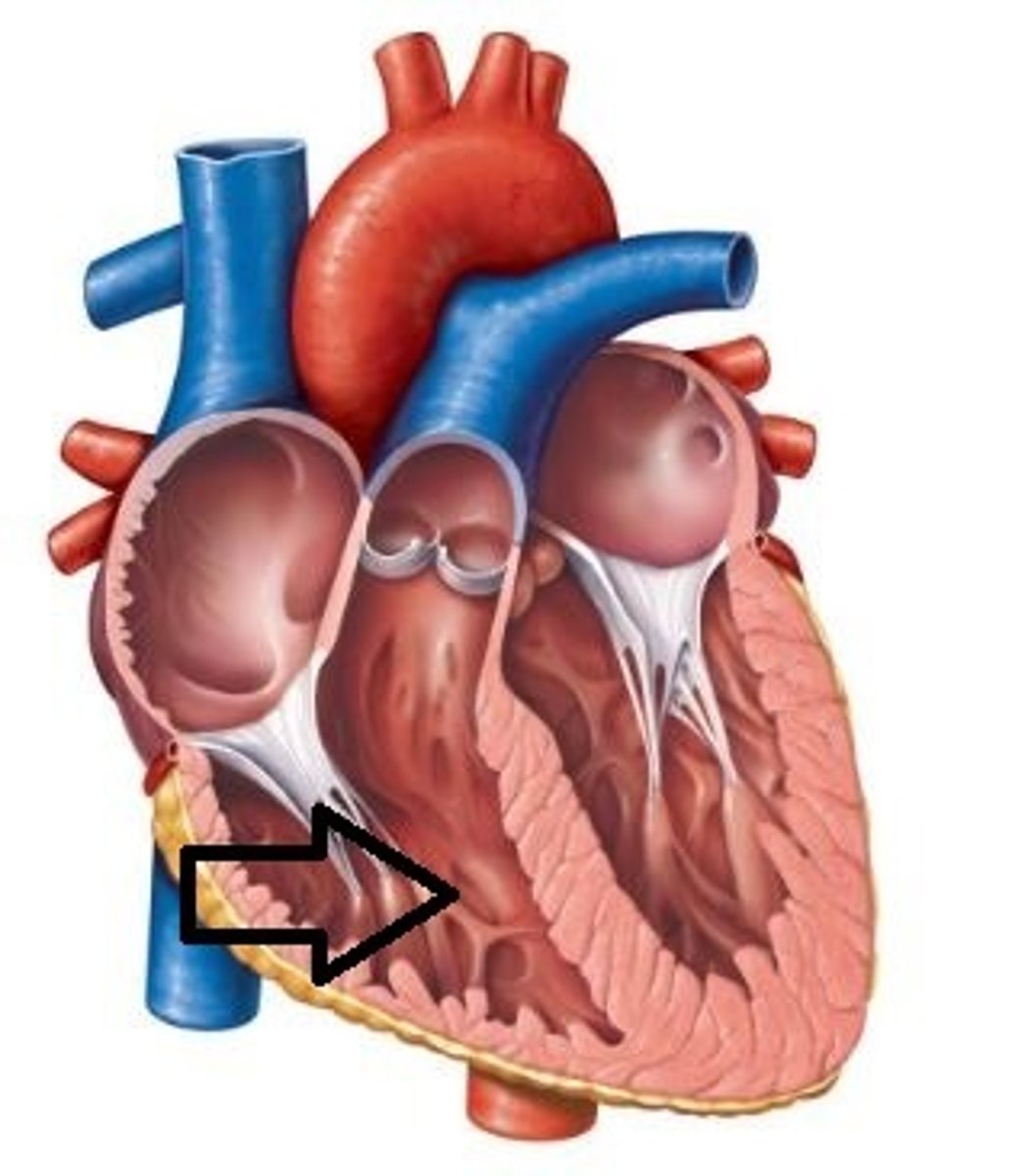
Left Atrium
4 pulmonary veins
Left atrial appendage (AFib thrombus site)
Mitral valve orifice
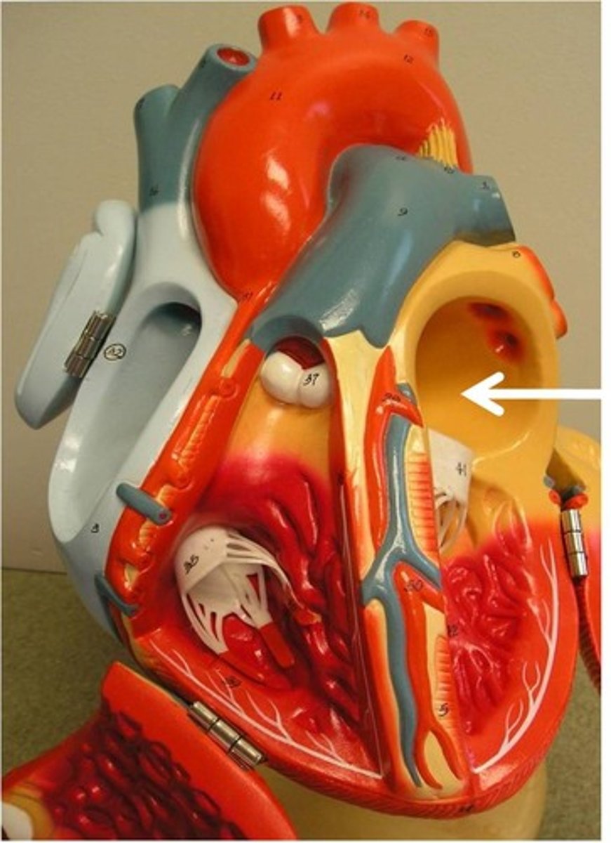
Left Ventricle
Thick myocardium
Papillary muscles
Chordae tendineae
Aortic valve
Aortic sinuses (coronary origins)
Blood Flow Through the Heart (In->Through-> Out)
1. SVC / IVC / Coronary sinus
2. Right atrium
3. Tricuspid valve
4. Right ventricle
5. Pulmonary valve
6. Pulmonary trunk
7. Pulmonary arteries
8. Lungs
9. Pulmonary veins
10. Left atrium
11. Mitral valve
12. Left ventricle
13. Aortic valve
14. Aorta → Body
Air Flow - Inhale
1. Nose/mouth
2. Pharynx
3. Larynx
4. Trachea
5. Main bronchi
6. Lobar bronchi
8. Segemntal bronchi
9. Terminal bronchioles
10. Respiratory bronchioles
11. Alveolar ducts
12. Alveoli
Exhale (Air Out - Reverse Order)
1. Alveoli
2. Ducts
3. Respiratory bronchioles
4. Terminal bronchioles
5. Bronchioles
6. Segmental bronchi
7. Lobar bronchi
8. Main bronchi
9. Trachea
10. Larynx
11. Pharynx
12. Nose/mouth
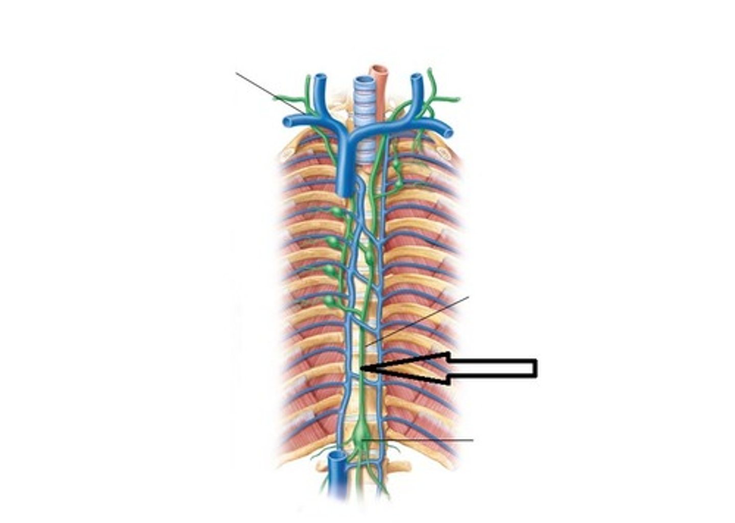
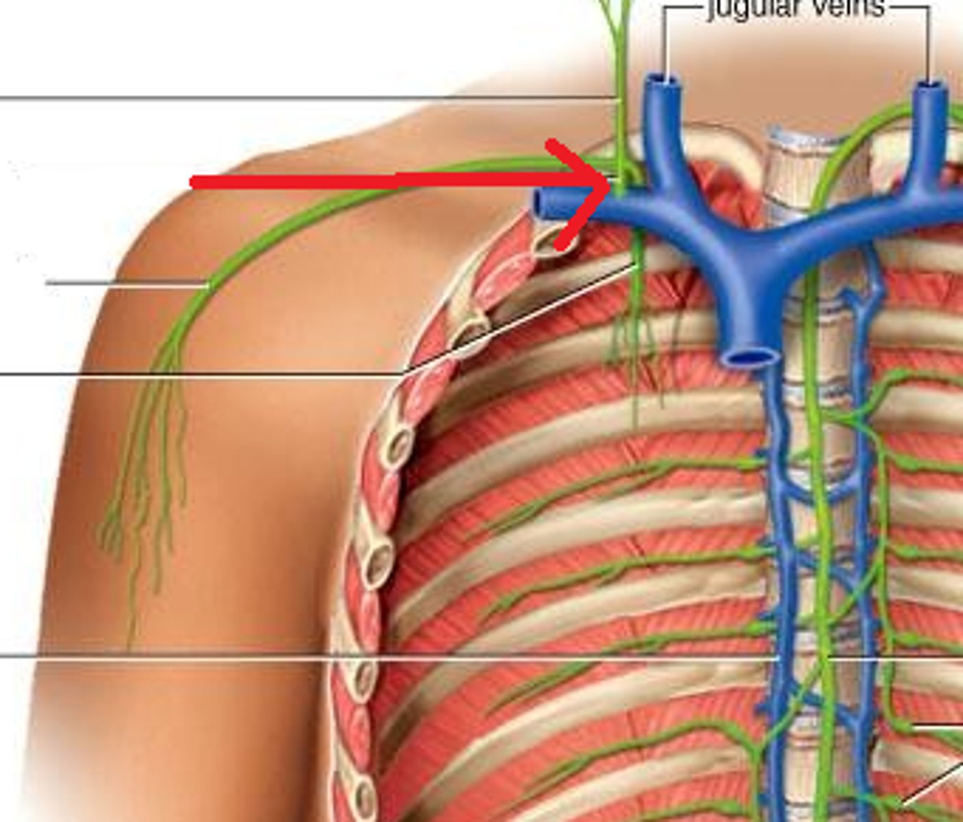
Major Lymphatic Ducts
Thoracic Duct
Drains entire body except right upper quadrant
Empties at left venous angle (left ij + left subclavian)
Passes through aortic hiatus (T12)
Most clinically important duct
Right Lymphatic Duct
Drains right upper quadrant only
Right head / neck
Right thorax
Right upper limb
Empties at right venous angle
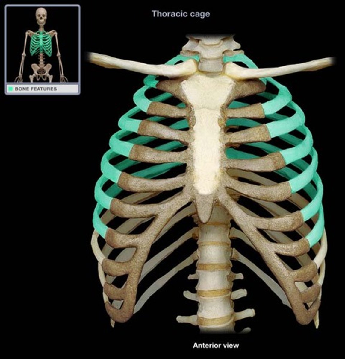
Rib Types
True ribs
Numbers: 1-7
Attachment: Directly to sternum
Clinical notes: Most mobile
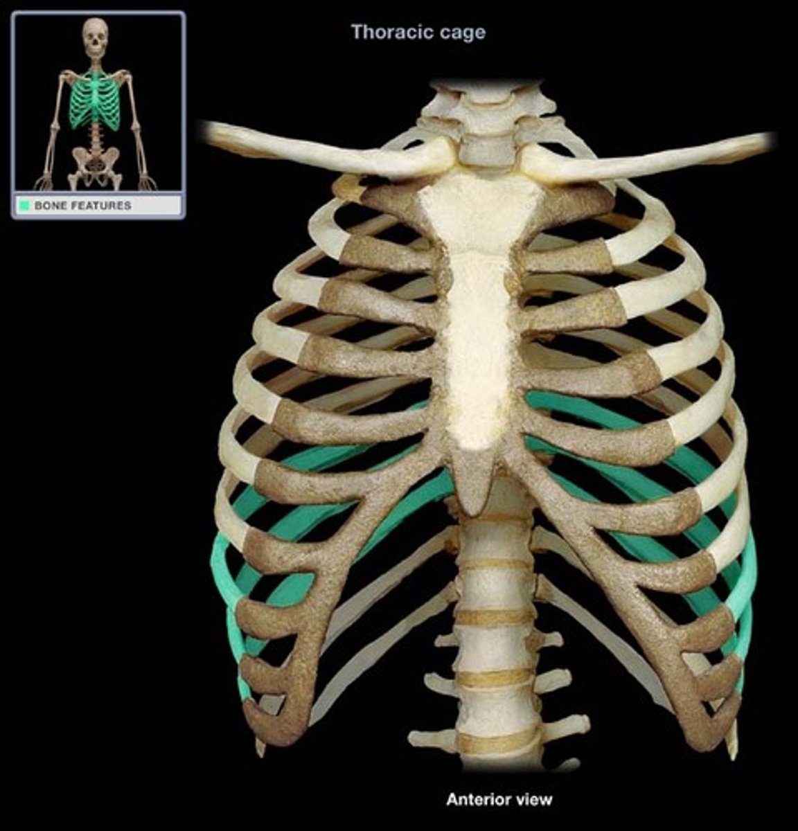
False ribs
Numbers: 8-10
Attachment: Indirect via costal cartilage
Clinical notes: Form costal margin
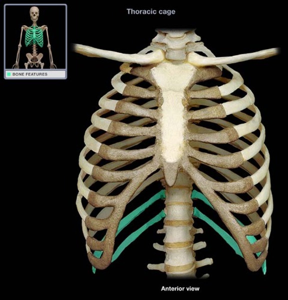
Floating ribs
Numbers: 11-12
Attachment: No anterior attachment
Clinical notes: Rib 12 -> kidney injury
Lung Structures, Fissures, Hilum, RALS
Right Lung
3 lobes: superior, middle, inferior
2 fissures: horizontal, oblique
Hilum: pulmonary artery anterior to bronchus (RALS)
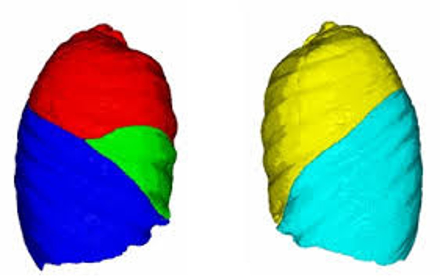
Left Lung
2 lobes: superior, inferior
1 fissure: oblique
Cardiac notch
Hilum: pulmonary artery superior to bronchus (RALS)
RALS Mnemonic
Right lung -> Artery is Anterior "RA"
Left lung -> Artery is Superior
"LS"