topic #11: sensory transduction lll
1/70
There's no tags or description
Looks like no tags are added yet.
Name | Mastery | Learn | Test | Matching | Spaced |
|---|
No study sessions yet.
71 Terms
what are rods
* scotopic vision
- non-color (achromatic) dim light vison; high light sensitivity
- photopigment = rhodopsin
- rods located outside fovea, used in peripheral vision
- slow responses, summate over 100 ms intervals , which helps in detection of small amounts of light
- low "flicker resolution" (12Hz) in humans (flicker solution is highest freq of light flicker that can be detected)
what are cones
* photopic vison
- in color (chromatic) day light vision; lower light - sensitive than rods
- 3 types of cone ; Short (S, blue) , medium (M, green), Long (L-red) wavelength sensitive cones
- fast responses
- high "flicker resolution " (55Hz) in humans
*3 types of cone photopigments (blue,green,red sensitive) all use same chromophore but have diff opsin proteins
what is chromosphere
chemical group capable of selectice light absorption producing color in compound
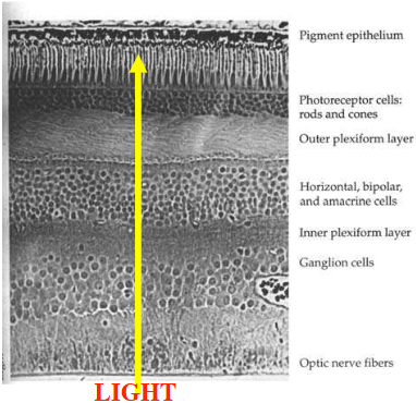
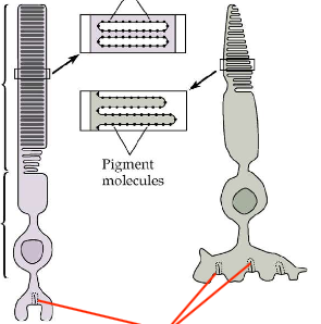
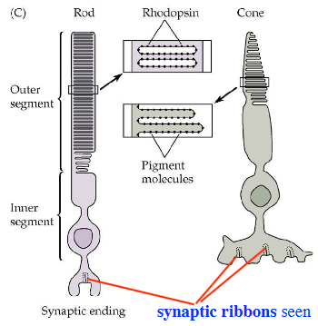
what is the structure of photoreceptors
- inner seg includes cell body
- outer seg, modified cilium where phototransduction takes place
- photopigments bound to folds of outer seg memb
in rod outer seg : memb disks are separate from outer memb (80% of disk protein = rhodopsin)
in cone outer seg : memb infoldings are continuous w/ outer menb
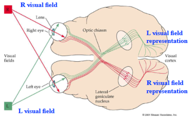
Draw the Visual System Pathways
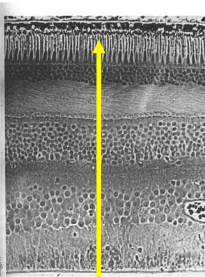
identify the section of a human retina: show the 5 principal cell types and the layers
Define Fovea
A small pit packed densely with cones, sharpest vision.
Blind Spot
The place where ganglion cell axons leave the eye to form the optic nerve, no rods or cones here.
Ganglion cell axons gather together → form the optic nerve → exit through the blind spot → no photoreceptors there, so you can’t see anything at that exact location.
what’s a 3rd type of photo-sensitive cell?
Intrinsically photosensitive ganglion cell
What is the function of Intrinsically photosensitive ganglion cell?
Contributes to entrainment of the circadian rhythm to the diurnal light cycle rather than to vision.
What is the molecular machinery associated with transmitter release adapted for?
Adapted for nearly constant output.
How are vesicles arranged for transmitter release in certain sensory cells?
Vesicles are tethered to “synaptic ribbons” like a conveyor belt.
In which cells are synaptic ribbons found?
Synaptic ribbons are seen in rods, cones, and bipolar cells (also in hair cells of the inner ear).
identify the structure
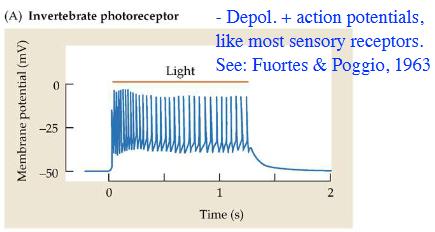
What type of electrical responses do vertebrate photoreceptors have to light?
Depolarized, + Action Potentials, like most sensory receptors.
What happens to a turtle rod when light is applied?
Turtle rod hyperpolarized by light.
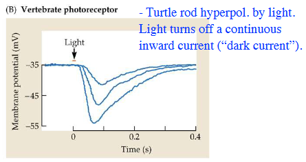
What does light do to the continuous inward current in photoreceptors?
Light turns off a continuous inward current (“dark current”).
What is the rod photopigment rhodopsin composed of?
Combination of retinal and opsin with retinal group sitting in middle of opsin, which has 7 transmembrane domains.
What is retinal a derivative of?
Retinal is a derivative of vitamin A (“Eat your carrots, or you’ll go blind!”)
Where is rhodopsin located in rods, and how abundant is it?
Rhodopsin embedded in disk membrane of rod outer segment; approximately 80% of membrane protein is rhodopsin in mammalian rods = 10^8 molecules per rod.
What do cones contain instead of rhodopsin, and what is their function?
In cones, there is also a rhodopsin-like combination of retinal and opsin, but the opsins are different, conferring differing wavelength sensitivity (blue, green & red “photopsins”).
What does the bleaching of rhodopsin trigger?
Triggers Metarhodopsin II which activates 2nd messenger pathway.
what is the pathway that activates 2nd messenger and triggers Metarhodopsin II?
GT (transducin)
Why can't photoreceptors regenerate rhodopsin on their own?
Photoreceptors lack the trans-cis isomerase needed for regeneration of rhodopsin.
What does rhodopsin regeneration depend on?
Rhodopsin regeneration depends on interaction between photoRs and cells of the pigment epithelium.
What effect does light have on rhodopsin?
Bleaching of rhodopsin by light.
What are the spectral sensitivity of cones: Blue, Red, Green?
455 nm
625 nm
530 nm
How many types of cones do humans have for color vision?
Three types of cones (trichromatic vision).
How many types of cones do horses have for color vision?
Two types of cones (dichromatic vision).
What happens to color perception when the number of cone types goes from three to two?
There is an enormous reduction in the number of different colors seen.
In dichromatic animals like horses or dogs, which colors are harder to distinguish?
Reds and greens often look similar or blend into dull shades.
What does the “dog’s view” spectrum show compared to the human’s view?
The dog’s view shows much fewer colors—mainly shades of yellow and blue, lacking rich variety.
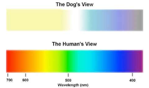
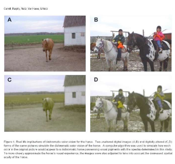
What is the purpose of the photographs A–D in the figure?
To demonstrate how the same scene looks different to trichromatic humans and dichromatic animals.
What is the photoreceptor Erest compared to most neurons?
Photoreceptor Erest less negative than in most neurons (only –30 to –40 mV).
What does light do to photoreceptors?
Light causes cells to hyperpolarized
What happens to membrane conductance (Gm) during the light response?
Gm decreases during light response, indicating that ion channels close.
What does hyperpolarization of a photoreceptor lead to?
Hyperpolarization of photoreceptor = decreased synaptic release = hyperpolarization of postsynaptic cells (bipolar & horizontal cells).
What causes the hyperpolarization of a photoreceptor?
Decrease in a depolarizing current that flows through the cell continuously at rest: “Dark Current”.
What is the other name mentioned for “Off-Center” cells?
“H-type” (“Off-Center”).
What ions contribute to the “dark current” in photoreceptors?
Na+ enters the outer segment and K+ leaves the inner segment. (See panel A in the figure.)
What happens to Na+ influx when light is present?
Light causes decreased Na+ influx, permitting the K+ efflux to hyperpolarize the cell. (See panel B in the figure.)
What is the result of hyperpolarization on neurotransmitter release?
In light, hyperpolarization leads to low release of glutamate onto bipolar cells.
What type of synapse continuously releases glutamate in the dark?
“Ribbon synapses” continuously release glutamate onto bipolar cells. (In dark: depolarized, high release.)
What protein mediates the decreased conductance potential caused by light?
A G-protein called transducin (also known as “GT”)
What is the sequence relationship between GT and gustducin?
GT has 80% sequence homology with gustducin in taste receptors.
In darkness, what is the electrical state of the photoreceptor and its release activity?
In the dark, the cell is depolarized and has high glutamate release.
In light, what is the electrical state of the photoreceptor and its release activity?
In the light, the cell is hyperpolarized and has low glutamate release.
What continously realease glutamate onto bipolar cells?
Ribbon Synapses
What makes Rhodopsin?
Opsin (Protein) & Retinal (Light-Sensitive molecule)
In how many structural forms does Retinal Exist?
2 Forms: 11-cis-retinal & All-trans-retinal
What did George Wald discovered in 1950s?
Light-induced isomerization of retinal
What are the steps in phototransduction?
light-mediated change in retinal causes a structural chnage in the protein (opsin) portion of rhodopsin. Rhodopsin → Meta-Rhodopsin.
Meta-Rhodopsin II acticates GT (transducin).
GT actiavtes PDE (Phosphodiesterase that decreases concentration of cGMP).
decreases [cGMP] causes cGMP-actiavted Na+ channels in outer segment to close.
Cell hyperpolarizes due to contonued K+ efflux from inner segment.
![<ol><li><p>light-mediated change in retinal causes a structural chnage in the protein (opsin) portion of rhodopsin. Rhodopsin <strong>→ Meta-Rhodopsin.</strong></p></li><li><p>Meta-Rhodopsin II acticates GT (transducin).</p></li><li><p>GT actiavtes PDE (Phosphodiesterase that decreases concentration of cGMP).</p></li><li><p>decreases [cGMP] causes cGMP-actiavted Na+ channels in outer segment to close.</p></li><li><p>Cell hyperpolarizes due to contonued K+ efflux from inner segment.</p></li></ol><p></p>](https://knowt-user-attachments.s3.amazonaws.com/db8f1f93-f9d1-4ef0-b7e2-a4eb356a0dd2.png)
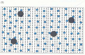
What are the ration in rod outer segment?
Rhodopsin= 1000 (black small dot)
Transducin = 100 (Blue medium dot)
PDE = 4 (Black Big dot)
What does Ca²⁺ inhibit in photoreceptor light adaptation?
Guanylate cyclase (GC)
What happens to intracellular Ca²⁺ ([Ca²⁺]ᵢ) when cGMP-gated Na⁺ channels close in light?
[Ca²⁺]ᵢ decreases
Why does [Ca²⁺]ᵢ decrease when cGMP-gated Na⁺ channels close?
Because Ca²⁺ is constantly removed by ion pumps and exchangers
What is the effect of decreased [Ca²⁺]ᵢ on guanylate cyclase (GC)?
GC is activated (relieved from Ca²⁺ inhibition)
What happens when GC is activated?
cGMP levels increase
What effect does increased cGMP have on the photoreceptor membrane?
Opens cGMP-gated Na⁺ channels
What ions pass through cGMP-gated channels?
Na+ & Ca2+
What is the purpose of Ca²⁺-mediated light adaptation?
To maintain photoreceptor responsiveness at different light levels
What is the summarized pathway of light adaptation in photoreceptors?
Light → cGMP-Na⁺ channels close → [Ca²⁺]ᵢ ↓ → GC activated → cGMP ↑ → cGMP-Na⁺ channels open.
What can rods respond to?
Rods can respond to single photons
What method was used for recording membrane currents of a rod outer segment?
Recordings were made by suction electrode from monkey rod outer segment.
What kind of light flashes were used to study quantal responses?
Very dim light flashes, corresponding to 1 or 2 photons each (red lines).
What did these dim flashes evoke in the recording traces?
Tiny “quantal” reductions in the inward dark current (upward deflections of blue traces).
What current reduction does transduction of a single photon evoke?
A 0.5 pA current reduction.
How many channels close in response to a single photon?
About 300 channels, or 3–5% of the rod channels that are open in the dark.
Where are Ribbon Synapses present?
In retinal Photoreceptordd and Bipolar cells
Characteristic of H Bipolar cells.
Off-Center
Depolarized by glut (iGLURs(
Characteristic of D Bipolar Cell
On center
Hyperpolarized by glut (mGLURs).