2093c First Lab Practical
 Call Kai
 Learn
Learn Practice Test
 Spaced Repetition
 Match
Match Flashcards
 Knowt Play
1/327

There's no tags or description
Looks like no tags are added yet.
Name | Mastery | Learn | Test | Matching | Spaced | Call with Kai | Chat |
|---|
No analytics yet
Send a link to your students to track their progress
328 Terms

Rat
Trachea

Rat
Trachea

Rat
Thymus

Rat
Thymus

Rat
Heart

Rat
Heart
Rat
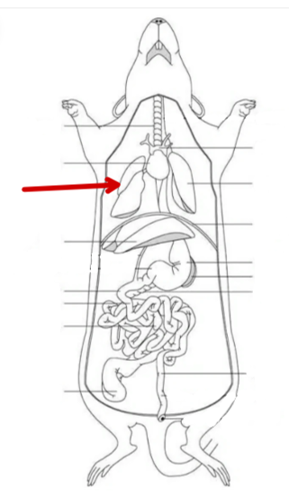
Lung

Rat
Lung

Rat
Diaphragm

Rat
Stomach
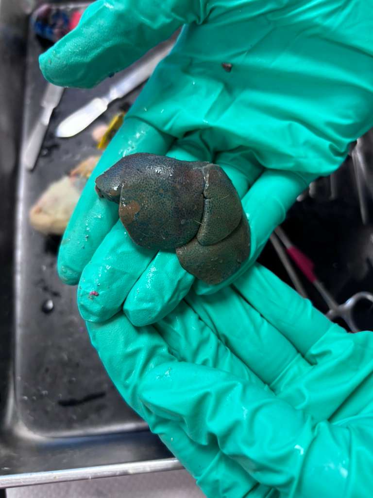
Rat
Liver
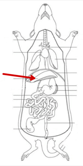
Rat
Liver

Rat
Spleen

Rat
Spleen

Rat
Pancreas

Rat
Pancreas

Rat
Duodenum
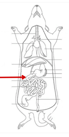
Rat
Duodenum

Rat
Jejunum

Rat
Jejunum

Rat
Ileum
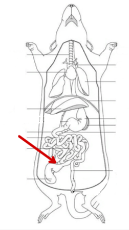
Rat
Illeum
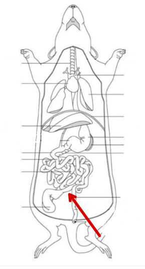
Rat
Ascending colon

Rat
Ascending colon

Rat
Transverse colon
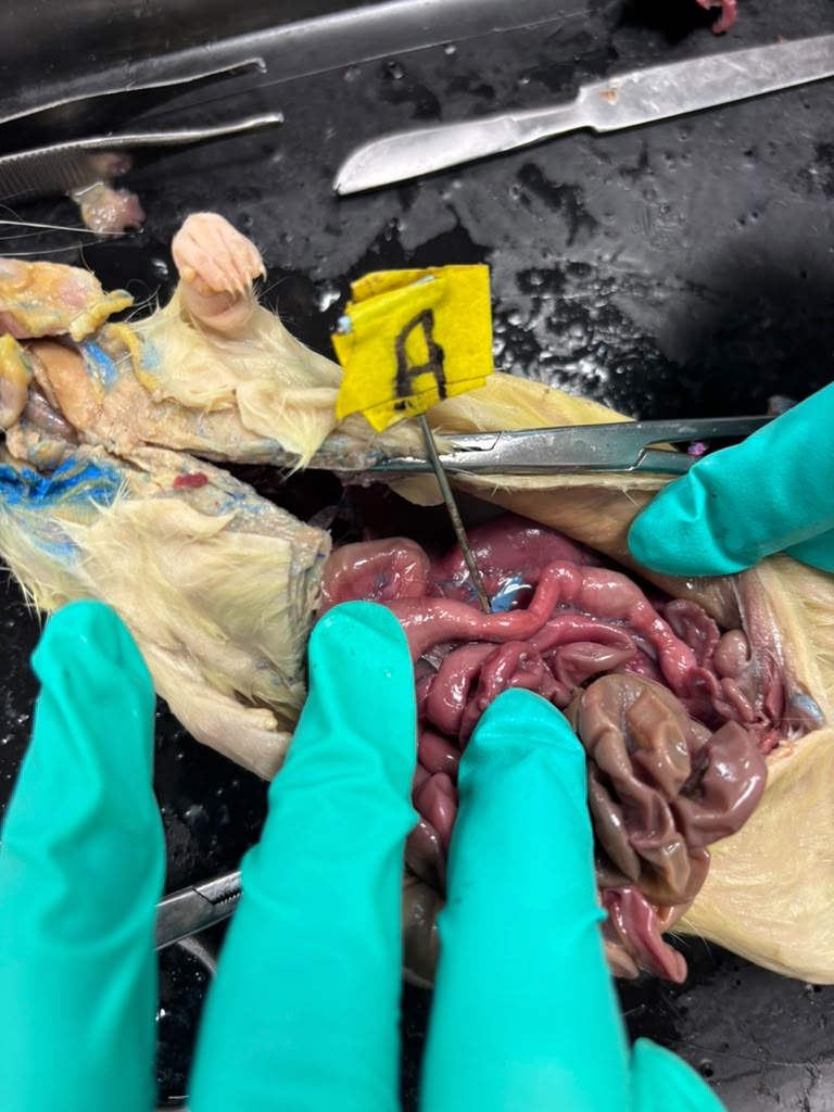
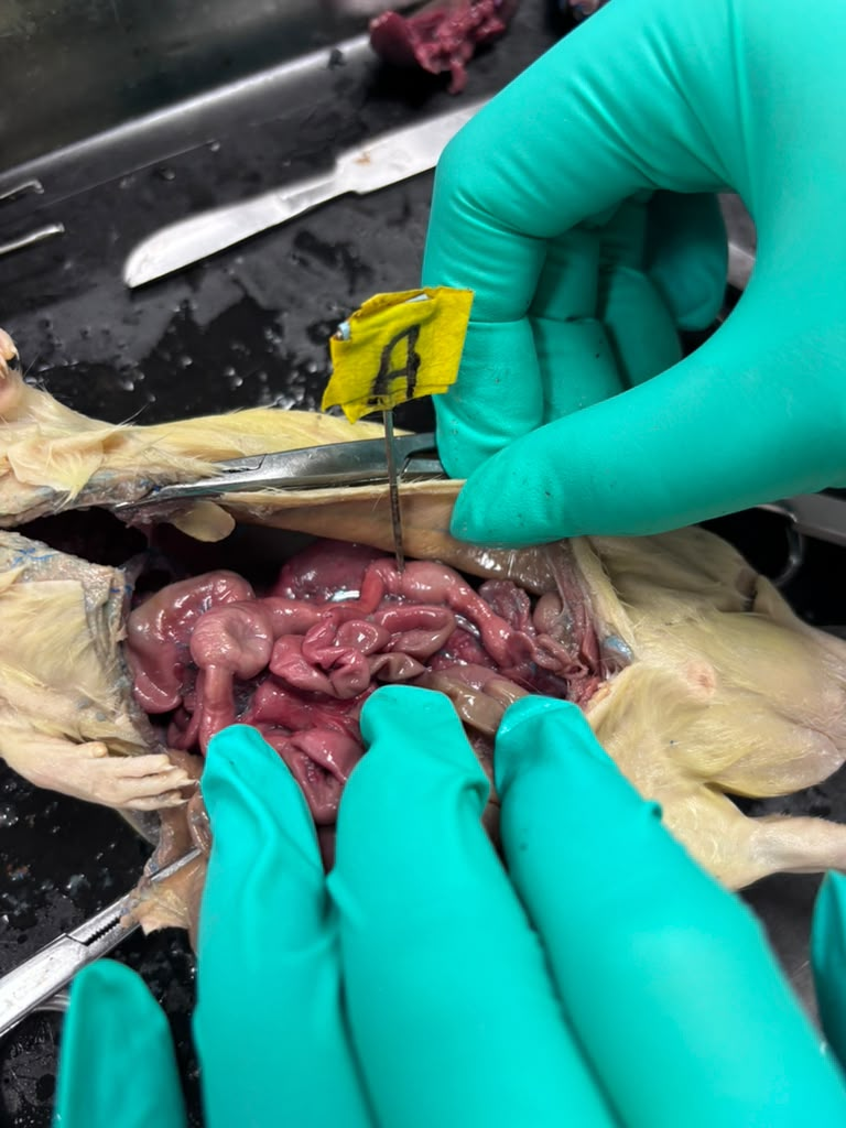
Rat
Transverse colon
Rat
Descending colon

Rat
Descending colon

Rat
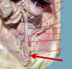
Cecum

Rat
Cecum
Rat
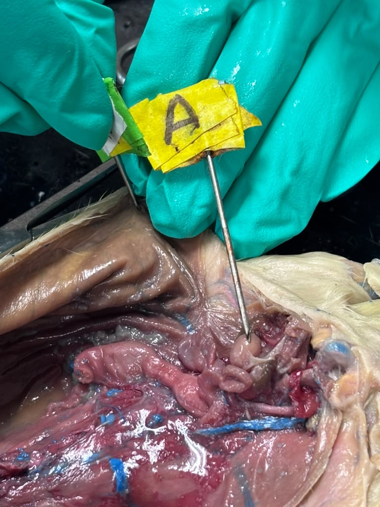
Urinary bladder
Rat
Urinary bladder
Rat
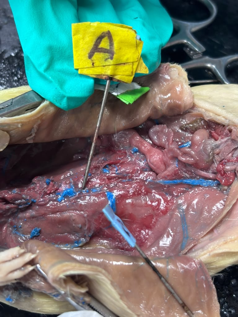
Ureter
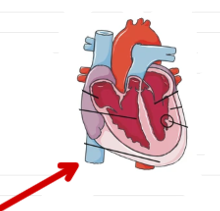
Rat
Inferior vena cava

Rat
Inferior vena cava

Rat
Descending aorta

Rat
Descending aorta

Rat
Kidney
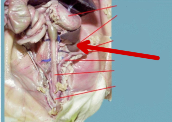
Rat
Kidney

Rat
Ovary

Rat
Uterus/uterine horns

Rat
Testis
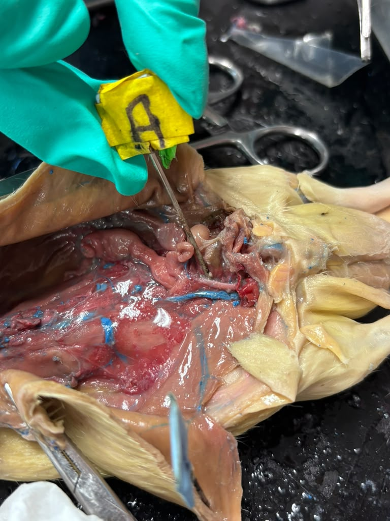
Rat
Vas (ductus) deferens

Rat
Adrenal Glands

Rat
Stomach

Rat
Diaphragm
Epithelial tissue
covers body surfaced or lines cavities
Simple squamous epithelium
Single layer of flattened cells with disc-shaped central nuclei and sparse cytoplasm; the simplest of the epithelia. Allows materials to pass by diffusion and filtration in sites where protection is not important; secretes lubricating substances in serosae. Located in kidney glomeruli; air sacs of lungs; lining of heart, blood vessels, and lymphatic vessels; lining of ventral body cavity (serosae).

Simple cuboidal epithelium
Single layer of cubelike cells with large, spherical central nuclei. Secretion and absorption. Located in Kidney tubules; ducts and secretory portions of small glands; ovary surface.
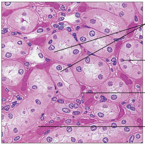
Simple columnar epithelium
Single layer of tall cells with round to oval nuclei; many cells bear microvilli, some bear cilia; layer may contain mucus-secreting unicellular glands (goblet cells). Absorption; secretion of mucus, enzymes, and other substances; ciliated type propels mucus (or reproductive cells) by ciliary action. Non-ciliated type lines most of the digestive tract (stomach to rectum), gallbladder, and excretory ducts of some glands; ciliated variety lines small bronchi, uterine tubes, and some regions of the uterus.

Pseudostratified columnar epithelium
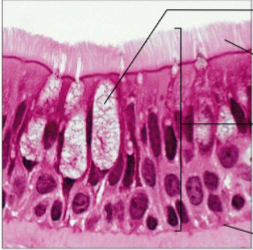
Single layer of cells of differing heights, some not reaching the free surface; nuclei seen at different levels; may contain mucus-secreting cells and bear cilia. Secrete substances, particularly mucus; propulsion of mucus by ciliary action. Non-ciliated type in males’ sperm-carrying ducts and ducts of large glands; ciliated variety lines the trachea, most of the upper respiratory tract.
Stratified squamous epithelium
Thick membrane composed of several cell layers; basal cells are cuboidal or columnar and metabolically active; surface cells are flattened (squamous); in the keratinized type, the surface cells are full of keratin and dead; basal cells are active in mitosis and produce the cells of the more superficial layers. Protects underlying tissues in areas subjected to abrasion. Nonkeratinized type forms the moist linings of the esophagus, mouth, and vagina; keratinized variety forms the epidermis of the skin, a dry membrane.

Transitional epithelium
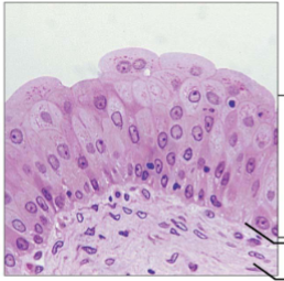
Resembles both stratified squamous and stratified cuboidal; basal cells cuboidal or columnar; surface cells dome shaped or squamous-like, depending on degree of organ stretch. Stretches readily, permits stored urine to distend urinary organ→ storage/increase flow. Lines the ureters, bladder, and part of the urethra.
Stratified cuboidal
Quite rare. Some sweat and mammary glands. Typically two cell layers thick

Stratified columnar
Limited distribution in the body. Occurs at transition areas between two other types of epithelia. Apical region is columnar. Pharynx, male urethra, lining some glandular ducts

Nervous tissue
Neurons are branching cells; cell processes that may be quite long extend from the nucleus-containing cell body; also contributing to nervous tissue are non-excitable supporting cells. Neurons transmit electrical signals from sensory receptors and to effectors (muscles and glands); supporting cells support and protect neurons. Located in the brain, spinal cord, and nerves.

Skeletal muscle
Long, cylindrical, multinucleate cells; obvious striations. Voluntary movement; locomotion; manipulation of the environment; facial expression; voluntary control. In skeletal muscles attached to bones or occasionally to skin.

Cardiac muscle
Branching, striated, generally uninucleate cells that interdigitate at specialized junctions (intercalated discs). As it contracts, it propels blood into the circulation; involuntary control. Located in the walls of the heart.

Smooth muscle
Spindle-shaped (elongated) cells with central nuclei; no striations; cells arranged closely to form sheets. Propels substances or objects (foodstuffs, urine, a baby) along internal passageways; involuntary control. Located mostly in the walls of hollow organs.

Connective tissue proper; loose connective tissue, areolar
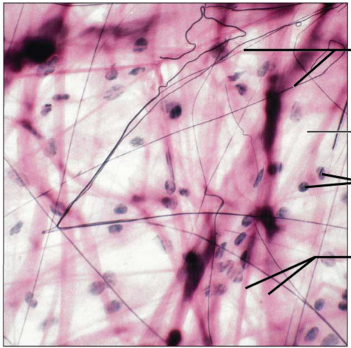
Gel-like matrix with all three fiber types; cells: fibroblasts, macrophages, mast cells, and some WBCs. Wraps and cushions organs; its macrophages phagocytize bacteria; plays important role in inflammation; holds and conveys tissue fluid.; edema. Supports and binds other tissue. Widely distributed under epithelia of body, e.g., forms lamina propria of mucous membranes; packages organs; surrounds capillaries.
Connective tissue proper: loose connective tissue, adipose
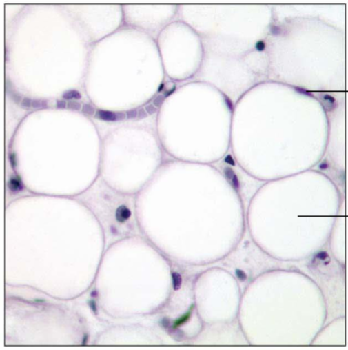
Matrix as in areolar, but very sparse; closely packed adipocytes, or fat cells, have nucleus pushed to the side by large fat droplet. Little matrix, cells are packed tight. Provides reserve food fuel; insulates against heat loss; supports and protects organs. Shock absorption, insulation, energy storage. Under skin in subcutaneous tissue; around kidneys and eyeballs; within abdomen; in breasts.
Connective tissue proper: loose connective tissue, reticular
Loose network of reticular fibers in a gel-like ground substance; reticular cells lie on the network. Fibers form a soft internal skeleton (stroma) that other cell types including white blood cells, mast cells, and macrophages. Lymphoid organs (lymph nodes, bone marrow, and spleen).

Connective tissue proper: dense connective tissue, dense regular
Primarily parallel collagen fibers; a few elastic fibers; major cell type is the fibroblast. Attaches muscles to bones or to muscles; attaches bones to bones; withstands great tensile stress when pulling force is applied in one direction. Wavy. Located in tendons, most ligaments, aponeuroses.
Connective tissue proper: dense connective tissue, dense irregular
Primarily irregularly arranged collagen fibers; some elastic fibers; fibroblast is the major cell type. Collagen bundles are thicker. Withstands tension exerted in many directions; provides structural strength. Located in fibrous capsules of organs and of joints; dermis of the skin; submucosa of digestive tract.
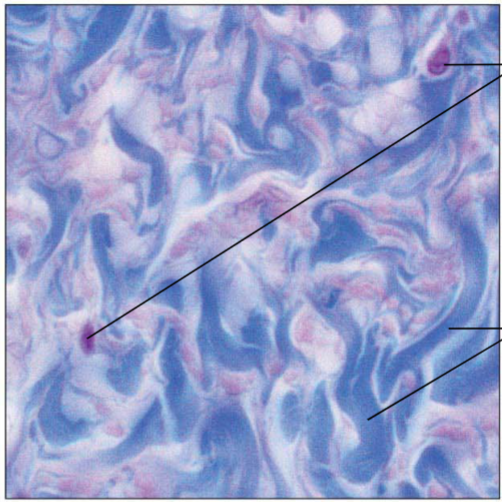
Connective tissue proper: dense connective tissue, elastic
Dense regular connective tissue containing a high proportion of elastic fibers. Allows tissue to recoil after stretching; maintains pulsatile flow of blood through arteries; aids passive recoil of lungs following inspiration. Walls of large arteries; within certain ligaments associated with the vertebral column; within the walls of the bronchial tubes.
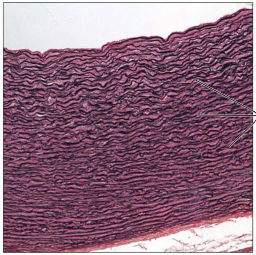
Cartilage: hyaline
Amorphous but firm matrix; collagen fibers form an imperceptible network; chondroblasts produce the matrix and when mature (chondrocytes) lie in lacunae. Most common. Supports and reinforces; serves as resilient cushion; resists compressive stress. Forms most of the embryonic skeleton; covers the ends of long bones in joint cavities; forms costal cartilages of the ribs; cartilages of the nose, trachea, and larynx.

Cartilage: elastic
Similar to hyaline cartilage, but more elastic fibers in matrix. Maintains the shape of a structure while allowing great flexibility. Supports the external ear (pinna); epiglottis.
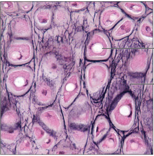
Cartilage: fibrocartilage
Matrix similar to but less firm than that in hyaline cartilage; thick collagen fibers predominate. Tensile strength allows it to absorb compressive shock. Intervertebral discs; pubic symphysis; discs of knee joint.

Bone (osseous tissue)
Hard, calcified matrix containing many collagen fibers; osteocytes lie in lacunae. Very well vascularized. Supports and protects (by enclosing); provides levers for the muscles to act on; stores calcium and other minerals and fat; marrow inside bones is the site for blood cell formation (hematopoiesis). Located in the bones.

Connective tissue: blood
Red and white blood cells in a fluid matrix (plasma). Fluid – no connection. Transport respiratory gases, nutrients, wastes, and other substances. Contained within blood vessels.

Scapula
shoulder blade

Suprascapular notch
a small, curved depression on the upper border of your shoulder blade that creates a protected tunnel for nerves and blood vessels traveling to your shoulder muscles

supraspinous fossa
a shallow, concave depression located on the upper, back side of the scapula (shoulder blade), directly above the scapular spine

infraspinous fossa
a broad, shallow depression located on the back surface of the scapula (shoulder blade) directly below the scapular spine

subscapular fossa
a broad, concave surface on the front (anterior or costal) side of the scapula, or shoulder blade, that faces the rib cage
Superior angle
the upper, smooth corner of the shoulder blade where its superior and medial borders meet

Inferior angle
bottom point of scapula

medial border
Inner edge of the scapula
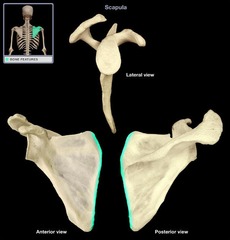
lateral border
Outer edge of the scapula.

spine of scapula
posterior ridge of scapula

Acromion
an extension of the scapula that forms the high point of the shoulder

Coracoid process
process above the glenoid cavity that permits muscle attachment

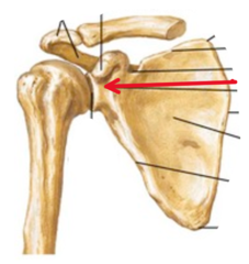
glenoid process
socket in the scapula for the arm bone
Clavicle
collar bone
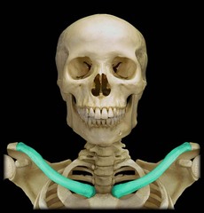
Acromial (lateral) end
Flattened lateral end that articulates with the acromion of the scapula to form the acromioclavicular (AC) joint

Sternal (medial) end
oval or triangular medial end that articulates with the sternum to form the lateral walls of the jugular notch

Humerus
upper arm bone

head of humerus
articulates with glenoid cavity of scapula

anatomical neck of humerus
distal to head of humerus; visible as an oblique groove

intertubercular sulcus of humerus
a deep depression on the upper part of the humerus bone that separates the greater and lesser tubercles. Located on front side.

Greater tubercles
a large bony prominence on the upper, outer side of the humerus (upper arm bone) that anchors key rotator cuff muscles

lesser tubercle
a small, prominent bump on the front and upper part of the humerus (the upper arm bone) that serves as the attachment point for the subscapularis muscle
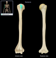
surgical neck
the narrowed part of the upper arm bone located just below the greater and lesser tubercles

deltoid tuberosity
a rough, raised, triangular landmark located on the middle of the outer (lateral) side of the upper arm bone (humerus)

radial fossa
a small, shallow depression on the front part of the lower humerus (upper arm bone) just above the capitulum. Located on front

medial epicondyle
prominent bony bump on the inner side of your elbow, located at the lower end of the upper arm bone

lateral epicondyle
The lateral epicondyle is the bony bump on the outer side of your elbow where your forearm tendons attach
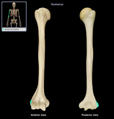
coronoid fossa
a small depression on the front part of the lower humerus (upper arm bone) just above the trochlea

olecranon fossa
a deep triangular depression on the back of the lower humerus (upper arm bone) that holds the tip of the ulna when you straighten your arm

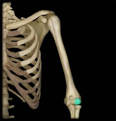
capitulum
a small head or rounded knob-like structure