Radiology: Alternate Imaging Modalities
 Call Kai
 Learn
Learn Practice Test
 Spaced Repetition
 Match
Match Flashcards
 Knowt Play
1/31

There's no tags or description
Looks like no tags are added yet.
Name | Mastery | Learn | Test | Matching | Spaced | Call with Kai | Chat |
|---|
No analytics yet
Send a link to your students to track their progress
32 Terms
an imaging technique commonly used to obtain real-time moving radiographic images of the internal structures
Fluoroscopy
Detectors determine the amount of radiation absorbed by the patient and the x-ray tube spins around the patient. 64 and 128 detector machines are most common. Thinner slices have higher resolution.
How do CT machines work?
wide WW best for imaging tissue types that vary greatly . Narrow WW best for imaging tissue types with similar densities.
Narrow vs. wide windowing
PROS:
More detailed view of an atomy without superimposition
Faster and less expensive than MRI
Best for bone, lung, abdomen
CONS
Radiation exposure
More expensive than radiographs
Poor soft tissue contrast
CT Pros and Cons
Hydrogen atoms line up along a magnetic field. An RF is pulsed causing the noncancelled out hydrogens to flip and then return to position after the pulse. The hydrogens induce the current in the receiver coil when returning. The hydrogens will relax at different rates depending on the tissue.
How does an MRI machine work?
Better Bone Detail
Good for thorax and abdomen
MRI
Better soft tissue detail
Good for neuroimaging and musculoskeletal
CT vs. MRI
2-20
Ultrasound wave are __MHz.
Signals from the moving RBCs are displayed in color as a function of their motion towards or away from the transducer
BART blue ←(away) red →(toward)
Color Flow Mapping
a picture element and has only two dimensions
Pixel
a volume picture element and has three dimensions
Voxel
attenuation value relative to water determining the shade of gray
Hounsfield Units
the range of HU represented on a specific image
Window
the central value of the HUs displayed
Level
white
Hyperattenuating on a CT
black
Hypoattenuating on a CT
the use of certain parameters to optimize the differences in relaxation rates to different tissues in order to provide contrast in the image
Weighting
Fluid is hypointense: black
Fat is hyperintense: white
T1 Weighted
Fluid is hyperintense: white
Fluid is hypointense: white
T2 Weighted
less bright than tissue you compare to
Hypointense in an MRI
brighter than tissue you compare to
Hyperintense in an MRI
same brightness as tissue you compare to
Isointense in an MRI
black
Anechoic in an US
darker (dark gray) than compared structure
Hypoechoic in an US
The same echogenicity to compared structure
Isoechoic in an US
Brighter than compared structure
Hyperechoic in an US
the perceived change in frequency that occurs as a result of the motion between the sound source and the observer
Doppler effect
CT
What is this an example of ?

x-ray
What is this an example of?

MRI
What is this an example of?
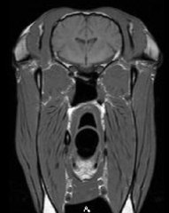
T1W
What is this an example of?
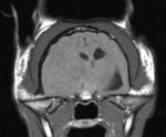
T2W
What is this an example of?

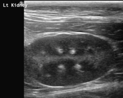
US
What is this an example of?