Looks like no one added any tags here yet for you.
conduction system and cardiac muscle cells
heart contraction involves what 2 events?
conduction system
initiates and propagates an action potential
cardiac muscle cells
what fire action potentials and contract?
false
*the cardiac muscles do this
true/false
the conduction system happens first in the atria and then the ventricles
SA node
what initiates action potential in conduction system?
action potential is propagated throughout the atria and conduction system
how is action potential spread in the conduction system?
across the sarcolemma of cardiac muscle cells
in cardiac muscle cells, where is action potential propagated?
thin filaments slide past thick filaments and sarcomeres shorten within cardiac muscle cells
how does muscle contraction work in cardiac muscle contraction?
Electrocardiogram (ECG/EKG)
Skin electrodes detect electrical signals of cardiac muscle cells
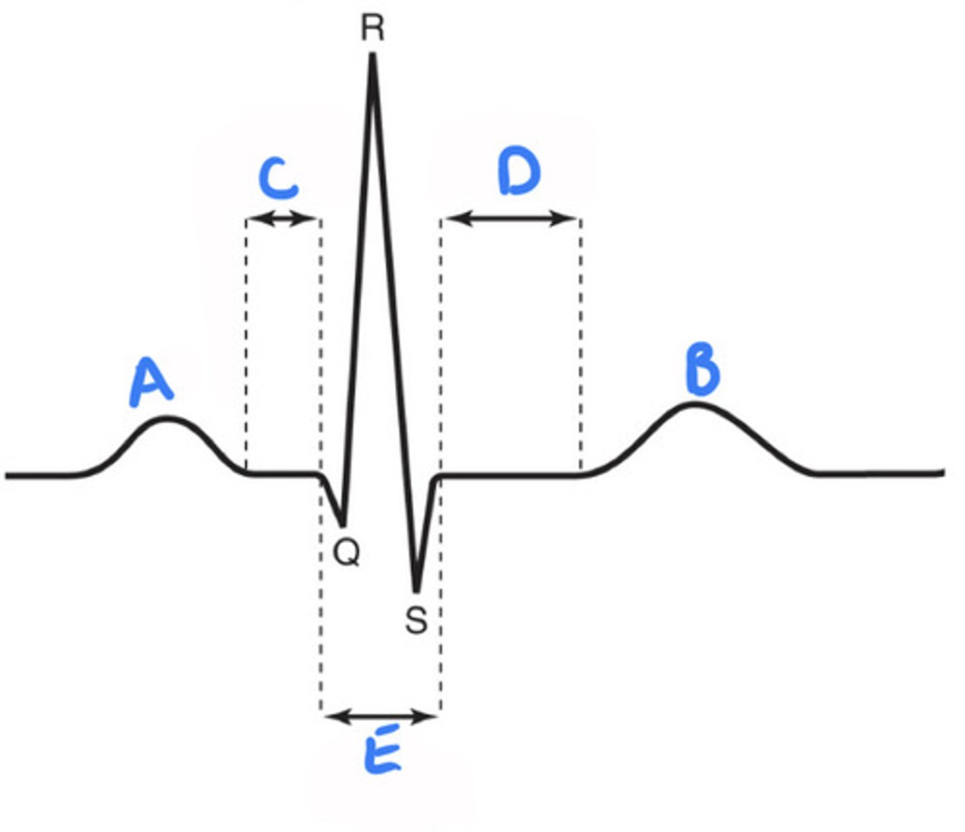
p wave, QRS complex, T wave
what are the waves and segments in an ECG recording?
P wave
electrical changes of atrial depolarization originating in SA node
A) p wave
B) t wave
C) p-q segment
D) s-t segment
E) QRS complex
A)
B)
C)
D)
E)
time of ventricular action potentials
how is time reflected in a Q-T interval?
QRS complex
Electrical changes associated with ventricular depolarization and the Atria also simultaneously repolarizing
T wave
Electrical change associated with ventricular repolarization
P-Q, S-t
what are the segments in ECG recording?
P-Q
Associated with atrial cells' plateau (atria are contracting
S-T
Associated with ventricular plateau (ventricles are contracting) The ECG Recording
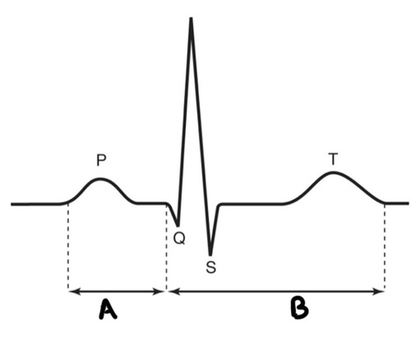
P-R interval
what is the time from beginning of P wave to beginning of QRS deflection?
P-R interval
what interval has time to transmit action potential through entire conduction system
it is from atrial depolarization to beginning of ventricular depolarization
how do you describe P-R interval
Q-T interval
what interval is the time from beginning of QRS to the end of T wave
A) p-r interval
B) q-t interval
A)
B)
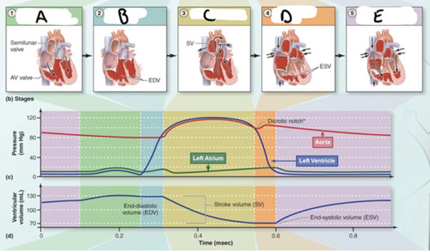
A) atrial contraction & ventricular filling
B) isovolumetric contraction
C) ventricular ejection
A)
B)
C)
D) isovolumetric relaxation
E) atrial relaxation and ventricular
D)
E)
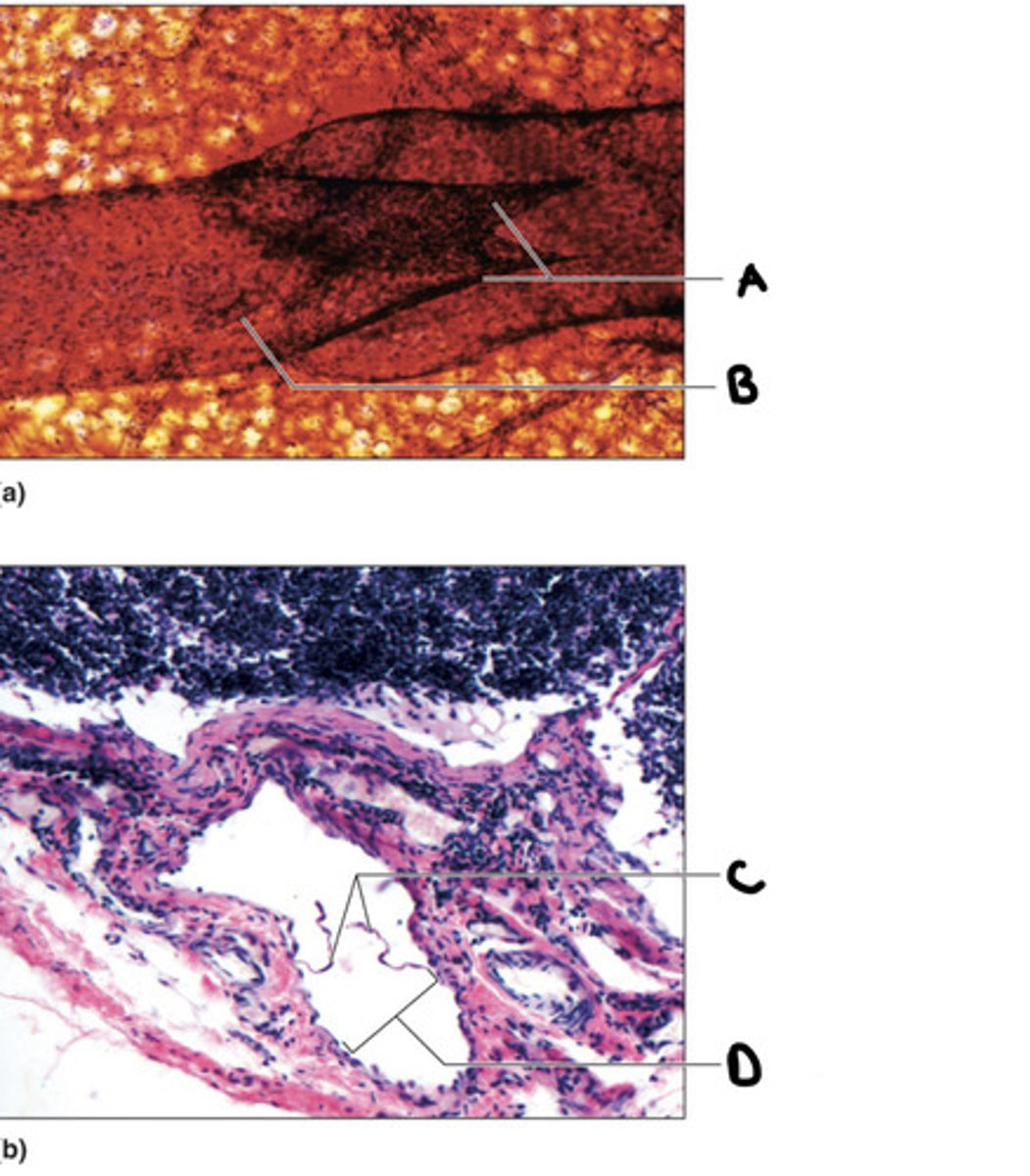
A) valve cusps
B) lymphatic vessels
C) valve cusps
D) lymphatic vessels
A)
B)
C)
D)
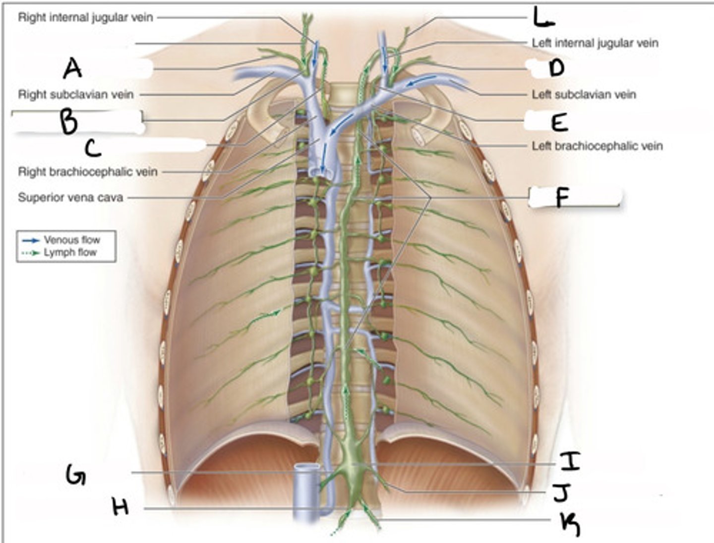
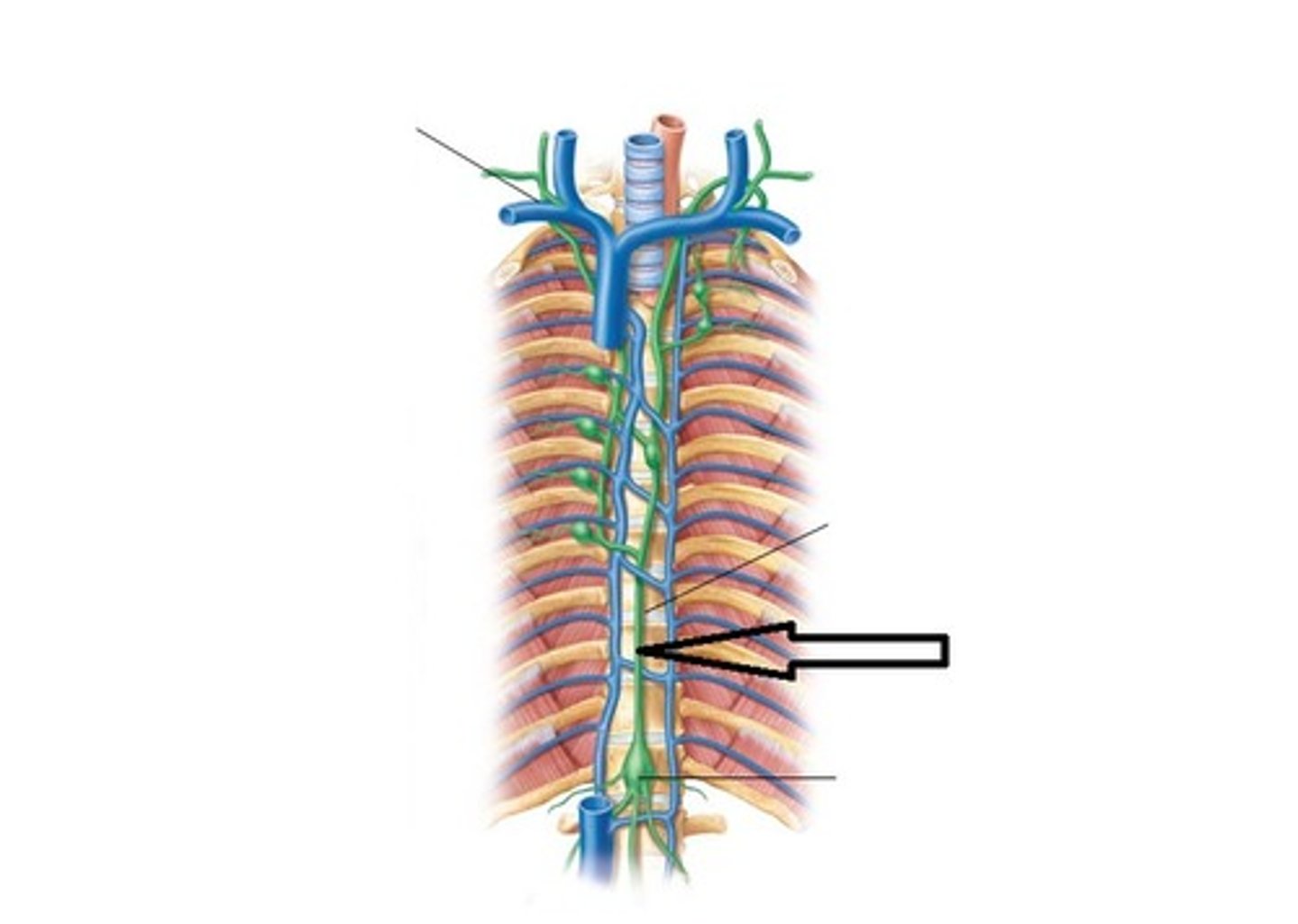
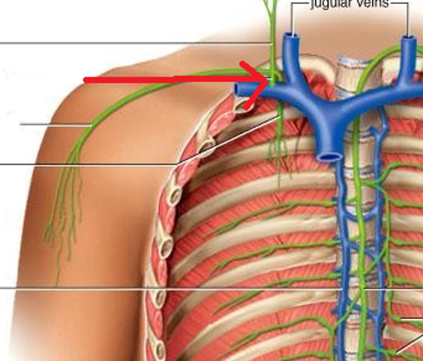
A) right subclavian trunk
B) right lymphatic duct
C) right bronchomediastinal trunk
A)
B)
C)
D) left subclavian trunk
E) left bronchomediastinal trunk
F) thoracic duct
D)
E)
F)
G) right lumbar trunk
H) right intestinal trunk
I) cisterna chyli
G)
H)
I)
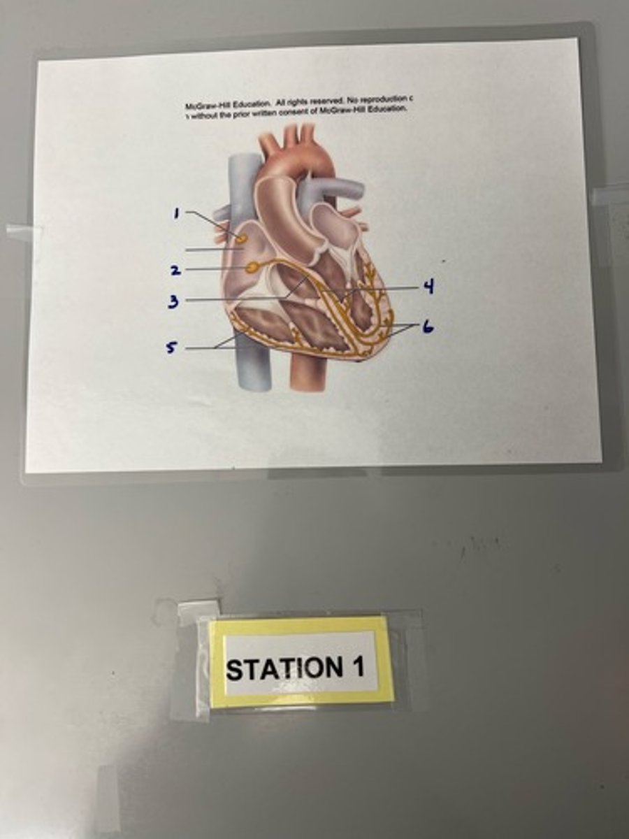
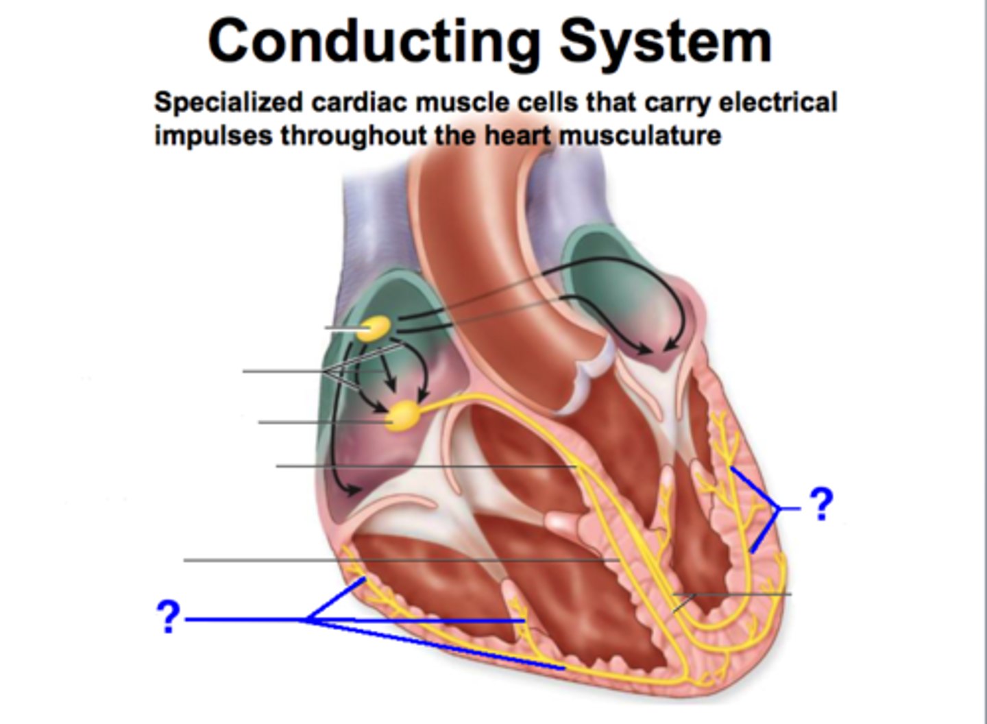
1) sinoatrial (SA) node
2) atrioventricular node
3) atrioventricular bundle
1)
2)
3)
J) left lumbar trunk
K) left intestinal trunk
L) left jugular trunk
J)
K)
L)
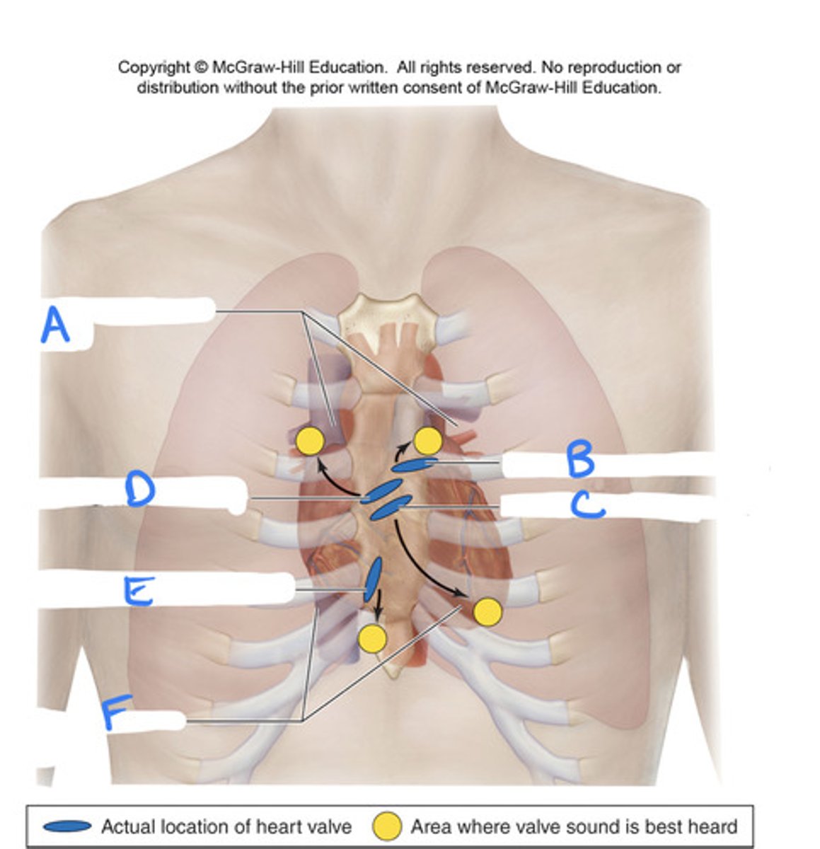
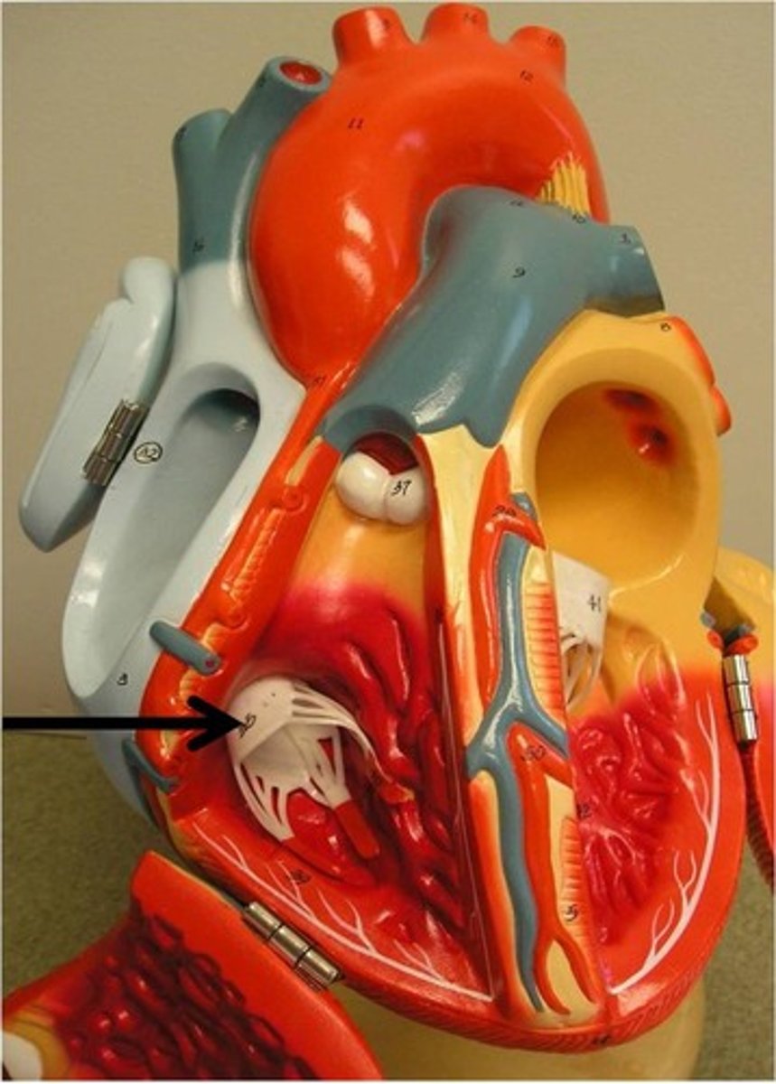
D) aortic semilunar valve
E) right atrioventricular valve
F) fifth intercostal space
D
E
F
4) right and left bundles
5) purkinje fibers
6) purkinje fibers
4)
5)
6)
A) second intercostal space
B) pulmonary semilunar valve
C) left atrioventricular valve
A
B
C
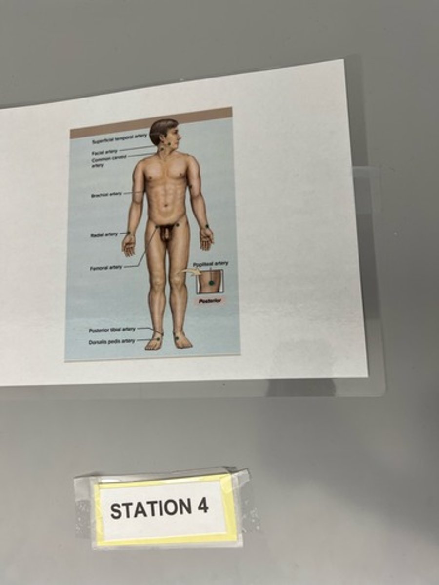
Brachial
Which pulse point are you using when measuring blood pressure?
Lymphatic vessel and cisterna chyli
What vessel and type receives lymph from multiple smaller ducts that drain the lower limbs and abdominal cavity?
Lymphatic vessel and right lymphatic duct
What vessel and type recieves lymph from the right side of the head, neck, and thorax?
What vessel and type receives lymph from the entire body below the diaphragm, and the left side of the head, neck, and thorax?
False
True/false
The lymphatic system has a pump
False
*lymphatic vessels fed by lymphatic capillaries
True/false
Lymphatic vessels are fed by lymphatic ducts
True
True/False
Lymphatic trunks are fed by lymphatic vessels
True
True/false
Lymphatic ducts are fed by lymphatic trunks and bring lymph to venous blood circulation
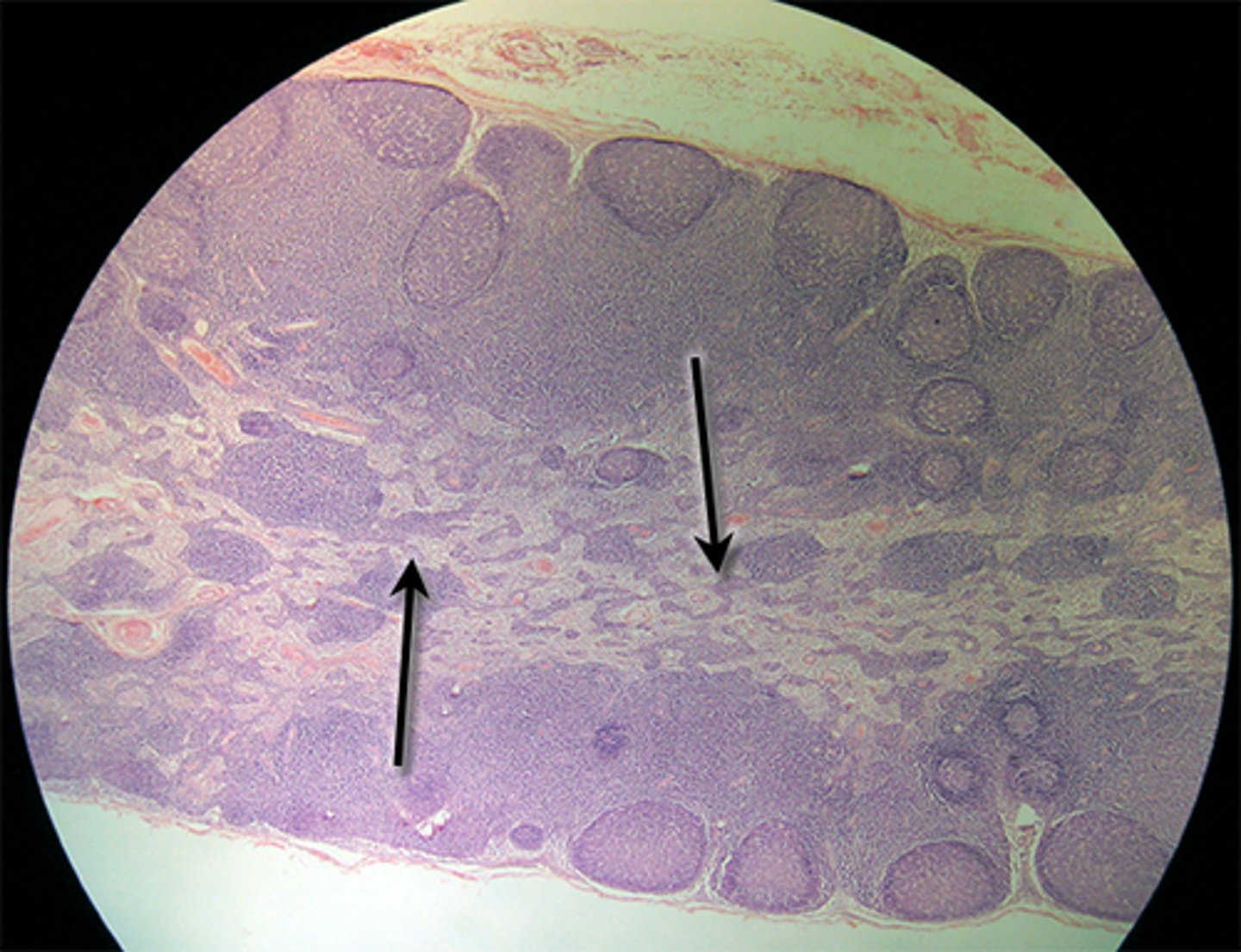
Lymph nodes for lymph filtration
Some vessels connect to _____ for _____
-Transport and house lymphocytes and other immune cells
-Return excess fluid in body tissues to blood for blood volume
What is a function of lymphatic system?
Lymph vessels, lymphatic tissues and organs
What are the lymphatic system components?
diastolic pressure + 1/3 pulse pressure e.g., if blood pressure is 120/80
MAP = 80 + 40/3 = 93
How to calculate MAP if blood pressure is 120/80?
True
*bc diastole lasts longer than systole
True/false
the mean is weighted closer to diastolic pressure
false
* MAP < 60 indicated insufficient blood flow
True/false
MAP > 60 may indicate insufficient blood flow
Facial, common carotid, brachial, radial, femoral, popliteal
Where are some of the best locations to detect pulse?
Pulse
throbbing of arterial wall
Diastolic pressure
occurs when ventricles relax, Lowest pressure generated in arteries (they recoil)
80 mm Hg, if blood pressure is 120/80
Systolic pressure
occurs when ventricle contracts or Highest pressure in arteries (they are stretched)
120 mm Hg, if blood pressure is 120/80
Pulse pressure
pressure in arteries added by heart contraction, difference between systolic and diastolic pressure (40 mm Hg if blood
pressure is 120/80)
Blood pressure gradient
Propels blood through vessels
mm Hg
*Ex: 120/80 mmHg
What is the unit measured when taking blood pressure?
Blood pressure
force of blood against vessel wall
Heart blocks
What abnormality in the heart is impaired conduction?
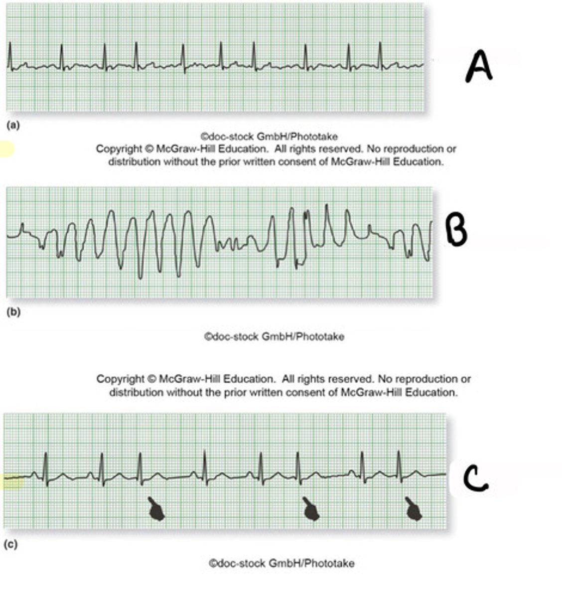
Atrial fibrillation
What abnormality in the heart has chaotic timing of atrial action potentials?
A) atrial fibrillation
B) ventricular fibrillation
C) premature ventricular contraction
A)
B)
C)
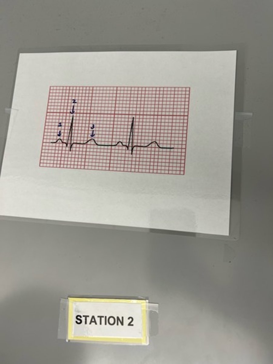
A) 2
B) QRS
A) Which number indicates ventricular depolarization?
B) what is the wave called?
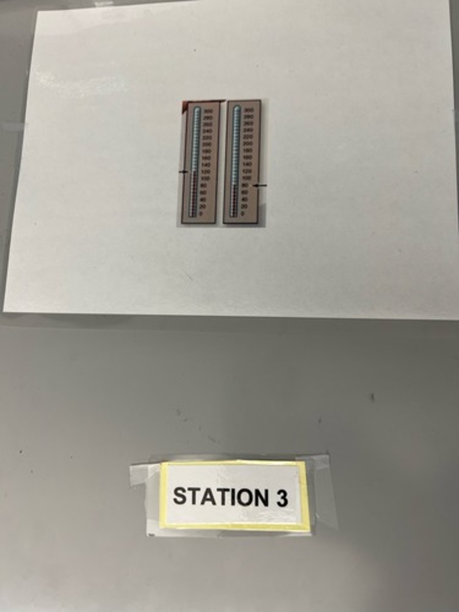
Systole, mm Hg
is the figure on the left the pressure measured during diastole or systole? Units?
A) 1
B) p wave
A) Which number indicates atrial depolarization originating in SA node?
B) what is the wave called?
A) t wave
B) 3
A) Which number indicates ventricular repolarization?
B) what is the wave called?
Ventricular fibrillation
What abnormality in the heart has chaotic electrical activity in ventricles typically with uncoordinated contraction and pump failure?
Premature ventricular contractions
What abnormality in the heart will have abnormal action potential within AV node or ventricles?
Purjinke fibers
specialized muscle fibers that conduct the cardiac impulse from the AV bundle into the ventricular walls
blood pressure
force of blood against vessel wall
blood pressure gradient
Propels blood through vessels; highest in arteries and lowest in veins
pulse
throbbing of arterial wall
lymph
fluid transported within lymph vessels
thoracic duct
receives lymph from the left side of the head, neck, chest, abdomen, left arm, and lower extremities
right lymphatic duct
receives lymph from the upper part of the body
lymph
what structure is this
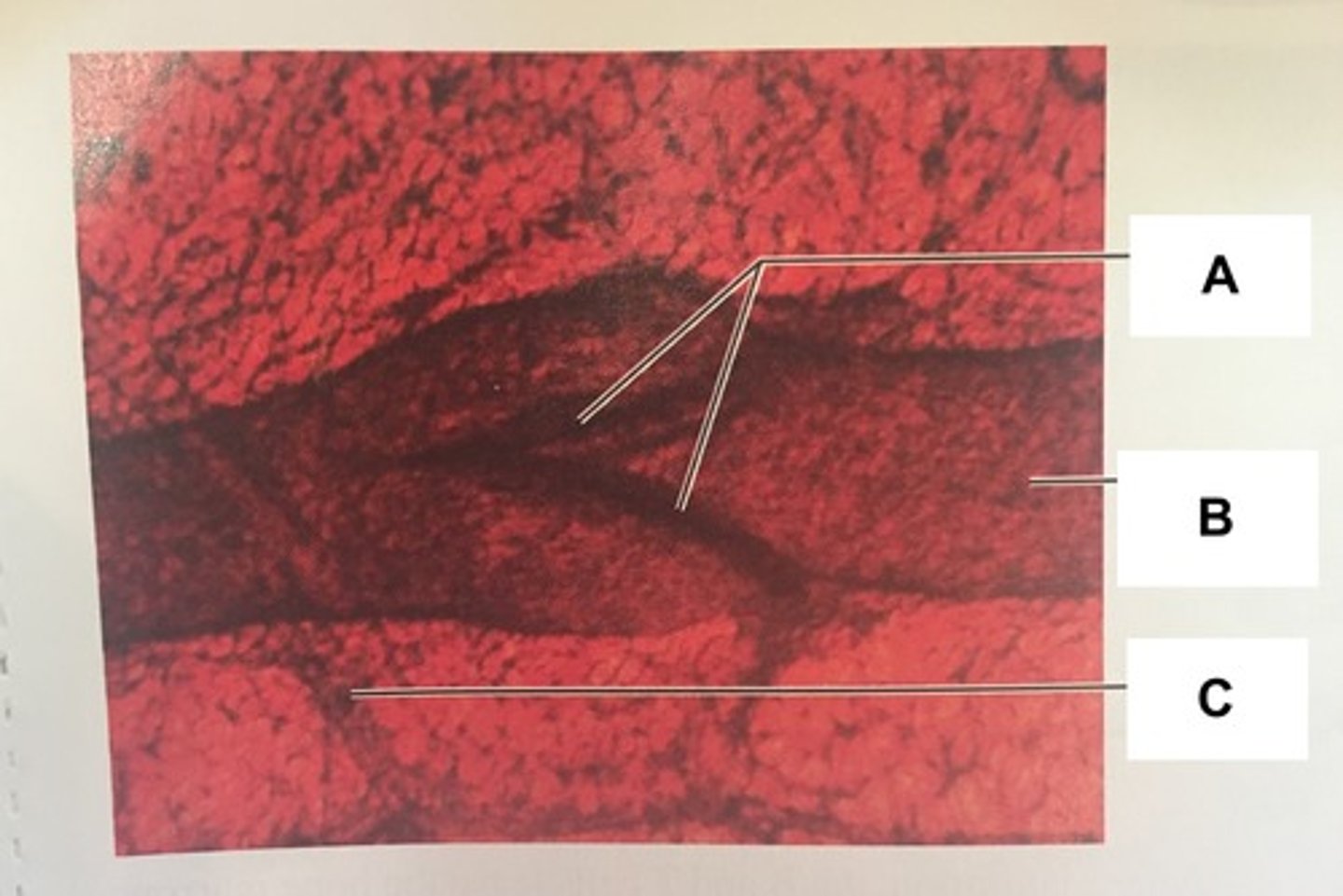
valve cusp
are found within the lumen of the veins. They close and block the flow to the heart when the heart isn't contracting
lymphatic vessel
Vessel that carries lymph