Antamoy week two
 Call Kai
 Learn
Learn Practice Test
 Spaced Repetition
 Match
Match Flashcards
 Knowt Play
1/100

There's no tags or description
Looks like no tags are added yet.
Name | Mastery | Learn | Test | Matching | Spaced | Call with Kai | Chat |
|---|
No analytics yet
Send a link to your students to track their progress
101 Terms
What is the lymphatic system composed of
lymph vessels and lymphoid structures
what is Lymph
fluid transported within vessels
interstitial space
hydrostatic pressure
Primary lymphoid structures
thymus & redbone marrow
lymphocytes born here
Secondary lymphoid structures
tonsils, lymph nodes, spleen, MALT (mucosa associated lymphoid tissue)
immune cells/ immune response
Lymph vessels
right lymphatic duct, thoracis duct, cisterna chyli, lymphatic trunk, lymphatic vessels
Characteristics of lymph
15% not reabsorbed (3L daily)
water, dissolved solutes, protein (100-200g)
cell debris, cancer, virus/bacteria
Lymphatic capillaries
Small
open-ended lymph vessels absorb interstitial fluid
areolar/ vascularized tissue
wall endothelium
no basement membrane/overlap endoth cells
Anchoring filaments
help hold endothelial cells to nearby structures
prevent collapse when fluid enters
GI lymphatic capillaries
lacteals
absorb lipid soluble
lymph lipid mixture=chyle
interstitial hydrostatic pressure
increase fluid capillary-lumen= closed
greater than lymph pressure
capillaries-vessels-trunk-duct
Lymphatic vessels
next to deep arteries/viens
valves in lumen
3 vessel tunics
what are the 3 vessel tunics
intima, media, externa
purpose of valves
prevent backflow
(crucial lower limbs)
mechanisms to move lymph
1. contraction skeletal muscle/ respiratory pump
2.ryhtmic contraction smooth muscle trunk/ducts
3.pulsate blood in arteries
Lymphatic trunk
left & right drain/remove lymph
Jugular trunk
drains head and neck
Subclavian trunk
drain upper limbs, breasts, superficial thoracic wall
bronchomediastinal trunks
drain deep thoracic wall, lung, heart
Intestinal trunk
drains stomach, intestines, pancreas, spleen, part of the liver
lumbar trunk
drains lower limbs, lower abdominal wall, and pelvic organs
Right lymphatic duct
clavicle, short 1-2cm
drain head, neck, upper limb, thorax
return to venous circulation @junction-subclavian & jugular vein
(Left) Thoracic lymphatic duct
large 37.5-45cm (15-18in)
drain head, neck, upper limbs, thorax, abdomen, both lower limb
diaphragm-junction-left subclavian & jugular vein
cisterna chyli
cisterna chyli
pouch thoracic duct
storage chyli
base thoracic duct L2- aortic open diaphragm- up vertebral midline
Lymphedema
swelling abnormal accumulation of lymph fluid in tissues
lymphatic filariasis
thread worms in vessels
Lymph SUPER LARGE swell extreme cases
elephantiasis
Red bone marrow
name 1 or 2, location & function?
1
location: spongey bone
function: B-lymphocyte mature
Thymus
name 1 or 2, location & function?
1
location: superior mediastinum (adults) & ant/sup (child)
function: T-lymp maturation & differentiation
Lymph nodes
name 1 or 2, location & function?
2
location: on length lymphatic vessels, cluster axillary, inguinal, cerv regions
function: filter lymph & immune response
Spleen
name 1 or 2, location & function?
2
location: left up quad abdomen, 9-11 ribs wraps
function: filter blood, immune response blood, reservoir/remove erythrocytes & platelets
Tonsils
name 1 or 2, location & function?
2
location: oral cavity/ pharynx (throat)
function: protect against inhaled/ ingested sub
Lymphoid nodules
name 1 or 2, location & function?
2
location: body organ wall of appendix
function: protect organ foreign sub
MALT (mucosa associated lymphoid tissue)
name 1 or 2, location & function?
2
location: clusters in GI, resp, urin, reproductive
function: protects mucosal memb foreign sub
Hematopoiesis or hemopoiesis
formed element production in red bone marrow
what are formed elements
erythrocyte, platelets, granulocytes (neutrophils, esinophils and basophils)
Thymus
bilobed organ in superior mediastinum
t-lymp maturation
grows until puberty 30-50g
replaced with adipose tissue
Thymus in children
2 fused thymic lobes surround connective tissue capsule
trabeculae/septa (fibrous) subdivide lobes into lobules (cortes & inner medulla)
why are they called secondary lymphoid structures?
they house lymphocytes & immune cells enmeshed in reticular connective tissue matrix
lymphoid organs or nodules
which contain complete capsules?
lymphoid organs
lymph nodes & spleen
which contain incomplete/absent capsules?
lymphoid structures
tonsils, lymphoid nodules & MALT
Lymph nodes
small oval clusters (0.1-2.5cm) lymphatic tissue @lymphatic vessels
500-700 through body
Afferent lymphatic vessels
bring lymph to a lymph node
Efferent lymphatic vessel
only 1 @hilum/hilus
drainer
Hilum/hilus
involuted portion of lymph node
Describe capsule of lymph node
dense irregular CT encapsulates nodes
internal extension trabeculae
blood vessels
lymph sinuses
cortex/inner medulla
Trabeculae
internal extension subdivide node into compartments
What's in the germinal center?
proliferating B-lymph & macrophages
surrounded by mantle zone
What's in the mantle zone?
T lymph, macrophages, & dendritic cells
Medulla
connective tissue for B/T lymph & macrophages
medullary chords
what are the structures in the cortex and medulla that allow lymph movement through the lymph node
cortical & medullary sinuses
Flow steps of lymph
1. afferent lymph vessel (inflow large)
2. lymph node sinus
3. efferent lymph vessel (outflow small)
Lymphoma
malignant tumor of lymph nodes and lymph tissue
1.Hodgkin lymphoma & 2 .Non-Hodgkin lymphoma
1. reedsternberg cell 2 nuclei owl eye cancer
2. aggressive/fatal malnigant
what are the functions of the spleen (white and red pulp)
White pulp: immune response in blood
Red pulp: venous phagocytosis of bacteria, old/defective erythrocytes & platelets & blood reservoir for eryth/platelets
Splenectomy, why, what happens after?
surgical removal of the spleen bc/ infection, injury, other body parts will take over function +chance of infection
Pharyngeal tonsil
posterior wall of nasopharynx, inflamed adenoids
Palatine tonsils
at posterior lateral end of oral cavity, lingual are 1/3
Blood flows through the spleen in order
1. splenic artery
2. central artery white pulp
3. splenic sinusoids red pulp
4. splenic vein
lymphatic nodules ilium small intestine called
peyer patches
What does the respiratory system consist of
respiratory tract (nose, nasal cavity, pharynx, larynx, trachea & bronchiole tree sacs alveoli)
Purpose of the passage for air
atmospheric air moves into alveoli breath in and out exhaled
Purpose of conditions inhaled air
mucous memb lining, warms/moist conditions & traps particles before reaching lungs
Purpose of sit for gas exchange
resp memb made alveolus/pulmonary capillaries
O2 alveoli diffuse into blood/ Co2 from blood to alveolis
Purpose of olfaction
olfactory receptors in nasal cav for smell sent by receptors to brain
Purpose of sound production
vocal chords larynx (voice box) vibrate as air moves in upper resp structures
Upper resp tract
nose, nasal cavity, pharynx, larynx
Lower resp tract
trachea, bronchi, brochioles (resp/terminal), alveolar ducts, alveoli
define conducting zone
passageway transport/ conduct air
nose-end bronchioles
define respiratory zone
structures gas exchange with blood
resp bronchioles, alveolar ducts, alveoli
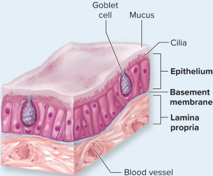
Respiratory tract
exposed external environment
lined internally by mucosa (mucous membrane or resp mucosa)
epithelium basement memb & under lamina propria areolar CT
cilia
Pseudostratified ciliated columnar epithelium lines what?
nasal cavity, parasanl sinus, nasopharynx, trachea, inferior larynx, main/lobar bronchi
Simple cilliated columnar epithelium lines what?
segmental, smaller and large bronchi
Simple cuboidal epithelium lines what?
terminal resp bronchi
Nonkeratinized stratified squamous epethelium lines what?
regions resp tract that get a abraission
oropharynx, laryngopharynx, vocal folds, superior larynx
Goblet cells
unicellular exocrine glands secrete mucus 1-7tsbp daily
Mucin
protien increase visosicty for trapping in mucus
has enzyme, antibodies, antimicrobial prop
sputum
Cystic fibrosis
defective chloride channels, ducts and musus begins to block causing infection
nose
bone, hyaline cartilage, dense irregular tiss
1 pair lateral cart & 2 pair alar cart
nares/nostrils
nasal cavity
oblonged opening chonae
floor is hard and soft palents
roof is frontal, ethmoid & sphenoid bone
nasal septum
divides r&l anterior is septal nasal cart
perpendicular plat & vomer bone