OB/GYN EOR Exam
 Call Kai
 Learn
Learn Practice Test
 Spaced Repetition
 Match
Match Flashcards
 Knowt Play
1/1242

There's no tags or description
Looks like no tags are added yet.
Name | Mastery | Learn | Test | Matching | Spaced | Call with Kai | Chat |
|---|
No analytics yet
Send a link to your students to track their progress
1243 Terms
granulosa cells
FSH stimulates what cells?
follicle growth
↑ aromatase → estrogen production
Actions of FSH
theca cells---> androgen production
LH stimulates what cells?
LH
what hormone triggers ovulation?
LH
hormone that maintains corpus luteum
FSH
hormone that increases aromatase --> estrogen production
negative feedback on GnRH/FSH/LH
how does low estrogen effect the HPO axis?
positive feedback → LH surge → ovulation
how does high sustained estrogen impact the HPO axis?
progesterone
______ has a negative feedback on GnRH
FSH
follicular phase is dominated by what hormone?
Begins with menses
Dominated by FSH
Ovarian follicles mature
Granulosa cells ↑ estrogen
main events of the follicular phase
LH surge --> ovulation
what does the follicular phase end with?
progesterone
what hormone dominates luteal phase?
Dominated by progesterone
CL forms
Prepares uterus for implantation
Fixed length (~14 days)
main events of the luteal phase
CL regresses → progesterone drops → menses
what causes menses if there is no pregnancy?
follicular and luteal
what are the phases of the menstrual cycle?
-proliferative phase (coincides with follicular)
-secretory phase (coincides with luteal)
what are the phases of the endometrium?
estrogen
the proliferative phase of the endometrium is driven by
Endometrium thickens
Glands are straight
Prepares for ovulation
main events of the proliferative phase
progesterone
the secretory phase is driven by
Endometrium becomes glandular & vascular
Glands become coiled
Ideal for implantation
main events of secretory phase
estrogen, progesterone
______ builds the endometrium, _______ stabilizes it
110-160 bpm
what is a normal baseline fetal heart rate?
<110 bpm
what is a bradycardia fetal heart rate?
>160 bpm
what is a tach fetal heart rate?
1. determine baseline fetal heart rate
2. determine HR variability (MOST IMPORTANT)
3. evaluate for accelerations
4. evaluate for decels
5. overall category
steps to interpreting a EFM pattern
0 bpm change
define absent variability on EFM
<5 bpm change
define minimal variability on EFM
moderate (6-25 bpm)
what is a reassuring variability on EFM?
>25 bpm
define a marked variability on EFM
reassuring
the presence of accelerations is
↑ ≥15 bpm for ≥15 sec
define an appropriate acceleration in a fetus >32 weeks
↑ ≥10 bpm for ≥10 sec
define an appropriate acceleration in a fetus <32 weeks
head compression (benign)
early decels indicate
cord compression
variable decels indicate
uteroplacental insufficiency
late decels indicate
≥2 min, <10 min
define a prolonged decel
Normal
a Category I EFM is
Indeterminate
a Category II EFM strip is
abnormal (requires immediate action)
a category III strip is
Baseline rate
Variability
Presence/absence of accelerations
Type & timing of decelerations
Category (I, II, or III)
Interventions and response
required documentation elements of a EFM strip
Normal baseline
Moderate variability
No late or variable decels
findings associated with a category I EFM pattern
Continue routine monitoring
No intervention needed
management of category I tracing
Category III: Abnormal
most common EFM category
Minimal variability
Recurrent variables
Tachycardia without decels
findings associated with Category II tracing
left lateral
maternal positioning indicated for category II tracing
Maternal repositioning (left lateral)
IV fluids
Stop oxytocin
O₂ if indicated
Consider amnioinfusion (for recurrent variables)
management options (intrauterine resuscitation) for category II tracing
Absent variability plus:
Recurrent late decels OR
Recurrent variable decels OR
Bradycardia OR
sinusoidal pattern
findings associated with a category III tracing
Immediate intrauterine resuscitation
Prepare for urgent delivery (C-section if unresolved)
management of category III tracing
maternal repositioning, amnioinfusion
management of variable decels d/t cord compression
O2, fluids, stop pitocin
management of late decels d/t placental insufficiency
Resuscitate, consider delivery
management of prolonged decels d/t hypoxia
Amnioinfusion
introduction of a solution into the amniotic sac; an isotonic solution is most commonly used to relieve fetal distress
late decels
d/t placental failure---> think oxygen delivery problem
amnioinfusion
key treatment of variable decels
O2 and fluids
key treatment of late decels
stop oxytocin
initial treatment of late decels
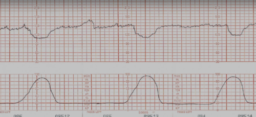
variable decels
name the decel

late decels
name the decel

early decels
name the decel
Gravida: pregnancies
Term
Preterm
Abortions/miscarriages
Living children
define GTPAL
Term/preterm deliveries
Miscarriages/abortions
Complications (PPH, preeclampsia, gestational diabetes)
Mode of delivery (vaginal vs C-section)
Neonatal outcomes
components of a thorough obstetric history
Menstrual history (LMP, cycle length, regularity, flow)
Contraceptive use (current/past)
Sexual history (partners, STI risk, pain with intercourse)
Pap smear history & results
STI history
Gynecologic surgeries or diagnoses (fibroids, endometriosis)
components of a thorough gynecologic history
Inspection → palpation (pads of fingers, circular motion)
Assess for masses, skin changes, nipple discharge
components of a breast exam
-pt supine
-locate symphysis pubis
-palpate top of the uterine fundus using the ulnar border of your hand
-measure with tape measure (in cm)
how to assess fundal height
45 degrees
insert the speculum at what angle?
monthly, ideally after menses
when is the ideal time to perform self exams?
left lateral tilt (esp after 20 weeks)
how do you avoid supine hypotension in pregnant patients?
gestational age (weeks) ± 2 cm
interpretation of fundal height (after 20 weeks only)
just above symphysis pubis
expected fundal height at 12 weeks
at umbilicus
expected fundal height at 20 weeks
xiphoid process
expected fundal height at 36 weeks
Multiple gestation
Polyhydramnios
Macrosomia
Incorrect dating
reasons why fundal height may be larger than expected
Fetal growth restriction (IUGR)
Oligohydramnios
Incorrect dating
reasons why fundal height may be smaller than expected
>2 cm discrepancy to dates
when does a fundal height result in reflex ultrasound?
anteverted, anteflexed
most common uterus position
cardinal (transverse cervical) and uterosacral ligaments
ligaments that support the uterus
Bladder – anterior to uterus
Rectum – posterior to uterus
organs anterior and posterior to the uterus
Blood supply from ovarian arteries (aorta)
blood supply of the ovaries
ureter
runs under the uterine artery
internal iliac
the uterine artery is a branch off of
broad ligament
The ligament extending from the lateral margins of the uterus to the pelvic wall; keeps the uterus centrally placed and provides stability within the pelvic cavity.
-peritoneal fold, not a true ligament
vesicouterine pouch (anterior cul-de-sac)
anterior to fundus between uterus and bladder
Rectouterine pouch (of Douglas)
-lowest point of female abdominal cavity
-accumulates blood/ascites
-drained from posterior fornix of vagina
3-8 weeks
critical period of embryonic development
urogenital ridge
the gonads originate from what embryonic structure?
the development of ovaries
absence of SRY leads to
Fallopian tubes
Uterus
Cervix
Upper ⅔ of vagina
structures that develop from the Mullerian (paramesonephric) ducts
Mullerian agenesis
Everything is normal except no uterus and upper vagina
Have ovaries, normal hair distribution, and are 46XX
uterine didelphys
Complete duplication of the uterus, cervix, and vagina
bicornuate uterus
a common uterine anomaly in which the endometrium divides into two horns
T10-L2
sympathetic innervation of the pelvic organs
Thoracolumbar soft tissue
Rib raising
Lumbar HVLA
main techniques to treat the sympathetics of the pelvic organs
S2-4, pelvic splanchnic
parasympthetics of the pelvic organs
motility, secretion
the parasymphathetics of the pelvic organs regulates
Sacral rocking
Sacral decompression
Pelvic diaphragm release
mainstays of parasympathetic treatment of pelvic organs
Levator ani (iliococcygeus, pubococcygeus, puborectalis)
Coccygeus
muscles of the pelvic diaphragm
pelvic diaphragm
Structure that separates pelvis and perineum
Cardiac enlargement (↑ LV mass)
Upward displacement of the heart
Anatomic cardiac changes in pregnancy
↑ CO, HR, SV
↓ Systemic vascular resistance (SVR)
cardiac output and SVR changes in pregnancy