RTT 220 Digestive System Exam (PART 2)
 Call Kai
 Learn
Learn Practice Test
 Spaced Repetition
 Match
Match Flashcards
 Knowt Play
1/68

Earn XP
Description and Tags
Colorectal cancer
Name | Mastery | Learn | Test | Matching | Spaced | Call with Kai | Chat |
|---|
No analytics yet
Send a link to your students to track their progress
69 Terms


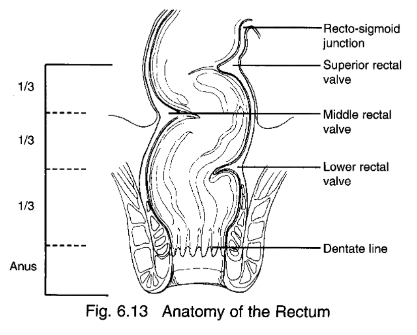
Dentate line
Transition from the columnar epithelium of the colon and rectum to the squamous epithelium of the anal canal
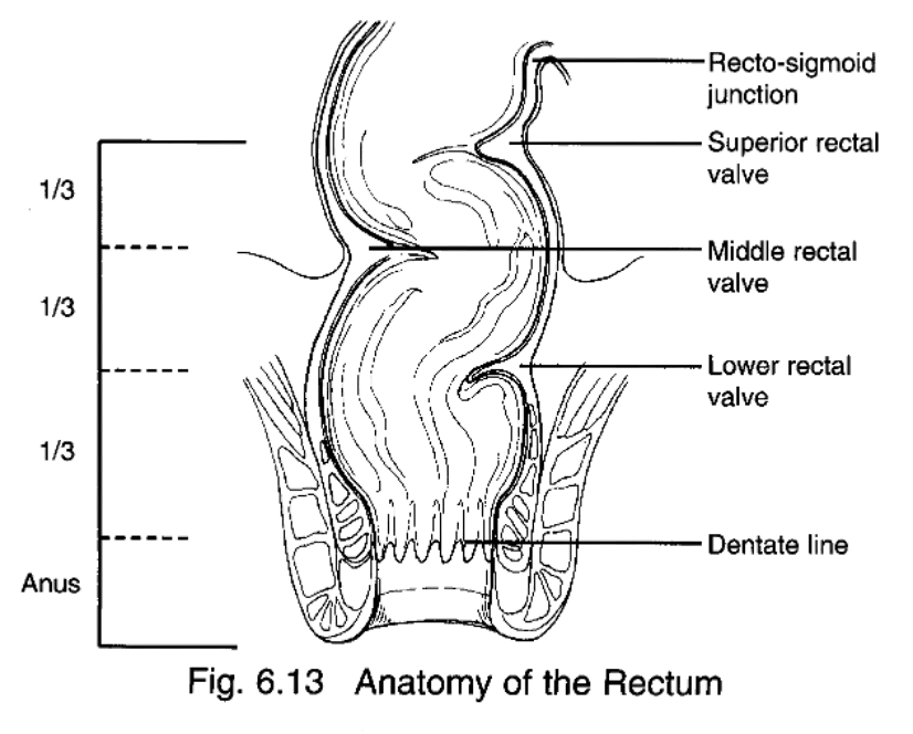
The Large Bowel
¡Functions
§Form and eliminate waste
¡Completion of absorption
§Water
§Electrolyte
§Vitamins
¡Manufacture certain vitamins
§Bacteria
large bowel anatomy
¡Length
§1.5 m or 5 ft
¡Begins with Cecum just beyond the ileocecal junction
¡Four major sections
1.Ascending colon
2.Transverse colon
3.Descending colon
4.Sigmoid colon
¡Eight subsections
Subsections of large bowel
¡Ascending colon
§Beginning at the cecum
§Terminating at the hepatic flexure
¡Transverse colon
§Terminates at the splenic flexure
¡Descending colon
¡Sigmoid colon
§Terminates at the rectum
Mucosa in large bowel
¡Contains no villi
¡Composed of simple columnar epithelium
¡Contains numerous goblet cells which secrete mucous to lubricate the intestinal contents as it passes through the colon
¡Lymphatic nodules are also found here
Submucosa in large bowel
¡Connective tissue
¡Provides the intestine with a network of:
§Vasculature
§Lymphatic support
¡Invasion into this layer is significant in regard to metastatic potential
Muscularis in large bowel
¡Composed of a circular layer of smooth muscle
¡3 bands of longitudinal smooth muscle that run the length of the colon
§Longitudinal muscles are broken up into 3 flat bands
§Each band (taenia coli) runs the entire length of the intestine
§Peristaltic contractions of these bands form pouches or haustra which give the colon a puckered appearance
Peritoneum (large bowel)
¡Covers a majority of the intestine
¡Considered a serosal layer
¡The rectum is not covered by peritoneum,therefore the rectum has no serosal layer to separate it from proximal organs
§Increased risk of direct tumor invasion to bladder and vagina in females and bladder, prostate, and seminal vesicles in males
Epidemiology of Colon Cancer
¡3rd most prevalent malignancy in the U.S.
¡Occurs equally in males and females
¡Most common >50 years old
¡Incidence and mortality rates are the greatest in developed Western nations
Etiology of Colon Cancer
¡High fat, low residue diet
§High in animal fat, low in fiber
¡Polyps
¡Ulcerative colitis
¡Crohn’s disease
¡Various genetic disorders
§Familial Adenomatous Polyposis (FAP)
§Gardner’s syndrome
§Hereditary Nonpolyposis Colorectal Syndrome (HNPCC)
Histopathology (colorectal)
¡Colorectal
§Adenocarcinoma most common
¡Anal
§Squamous Cell Carcinoma most common

Histology (colorectal)
¡Dentate line
§Transition from the columnar epithelium of the colon and rectum to the squamous epithelium of the anal canal
Wall is made of the 3 layers
1.Mucosa
2.Submucosa
3.Muscularis
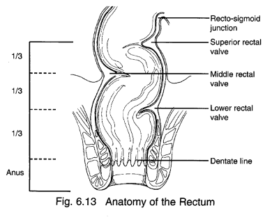
Tumor Location of Incidence (colorectal)
¡Statistically, the sigmoid and rectum are combined
¡When evaluated separately:
§Ascending colon has highest incidence followed by
▪Sigmoid
▪Rectum
Routes of Spread (colorectal)
¡Direct Extension
¡Regional
¡Lymphatic
¡Hematogenous – most common to liver
¡Accidental transplant implantation (occurs during surgery)
¡Superior mesenteric nodes
§Drain ascending and transverse parts of the large intestine
§They communicate with celiac and lumbar nodes which drain to the cisterna chyle
¡Inferior mesenteric nodes
§Drain the left end of the transverse colon to the rectum
§These nodes go to the lumbar nodes and the cysterna chyle

Cisterna chyle
the dilated part of the thoracic duct at its origin in the lumbar region that drains majority of lymph nodes


Lymphatic Drainage of the Rectum
¡Various routes of lymphatic drainage depending on how distal the lesion is
¡Upper portion drains to inferior mesenteric, sacral, and possible hypogastric nodes or ischiorectal nodes which drain to the inguinal nodes
¡Mid portion drains to internal iliac and sacral nodes
¡Lower portion drains to external iliac nodes, rectal wall nodes, and possibly inguinal nodes

Lymphatic Drainage of the Anal Canal
¡Tumors occurring in the upper region will drain to internal iliac, external iliac, and possibly superficial inguinal nodes
¡Tumors of the mid and lower region will drain to superficial inguinal nodes
¡Superficial inguinal nodes are more at risk when the tumor location is distal
Direct Extension (colorectal cancer)
¡Vagina
¡Prostate
¡Bladder
Local Spread (colorectal cancer)
¡Few colon cancers spread through the bowel wall (25%)
¡Rectosigmoid cancers commonly spread through the bowel wall with mets to lymph nodes (50% - 70%)
Metastatic Sites (colorectal cancer)
¡Liver
§Large intestine has direct access to the portal system
¡Lungs
Symptoms Of colorectal cancer
¡Change in bowel habit
¡Hematochezia
¡Melena
¡Mucous in rectum
¡Tenesmus
¡Abdominal mass
¡Pain due to obstruction
¡Anemia
¡Nausea
¡Vomiting
Signs & Symptoms (colorectal)
¡Tumor at any location in the bowel may cause blood in stool
¡Coloration of blood may indicate location
§Bright red blood – tumor is distal
§Dark or tar colored stool – tumor is proximal
¡Pain associated with a proximal tumor may be indicative of a larger lesion
Screening (colorectal)
¡Asymptomatic
¡Male or female >50 without personal family history of polyps or cancer
¡Optical colonoscopy is currently the most sensitive screening tool
Diagnostic Work Up (colorectal)
¡Colonoscopy – examines entire length of the colon
¡Sigmoidoscopy – examines only the lower third
¡Air contrast barium enema
¡Pelvic CT
¡DRE for rectal tumors
¡Endorectal US
¡MRI
Lab Studies (colorectal)
¡Typical blood panels
§CBC
¡Liver function evaluation
¡CEA screening
Prognosis colorectal cancer
¡Disease stage at diagnosis is the most important prognostic factor for patients suffering with colorectal cancer
¡CEA
¡Obstruction & perforation
¡Diabetes
Staging colorectal cancer
¡Colorectal tumors tend to grow at an angle through the bowel wall
¡Staging is dependent on tumor infiltration of mucosal layers
¡The Astler-Duke staging system has been widely used (historically)
¡AJCC is commonly used now
T-Stage (colorectal)
¡T1: The cancer has grown through the muscularis mucosa and extends into the submucosa
¡T2: The cancer has grown through the submucosa and extends into the muscularis propria
¡T3: cancer has grown through the muscularis propria and into the outermost layers of the colon or rectum but not through them. It has not reached any nearby organs or tissues
¡T4: Cancer has grown through the serosa
Treatment for Colon Cancer
¡Surgery is treatment of choice for most lesions
§Colectomy: Removal of affected bowel and associated mesentery
§Temp. colostomy for most
§Perm. Colostomy for extensive involvement
▪Lesions within 6cm of anal verge
Radiation Therapy (colorectal)
¡Alternative to surgery for small T1 or T2 lesions with no nodal involvement
¡Considered when the tumor is near the anal canal
§Surgery in this region can impair sphincter function and result in unnecessary permanent colostomy
Radiation Therapy & Chemo (colorectal)
¡Preoperative:
§Improve surgical outcome
§Can reduce surgical margin and improve functional outcome
§Dose should be approximately 45 Gy with no boost
¡Post-operative:
§Improve local control and reduce risk of metastasis
§3 or 4-Field ports to 45 Gy
§Post-op boost of 10-15 Gy to tumor site only
§Post-op total dose 55 or 60 Gy
§Initial radiation portals should include surgical scar and perineum
Chemotherapy (colorectal)
¡Often given concomitantly with RT
¡Can be used as adjuvant therapy to help control metastatic disease
¡Fluorouracil (5-FU) alone or with Mitomycin C or Leucovorin
§FOLFOX (most widely used adjuvant therapy.)
▪5-FU, leucovorin and oxaliplatin
¡Care must be taken to schedule RT appropriately with chemo
Simulation (rectal)
¡In order to assess small bowel dose the therapist may be required to perform a small bowel follow through during simulation
¡If contrast is to be used, ample time must be scheduled to complete the exam correctly
¡When simulating the digestive tract, the patient may be required to complete a bowel prep
¡A full bladder or belly board (prone pillow)
¡Patients with distal lesions will likely be positioned prone
¡Always speak to oncologist or dosimetrist prior to sim to confirm set up
Treatment Portals (colorectal)
¡3 field approach:
§Tumors in the recto-sigmoid or anal region
§PA + R/L laterals
§Eliminates some dose to small bowel and bladder
§Better coverage of area of concern with laterals
¡4 field approach
§For tumors occurring in other portions of the large bowel
Treatment Design (colorectal)
¡All initial fields must include the tumor + internal iliac nodes + presacral nodes
¡Lower rectum, bladder, vagina, uterus or prostate involvement requires the external iliac nodes to be included
General Portal Margins (colorectal)
PA field
§Bottom of L5 to bottom of obturator foramen
§Lateral margins must extend 1-2 cm beyond pelvic rim
Lateral fields
§Superior and inferior borders match PA
§Anterior margin just anterior to acetabulum
§Posterior margin should include entire sacrum
Portal Design for Anal Region
Patient supine
2 field approach
§AP/PA
▪Superior border should be near the inferior margin o the SI joints
▪Inferior border should be 3 cm inferior to the tumor
▪Lateral margin should extend to cover the inguinal region
Initial fields include tumor + internal & external iliac nodes + inguinal nodes
Radiation Dose (anal cancer)
¡Initial portals should deliver approximately 45 Gy to the tumor and nodal regions
¡Large tumors may require a “cone down” of the AP/PA portals to boost the tumor dose to 50 or 60 Gy
¡Smaller tumors may be boosted to 65 Gy by using a perineal field, brachytherapy, or multifield technique
¡Separate electron fields may be utilized to boost the dose to the superficial inguinal nodes to 65 Gy if involvement is confirmed
Side Effects of RT (colorectal)
¡Diarrhea is the most common acute toxicity
Follow Up ¡Two primary goals: (colorectal)
§Patients with a history of colorectal cancer are at increased risk of a second colorectal primary – colonoscopic screening is indicated
§Second, surveillance may increase the chance of identifying local-regional or distant recurrence that is potentially curable by surgery
Anal Cancer Facts
¡Increased risk for women with ≥ 10 lifetime sexual partners
¡History of anal or genital warts
¡HPV
¡History of gonorrhea or cervical dysplasia
¡Engaging in anal receptive intercourse before the age of 30 or with multiple partners
Clinical Presentation of Anal Cancer
¡Bleeding
¡Anal discomfort
Anal Cancer Histology
¡80% are Squamous cell
Treatment ANal cancer
¡Multi-modality therapy
§RT
§5-FU
§Mitomycin C
Radiation Therapy anal cancer
¡RT alone for patients unable to receive combination therapy
Pancreas Anatomy
¢Located in the retroperitoneum
Head- midline at level of L-1 & L-2 and is overlapped by the duodenum
Body
Tail- Extends to splenic hilum

Epidemiology of Pancreatic cancer
¢Slight male predominance
¢Most common in >60 years of age
¢Incidence higher in African American males
Etiology of pancreatic cancer
¢Possible alcohol and cigarettes
¢Chronic pancreatitis
¢Fatty diets
¢Obesity
Symptoms of pancreatic cancer
¢> 90% of patients present with symptoms
¢Most common presenting symptoms are:
Weight Loss (most common)
Anorexia – loss of appetite
Jaundice – due to obstruction
¢Especially if tumor is in head of pancreas
Pancreatitis
Abdominal and back pain
Routes of Spread for pancreatic cancer
¢Direct Extension
¢Lymphatic
¢Hematogenous
Direct Extension: pancreatic cancer
¢Spreads to:
Bile duct system
Duodenum
Stomach
Spleen
Lymphatic Spread (pancreatic cancer)
¢Nodal involvement in 80-90% of patients
¢Pancreas primarily drains to the:
Peripancreatic nodes
Hepatic nodes
Para-aortic nodes (PAN)
Hematogenous Spread pancreatic cancer
most common are:
¢Liver
¢Lung
¢Pleura
Diagnostic Work Up: pancreatic cancer
¢History and physical exam with special attention to supraclavicular nodes and liver
¢Abdominal ultrasound
¢Pancreatic function tests
¢CT with contrast for defining extent of disease and detecting liver involvement
Histopathology:pancreatic cancer
¢Adenocarcinomas most common (85%)
Specifically duct cell adenocarcinoma
¢Effects exocrine function
¢Most common in the head of the pancreas
Surgery for a Cure: pancreatic cancer
¢Treatment of choice is surgery
¢Only 10%-25% present with localized disease
¢Pancreatduodenectomy- Whipple’s procedure
Remove head of pancreas, duodenum, gastric pyloric antrum, bile duct, and gallbladder
¢Total pancreatectomy
Removal of pancreas, spleen, nodes, and whipple
¢High morbidity and mortality with both
Useful Palliative Surgery (pancreatic cancer)
¢Pancreatic biopsy
¢Regional nodal biopsy
¢Interstitial radioactive iodine
¢Placement of metallic clips
¢Bypass procedures
Pancreatduodenectomy- Whipple’s procedure
Remove head of pancreas, duodenum, gastric pyloric antrum, bile duct, and gallbladder
Radiation Therapy (pancreatic cancer)
¢Survival comparable to Whipple’s procedure
¢Local control is achieved in <50%
¢EBRT to 45-50 Gy with split-course chemotherapy using 5-FU
¢Patient supine
¢3-Field ports (AP + Laterals with wedges with heels anterior)
Boost primary with interstitial brachytherapy using Iodine 125
¢Palliation of pain achieved in most cases
Liver Cancer
¢Primary liver cancer extremely rare
¢More common in males
¢More common in Africa and Asia
¢Most liver cancer are metastatic from another primary
Etiology: liver cancer
¢Hepatitis B is implicated
¢Thorotrast contrast may lead to hepatoma
¢Macronodular cirrhosis
¢Androgenic anabolic steroid abuse
¢Death is usually from liver failure
Treatment: liver cancer
¢Treatment of choice is surgical resection
¢Chemo –5-FU and doxorubicin
¢Radiation- Palliative role only
25-30 Gy in 3-4 weeks
Gallbladder Epidemiology & Histology
¢More common in women
¢Most common in >60 years of age
¢Adenocarcinoma most common histology
Etiology: Gallbladder cancer
¢Gallstones
¢Typhoid Carriage- Salmonella Typhi
Bacteria in gallbladder
Routes of Spread: gallbladder cancer
¢Direct extension to liver
¢Lymphogenous (most common)
¢Transperitoneal seeding
Treatment & Prognosis: gallbladder cancer
¢Treatment of choice is surgery
Cholecystectomy
¢Radiation therapy is rarely used
¢Only 5% survive > 1 year