Renal Transplant and Postsurgical Complications
 Call Kai
 Learn
Learn Practice Test
 Spaced Repetition
 Match
Match Flashcards
 Knowt Play
1/19

There's no tags or description
Looks like no tags are added yet.
Name | Mastery | Learn | Test | Matching | Spaced | Call with Kai | Chat |
|---|
No analytics yet
Send a link to your students to track their progress
20 Terms
— is often the best indicator of renal transplant rejection
renal biopsy
the transplanted kidney is also referred to as the
allograft
the allograft is often placed within the
right or left lower quadrant
the donor artery is typically anastomosed to the
external iliac artery and external iliac vein of the recipient
the donor ureter is attatched to the recipient
urinary bladder
the donor kidney should appear — to the normal kidney
sonographically similar
— is considered normal in the transplanted kidney
mild pelviectasis
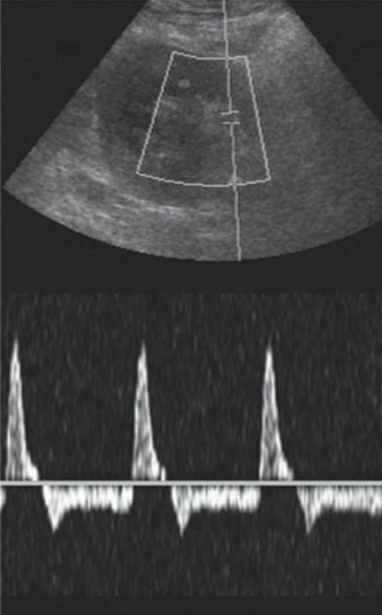
Renal vein thrombosis. A spectral Doppler waveform from a segmental renal artery in the renal sinus demonstrating reversed diastolic flow in a patient presented with abrupt anuria 3 hours postsurgery. Thrombosis of the main renal vein was found at surgery.
the normal waveform should yeild low resistance with —
continuous diastolic flow
the resistive insitive indices should be —
0.6 to 0.8
anything over — is considered abnormal
0.9
the renal vein should demonstrate — and may be examined at the anastomosis site eith the iliac vein as well
normal continous flow away from the kidney
postrenal transplant fluid collections are commom and could be a
lymphocele, urinoma, hematoma, or abscess
these fluid collections may appear similar with sonogeaphy, with most
echogenic, complex, or anechoic
clinical signs of transplant rejection include
anuria, oliguria, elevated creatinine nad BUN, proteinuria, hypertension, and enlargement of the transplant
complications of a renal transplant include
rejection, infection, obstruction, vascular stenosis, renal artery stenosis, renal artery thrombosis, renal vein thrombosis, pseudoaneurysm, and fistulas
— is the most common vascular complication following renal transplantation
renal artery stenosis
the sonographic indication of renal artery stenosis includes — at the level of the anastomosis site
color aliasing and turbulent flow
the diagnosis of renal artery stenosis in a renal transplant includes an elevated peak systolic velocity greater than
200 to 250 cm per second
the diagnosis of renal artery stenosis in a renal transplant includes an artery to external iliac ratio greater than — and post stenostic turbulence2.0 to 3.0