6.2.1 nerve impulses
1/24
There's no tags or description
Looks like no tags are added yet.
Name | Mastery | Learn | Test | Matching | Spaced |
|---|
No study sessions yet.
25 Terms
structure of cellulose
chain of beta glucose - unbranched - 1,4 glycosidic bonds to make unbranched
every other glucose is inverted to make sure hydroxyl groups are in right position for reaction
describe the function of the capsule in biology
protection also can help bacterial cells stick together (fro further protection)
state what is happening during prophase in mitosis
chromosomes condense
chromosomes become visible - this differentiates from interphase and mitosis
nuclear envelope disappears
centriole moves to opposite ends of the cell
brain structure
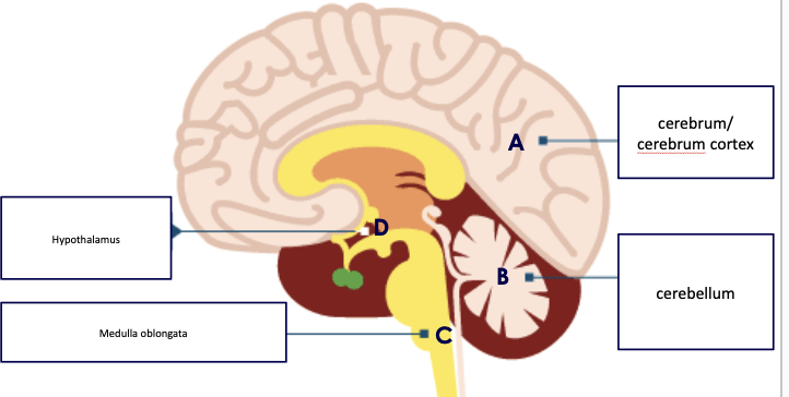
cerebrum / cerebral cortex
Controls intelligence, personality, conscious thought and high-level functions, such as language and verbal memory.
The outer layer is called the cerebral cortex, which is split into two hemispheres and is highly folded.
cerebellum
Controls balance, coordination of movement and muscular activity.
medulla oblongata
Controls unconscious activities such as heart rate and breathing rate,
hypothalamus
Regulating centre for temperature and water balance within the body.
neurones
specialised cells adapted to rapidly carrying electrochemical changes called nerve impulses from one part of the body to another.
neurone = single nerve cell
nerve = bundle of neurones (nerve cells) surrounded by tough connective tissue
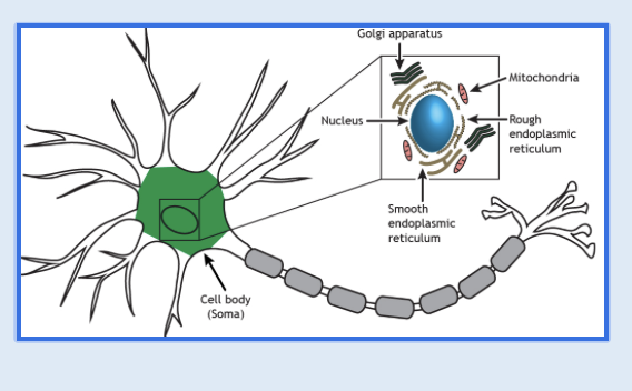
cell body of a neurone cell
cell body contains all the usual cell organelles including
nucleus
large amounts of RER
the large RER is associated with the production of proteins and neurotransmitters
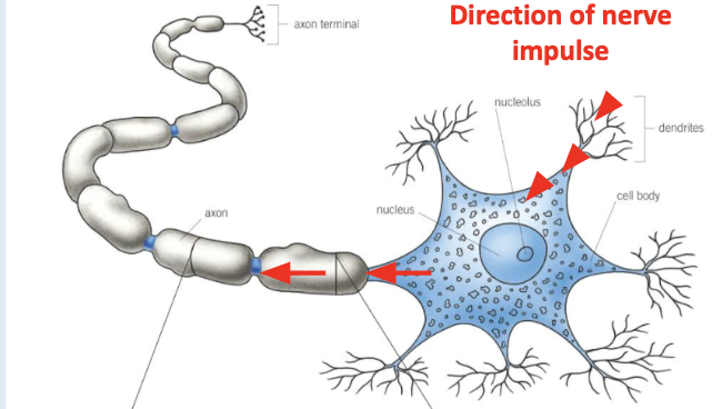
Dendrons
are extension of cell body which subdivide into smaller branched fibres called dendrites
dendrites carry nerve impulses towards the cell body
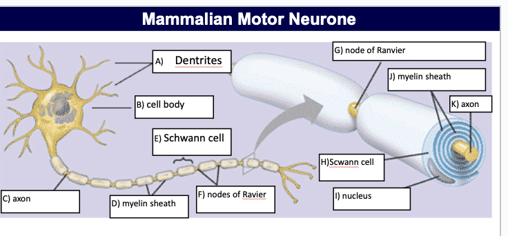
myelin sheath - fatty ;layer around axon
axon and Schwann cells
an axon í a single long fibre that carries nerve impulses away from the cell body
the axon is surrounded by Schwann cells which protect it and provide electrical insulation
salutary conduction takes place - iron channels open and impulse can jump from uninsulated area to uninsulated area - increasing the speed the impulse moves down
Schwann cells and myelin sheath
Schwann cells carry out phagocytosis to remove cell debris and play a part in nerve regeneration
the Swann cells wrap around the axon many times - layers of their membranes build up around it
Schwann cell membranes are rich in a lipid called myelin
a Schwann cell envelops and rotates around the axon forming myelin sheath
neurones with a myelin sheath are called myelinated
node of Ranvier
between adjacent Schwann cells there are constrictions where there is no myelin sheath, these are called nodes of Ravier
the constrictions are 2-3um long and occur every 1-3mm in humans
diagram of mammalian motor neurone
active transport defined
the movement of molecules and ions through a cell membrane against their concentration gradient form a region of low concentration to a region of high concentration using ATP
describe the difference between a channel protein and a carrier protein
channel proteins are water filled pores that go straight through the membrane, structured shape allowing charged substances (ions) to diffuse through cell membrane, - facilitated diffusion for specific ions
carrier proteins can switch between 2 shapes, allowing the binding site of the carrier to be open on different sides of the membrane.
neurones are cells with long projections called axons. describe the structure of an axon membrane
normal cell membrane - cell membranes are a phospholipid bilayer and are important as they establish the boundary of each cell
L2 - maintaining the resting potential
nerve impulse
a self-propagating (self sustaining) wave of electrical activity that travels along the axon membrane
it is a temporary reversal of the electrical potential difference across the axon membrane
the reversal is between two states
the resting potential - axon not transmitting an impulse
the action potential - axon is transmitting an impulse
what is electrical potential difference due to?
across cells membrane is due to unequal ion concentrations
resting potential
resting potential occurs in resting axons - neurons that are not transmitting impulse
the inside of the axon always has a negative electrical potential compared to the outside of the axon
there’s always something happening inside it - it isn’t actually resting.
at resting potential (-70mV) the axon is said to be polarised
it is more negative inside the axon than outside
the -70mV resting potential is achieved through the movement of ions (specifically Na+ and K+) across the axon membrane
movement of ions across axon membrane
the phospholipid bilayer of the axon prevents sodium Na+ and potassium K+ ions diffusing across it and can cross the membrane in one of two ways
facilitated diffusion via channel proteins - both Na and K moving down their respective concentration gradients
active transport via a carrier protein with K+ are actively transported into the axon and Na+ out of the axon via a special carrier called a sodium potassium pump
establishing and maintaining the resting potential
two factors contribute to establishing and maintaining the resting potential
differential membrane permeability
the active transport of sodium ions and potassium ions (via the Na+/ K+) pump
what does differential membrane permeability mean?
the axon membrane is not equally permeable to all ions
it allows some ions to pass through more easily than others due to distribution of (open) ion channel proteins in the membrane so it differs
at resting - more permeable to potassium ions and sodium ions - effecting the amount of ions that are entering and leaving the axon