Comp vert anatomy lab
 Call Kai
 Learn
Learn Practice Test
 Spaced Repetition
 Match
Match Flashcards
 Knowt Play
1/218

Earn XP
Description and Tags
Lab 1
Name | Mastery | Learn | Test | Matching | Spaced | Call with Kai | Chat |
|---|
No analytics yet
Send a link to your students to track their progress
219 Terms
Two types of vertebrae that make up the shark’s vertebral column
Trunk and caudal
What is the shape and structure of a shark’s vertebral centrum
Cylindrical and biconcave (amphicoelous)

Vertebral centrum
The thick, solid front part of a vertebra that forms the main vertebral body

Notochord
A flexible, cylindrical structure found in the embryos of all chordates
Chordate key characteristic
Notochord, a nerve tube along the back that becomes the central nervous system, openings in the pharynx used for filter feeding
Foramen
Natural opening, hole or passage found in bones
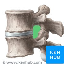
Which foramen allows the passage of the dorsal root of a spinal nerve
Intervertebral foramen
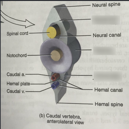
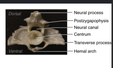
What structure extends ventrally from the centrum in caudal vertebrae
The hemal arch which terminates in a hemal spine
What are basapophyses and what attaches to them
Short ventrolateral projections on trunk centra (homologous to the base of hemal arches) that articulate with short cartilaginous ribs
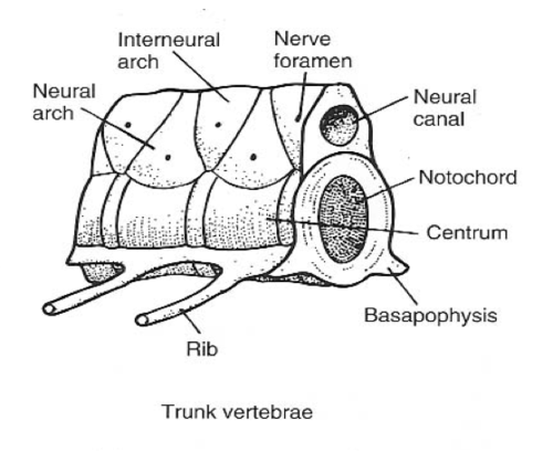
What structures are located between the basapophyses of adjacent trunk vertebrae
Very small triangular ventral intercalary plates
Heterocercal caudal fin
A tail type with two unequal lobes (larger dorsal lobe, smaller ventral lobe) where the vertebral column tilts upward

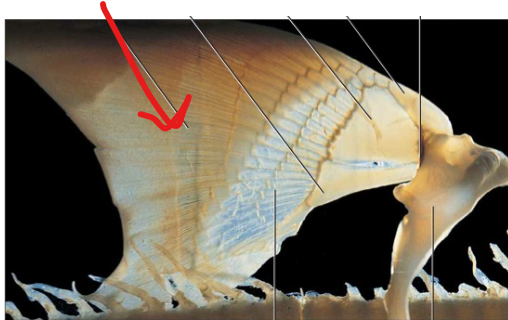
Ceratotrichia
Fibrous fin rays that extend into and stiffen the fin web on the caudal and dorsal fins

Dorsal Fin Spines
Protective skeletal spines that precede each of the dorsal fins (anterior and posterior)

Basal Pterygiophore
A large, proximal cartilaginous skeletal element that supports the bases of each dorsal fin

Caudal fin skeletal support
Elongated neural and hemal arches paired with fibrous ceratotrichia stiffening the fin web
Dorsal root Foramen
The opening located in each dorsal intercalary plate for the passage of the dorsal root of a spinal nerve
Ventral Root foramen
The opening located in each neural plate/arch for the passage of the ventral root of a spinal nerve
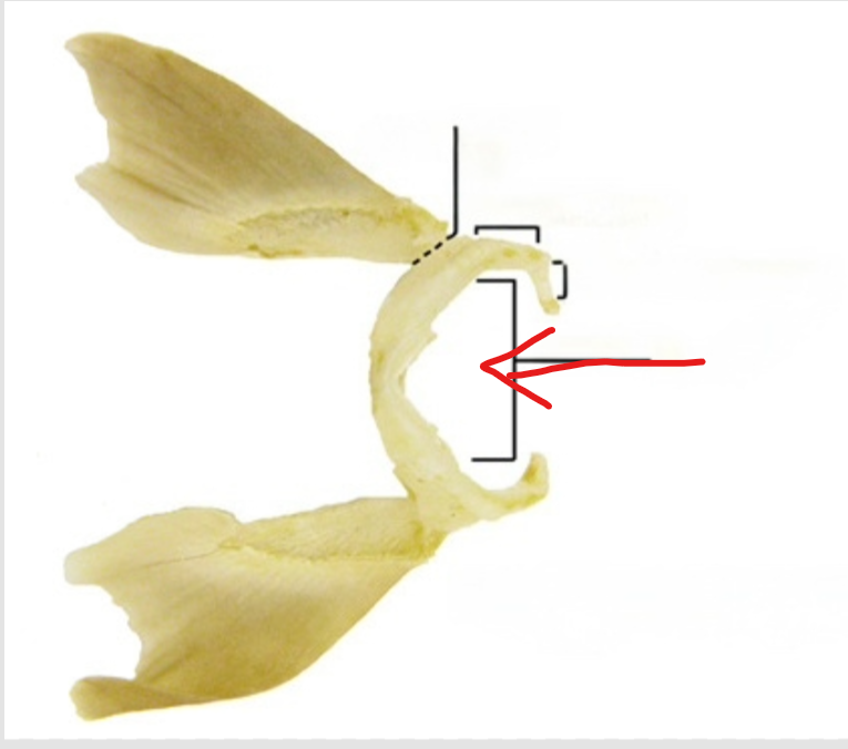
Scapulocoracoid bar
The U-shaped skeletal complex forming the pectoral girdle, situated just posterior to the last gill slit
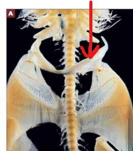
Coracoid Bar
The ventral portion of the U-shaped scapulocoracoid bar
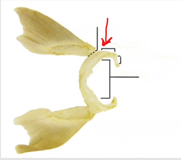
Scapular Cartilage
The dorsal, distal part of the pectoral girdle that articulates with the pectoral fin
Glenoid surface
The dorsolateral smooth surface on the scapular cartilage that accommodates articulation of the pectoral fin

Suprascapular Cartilage
A small attenuated medially curved structure articulating dorsally with the scapular cartilage to complete the U of the pectoral girdle

Propterygium
The anterior basal pterygiophore supporting the proximal end of the pectoral fin (also found in pelvic fins)

Mesopterygium
The middle basal pterygiophore supporting the proximal end of the pectoral fin
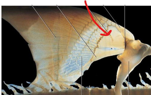
Metapterygium
The posterior (or elongated) basal pterygiophore supporting the proximal end of pectoral and pelvic fins
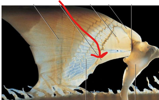
Radial Pterygiophores
Rows of cartilages articulating with and radiating from the basal pterygiophores to form the major support of the fin

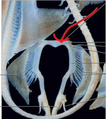
Puboischiadic bar
The short, horizontal bar forming the pelvic girdle, located just anterior to the cloaca
Iliac Processes
Small processes projecting from the anterior ends of the puboischiadic bar.
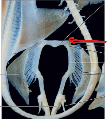
Acetabular surface
A smooth surface on the posterolateral aspect of the puboischiadic bar where the pelvic fins articulate.
Clasper
A modified sperm transfer organ present on the pelvic fins of male sharks, supported by cartilages derived from radial pterygiophores.
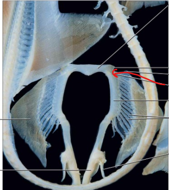
Splenial
A dermal bone on the medial surface of the mandible in Necturus
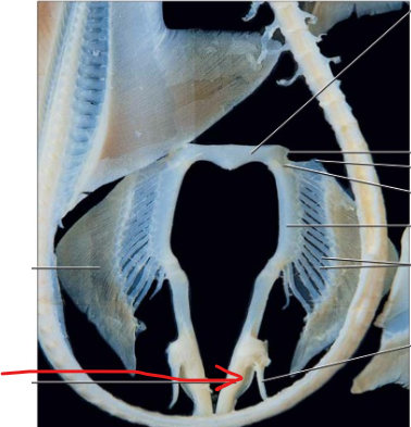
Atlas/Cervical
The single first vertebra that articulates with the two occipital condyles of the skull, allowing head movement independent of the vertebral column
Occipital condyles
Paired bony projections on the posterior aspect of the skull that articulate with the atlas
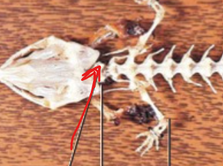
Neural arches
Arches arising dorsally from the centrum to form the vertebral canal housing the spinal cord

Neural spines
A process projecting posteriorly from the neural arch of a vertebra.
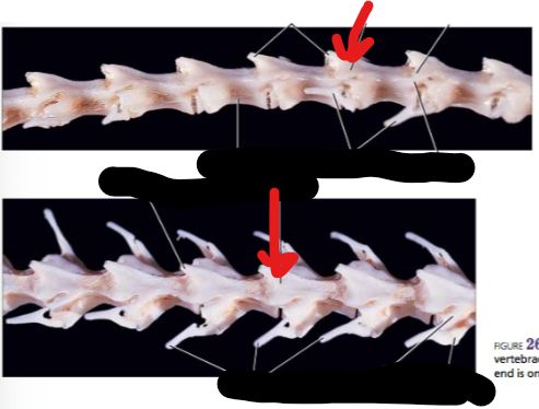
Prezygapophyses
A pair of anteriorly located articular processes on the neural arch that overlap with the postzygapophyses of an anterior adjacent vertebra to reduce vertical bending.

Postzygapophyses
A pair of articular processes projecting from the posterior end of the neural arch that articulate with prezygapophyses.
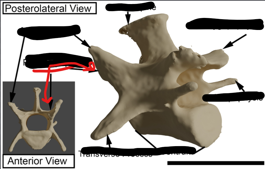
Transverse process
A caudally projecting process consisting of a dorsal diapophysis and a ventral parapophysis where ribs articulate

Diapophysis
The dorsal portion of the transverse process arising from the neural arch.
Parapophysis
The ventral portion of the transverse process arising from the centrum.

Caudal vertebrae
Vertebrae following the sacral region that lack ribs, feature prominent neural spines, and have a ventral hemal arch with a spine.

Hemal arch
An arch with a spine attached to the ventral surface of the caudal centrum through which the caudal artery and vein pass.
Ribs (Bicipital ribs)
Two-headed tetrapod ribs that articulate with the transverse processes of the trunk vertebrae.
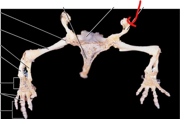
Sternum
A breastbone structure that is absent as a solid element in Necturus, though small cartilage pieces embedded in the thoracic myosepta may represent it.
Coracoids
Broad plates of cartilage that project medially to overlap in the midline of the pectoral girdle.

Coracoid foramen
An opening piercing the coracoid plate that permits the passage of blood vessels and nerves.
Procoracoid
An ossified element projecting dorsally from the rim of the glenoid fossa with a distal suprascapular cartilage.


Suprascapular cartilage
A distal cartilaginous structure projecting dorsally from the ossified scapula.
Glenoid fossa
The depression on the pectoral girdle with which the head of the humerus articulates.
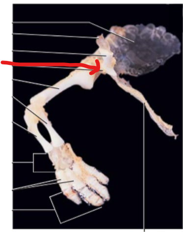
Brachium
The upper arm segment of the forelimb containing a single bone, the humerus.

Antebrachium
The forearm segment containing two bones: the medial radius and the lateral ulna.
Manus
The wrist and hand segment of the forelimb, consisting of the carpus, metacarpals, and phalanges terminating in four digits.
Humerus
The single bone found in the brachium (upper arm).

Radius
The medial bone of the antebrachium (forearm).
Ulna
The lateral bone of the antebrachium (forearm).
Carpus
The proximal portion of the manus (wrist) consisting of six small bones arranged in three rows, serving as the distal point of flexure.
Metacarpals
The intermediate series of bones in the manus, with one metacarpal per digit.
Phalanges
The distal bones of the digits
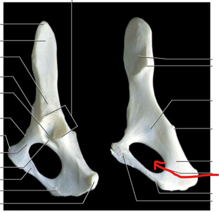
Pubis
The anterior cartilaginous element of the pelvic girdle that projects anteriorly and is pierced by the obturator foramen.
Ischium
The bony, posteriorly projecting element of the pelvic girdle.

Ilium (Ilia)
The dorsal bone of the pelvic girdle that extends to articulate with the modified rib of the sacral vertebra.

Acetabulum
The depression at the junction of the pubis, ischium, and ilium that accommodates the head of the femur.
Puboischiadic plate
The ventral plate formed by the medioventral symphysis of the paired anterior cartilaginous pubes and posterior bony ischia.

Obturator foramen
An opening piercing the pubis to permit the passage of the obturator nerve.
Pelvic canal
The space enclosed by the pelvic girdle that permits digestive and urogenital organs to access the cloaca.
Thigh
The upper leg segment containing a single bone, the femur.
Shank
The lower leg segment containing the tibia and fibula.
Pes
The foot segment of the hindlimb, consisting of the tarsus (ankle), metatarsals, and phalanges terminating in four digits.
Femur
The single bone occurring in the thigh (upper leg) that articulates proximally with the acetabulum.
Genu
The proximal portion of the pes (ankle), consisting of six small tarsal bones in three rows that articulate with the shank.
Hallux
The medial bone of the shank (lower leg). Big toe.
Fibula
The lateral bone of the shank (lower leg).
Tarsals
The six small bones arranged in three rows within the tarsus (ankle).
Metatarsals
The intermediate series of bones in the pes.
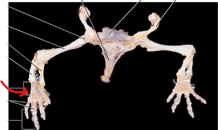
Tympanohyal
The most dorsal element of the cranial horn of the hyoid apparatus.

Stylohyal
A bony element of the cranial horn located ventral to the tympanohyal and dorsal to the epihyal.
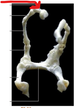
Epihyal
A bony element of the cranial horn positioned between the stylohyal and the ceratohyal/body of the hyoid.
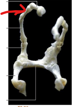
Cranial horn (Hyoid)
The anterior/cranial projection of the hyoid apparatus.

Caudal horn (Hyoid)
The posterior projection of the hyoid apparatus extending from the body (basihyal).

Body (Hyoid)
The central transverse portion of the hyoid apparatus that connects the cranial and caudal horns.

Lumbar vertebrae
The region of the vertebral column located in the lower back between the thoracic and sacral regions.

Caudal vertebrae
The vertebrae forming the tail region posterior to the sacrum.
Fibrocartilaginous intervertebral discs
Cushioning discs located between adjacent individual vertebrae in the mammalian column.
Centrum (Body)
The solid body of a vertebra forming the primary ventral support for the spinal cord.
Vertebral canal
The opening formed dorsally by the neural arch/laminae and ventrally by the centrum through which the spinal cord passes.

Demifacets
Articular facets occurring on the centrum of adjacent thoracic vertebrae that articulate with the capitulum of a rib.
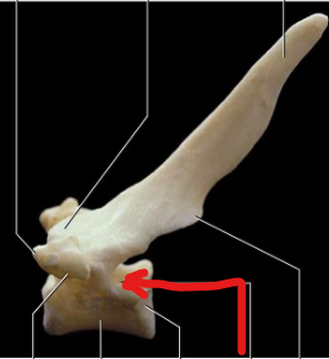
Laminae
A pair of dorsal plates that complete the vertebral canal, whose dorsocaudal extensions form the spinous process.

Spinous process
The dorsocaudal projection formed by the fusion of the laminae, homologous with the neural spine.
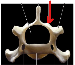
Pedicles
A pair of bony segments extending between the laterally projecting transverse processes (diapophyses) and the centrum.

Transverse processes (Diapophyses)
Lateral projections on a vertebra; in thoracic vertebrae, their ventral surface bears an articular facet for the rib tuberculum.

Articular facet (Thoracic transverse process)
A smooth facet on the ventral surface of the transverse process that articulates with the tuberculum of a rib.

Transverse foramen
A characteristic opening passing through the transverse process of the first six cervical vertebrae in cats, transmitting the vertebral arteries and veins.

Atlantal foramen
The first intervertebral foramen located dorsal to the prezygapophysis of the atlas, through which the first spinal nerve and vertebral vein exit and the vertebral artery enters.

Odontoid process (dens)
A prominent cranial projection of the axis (formed by the fusion of the centrum of the atlas) flanked by prezygapophyses to articulate with the atlas.

Lumbar Vertebrae
The seven largest vertebrae in the cat, increasing in size caudally and featuring prominent pleurapophyses, accessory processes, and mammillary processes.

Pleurapophysis
A prominent, cranially projecting landmark on lumbar vertebrae representing a transverse process fused with an embryonic rib.
Accessory processes
Processes evident on all but the last two lumbar vertebrae that attach ligaments and muscles to stabilize the lower back against rotation.
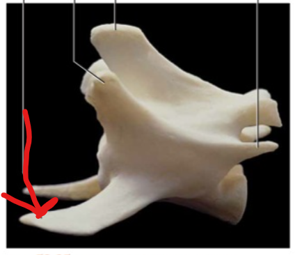
Mammillary processes
Processes present on all lumbar vertebrae that work with accessory processes to attach stabilizing ligaments and muscles.

Sacrum
A complex of three fused vertebrae in the adult cat (decreasing in size caudally) that acts as a solid brace for the pelvic girdle.

Caudal Vertebrae
The vertebrae of the tail, being the smallest and most variable in number; more cranial caudal vertebrae show typical vertebral features, while more caudal ones resemble simple cylinders (centra).
