Topic 1 - Blood Vessels
1/40
There's no tags or description
Looks like no tags are added yet.
Name | Mastery | Learn | Test | Matching | Spaced |
|---|
No study sessions yet.
41 Terms
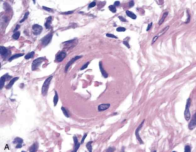
Hypertensive vascular disease. (A) Hyaline arteriolosclerosis. The arteriolar wall is thickened with the deposition of amorphous proteinaceous material (hyalinized), and the lumen is markedly narrowed.
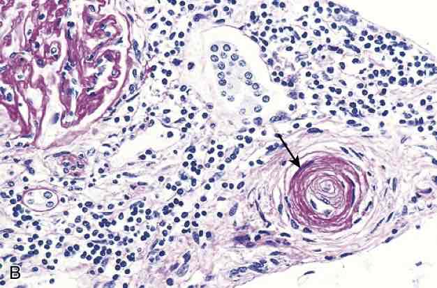
Hypertensive vascular disease. (B) Hyperplastic arteriolosclerosis (“onion-skinning”) (arrow) causing luminal obliteration (periodic acide Schiff stain)
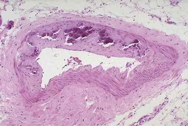
Mönckeberg medial sclerosis. Note the purplish blue amorphous densities representing calcification. Lumen is unaffected.
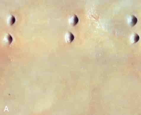
Fatty streaks. (A) Aorta with yellowish fatty streaks mainly near the ostia of branch vessels.
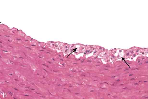
Fatty streaks. (B) Fatty streak in an experimental hypercholesterolemic rabbit, demonstrating intimal, macrophage-derived foam cells (arrows).
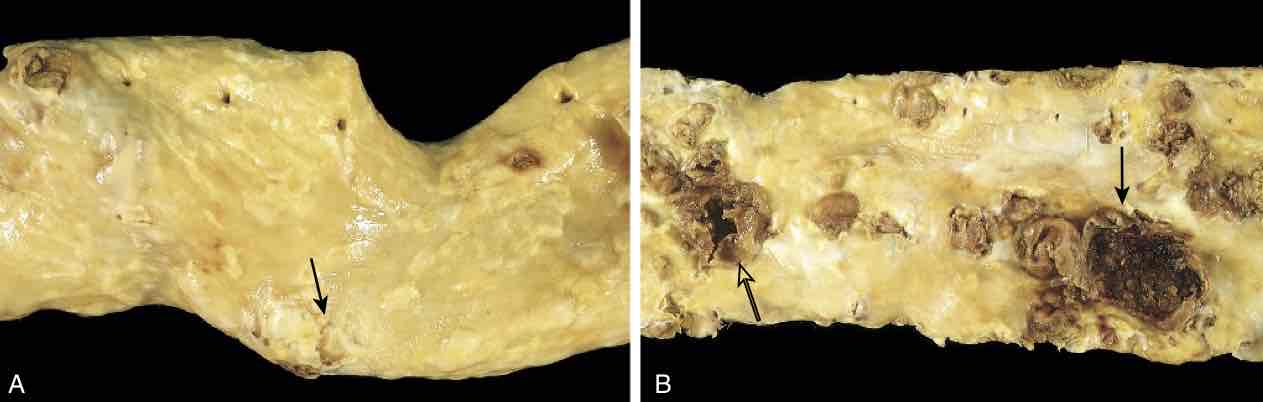
Atherosclerotic lesions. (A) Aorta with mild atherosclerosis composed of fibrous plaques, one denoted by the arrow. (B) Aorta with severe diffuse complicated lesions, including an ulcerated plaque (open arrow), and a lesion with overlying thrombus (closed arrow).
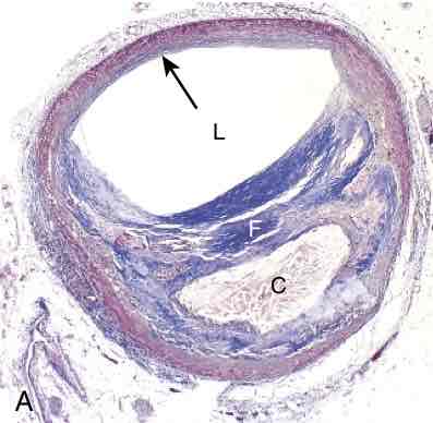
Atherosclerotic plaque, coronary artery. (A) Overall architecture demonstrating fibrous cap (F) and a central necrotic (largely lipid) core (C); collagen (blue) is stained with Masson trichrome. The lumen (L) is moderately narrowed by this eccentric lesion, which leaves part of the vessel wall unaffected (arrow).
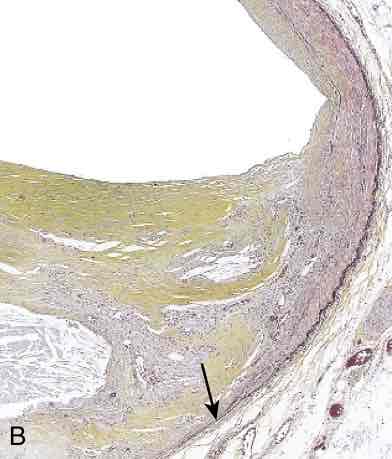
Atherosclerotic plaque, coronary artery. (B) Medium-power view of the plaque shown in A, stained for elastin (black); the internal and external elastic membranes are attenuated, and the media of the artery is thinned under the most advanced plaque (arrow).
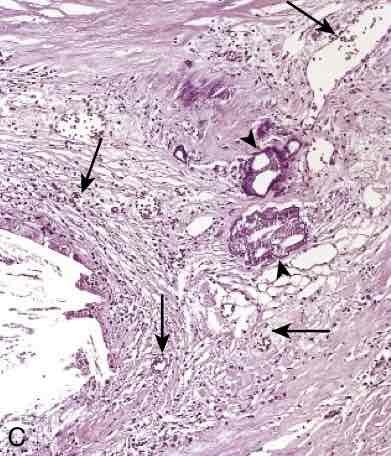
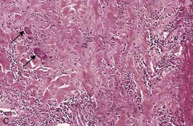
Atherosclerotic plaque, coronary artery. (C) High-power view of the junction of the fibrous cap and core, showing scattered inflammatory cells, calcification (arrowheads), and neovascularization (small arrows).
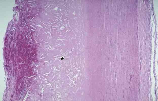
Atherosclerosis. This cross-section of aorta shows advanced atheroma with numerous thin cholesterol clefts (asterisk). The luminal surface on the far left shows ulceration of the fibrous cap with hemorrhage. Ulceration is likely to be complicated by thrombosis. The media and adventitia are normal.
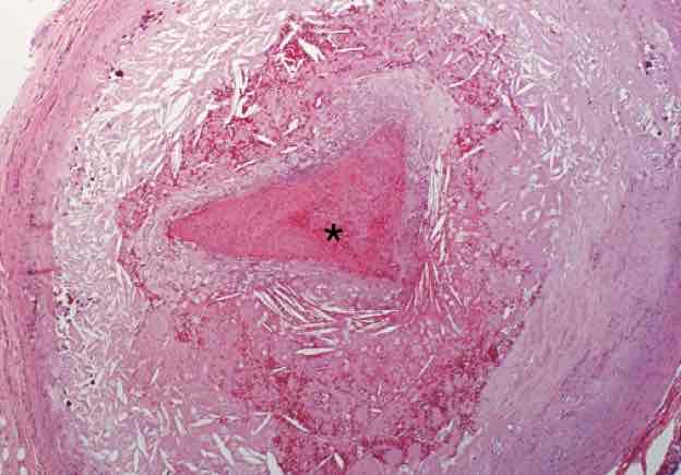
Atherosclerosis with thrombus. This coronary artery cross-section shows advanced atherosclerosis complicated by an occlusive thrombus.
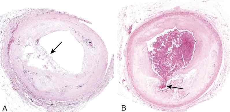
Atherosclerotic plaque rupture. (A) Plaque rupture without superimposed thrombus in a patient who died suddenly. (B) Acute coronary thrombosis superimposed on an atherosclerotic plaque with focal disruption of the fibrous cap, triggering fatal myocardial infarction. In both A and B, an arrow points to the site of plaque rupture.
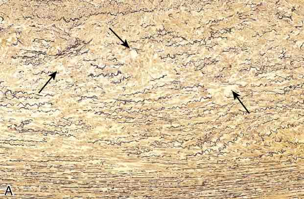
Cystic medial degeneration. (A) Cross-section of aortic media from a patient with Marfan syndrome, showing marked elastin fragmentation and areas devoid of elastin that resemble cystic spaces (arrows). In both (A) and (B), elastin is stained black.
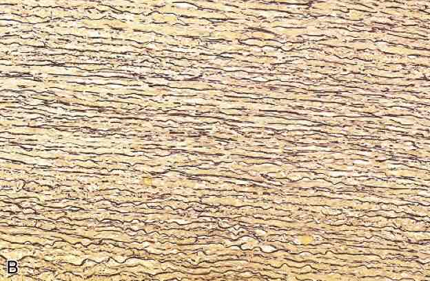
Cystic medial degeneration. (B) Healthy media for comparison, showing the regular layered pattern of elastic tissue. In both (A) and (B), elastin is stained black.
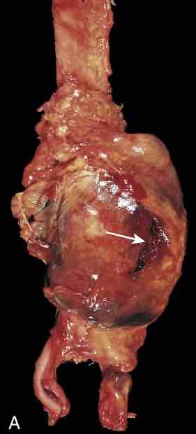
Abdominal aortic aneurysm. (A) External site of rupture of a large aortic aneurysm is indicated by the arrow.
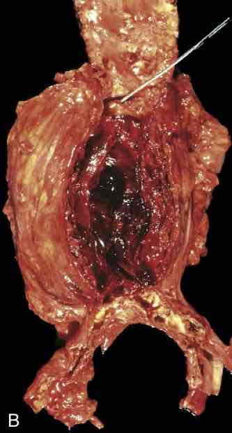
Abdominal aortic aneurysm. (B) Opened aorta, with the location of the rupture tract indicated by a probe. The wall of the aneurysm is attenuated, and the lumen is filled by a large, layered thrombus.
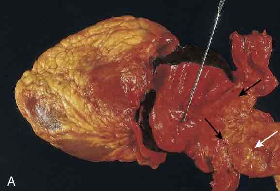
Aortic dissection. (A) An opened aorta with a proximal dissection originating from a small, oblique intimal tear (identified by the probe) associated with an intramural hematoma. Note that the intimal tear occurred in a region largely free of atherosclerotic plaque. The distal edge of the intramural hematoma (black arrows) lies at the edge of a large area of atherosclerosis (white arrow), which arrested the propagation of the dissection. The heart is on the left.
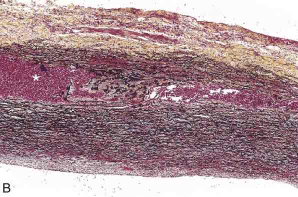
Aortic dissection. (B) Histologic preparation showing the dissection and intramural hematoma (asterisk). Aortic elastic layers are black, and blood is red in this section, stained with Movat stain.
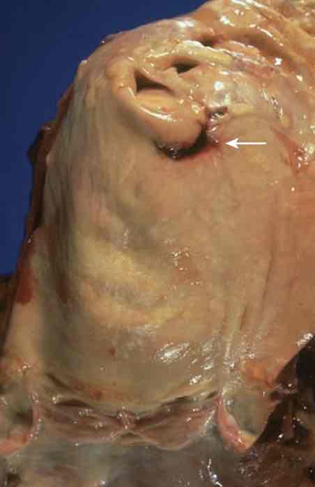
Aortic dissection. The aorta has been opened to show an intimal tear (arrow) located 7 cm above the aortic valve and just below the origin of the great vessels.
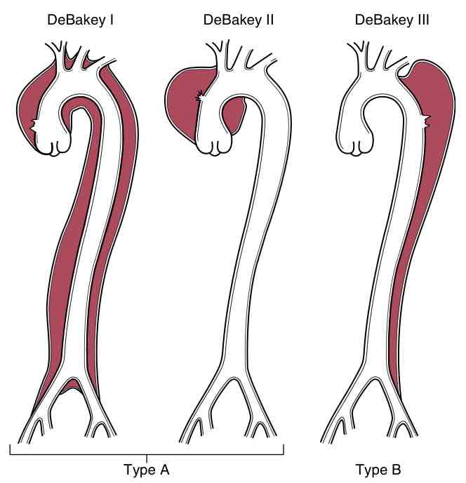
Classification of dissections. Type A dissections (proximal) involve the ascending aorta, either as part of a more extensive dissection (DeBakey type I), or in isolation (DeBakey type II). Type B dissections (distal, or DeBakey type III) arise after the takeoff of the great vessels. Type A dissections typically have the most serious complications and the greatest associated mortality.
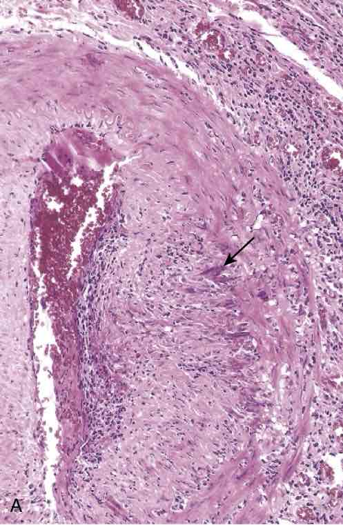
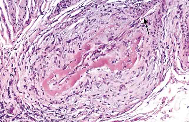
Giant cell arteritis. (A) Hematoxylin-eosin-stained section of a temporal artery showing giant cells near the fragmented internal elastic membrane (arrow), along with medial and adventitial inflammation.
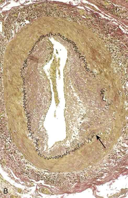
Giant cell arteritis. (B) Elastic tissue stain demonstrating focal destruction of the internal elastic membrane (arrow) and medial attenuation and scarring.
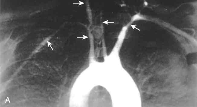
Takayasu arteritis. (A) Aortic arch angiogram showing reduced flow of contrast material into the great vessels and narrowing of the brachiocephalic, carotid, and subclavian arteries (arrows).
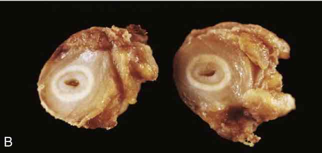
Takayasu arteritis. (B) Cross-sections of the right carotid artery from the patient shown in A demonstrating marked intimal thickening and luminal narrowing. The white circles correspond to the original vessel wall; the inner core of tan tissue is the area of intimal hyperplasia.
Takayasu arteritis. (C) Histologic appearance in active Takayasu aortitis illustrating destruction and fibrosis of the arterial media associated with mononuclear infiltrates and giant cells (arrows).
Polyarteritis nodosa, associated with segmental fibrinoid necrosis and thrombotic occlusion of a small artery. Note that part of the vessel (upper-right, arrow) is uninvolved.
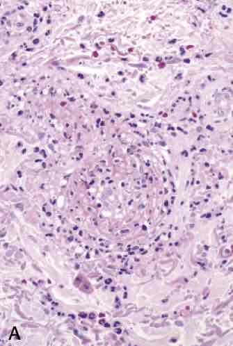
ANCA-associated small vessel vasculitis. (A) Microscopic polyangiitis (leukocytoclastic vasculitis) with fragmented neutrophils in the thickened vessel wall.
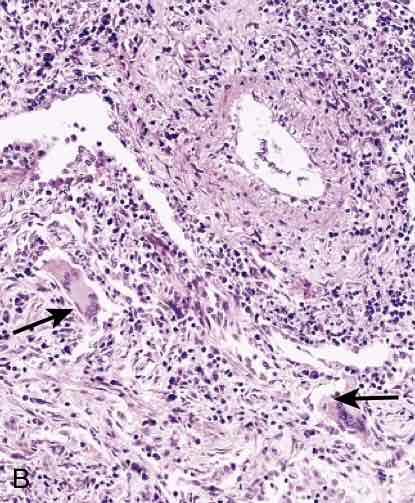
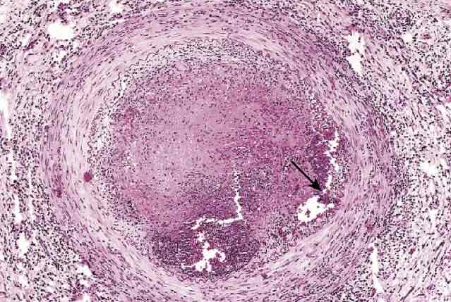
ANCA-associated small vessel vasculitis. (B and C) Granulomatosis with polyangiitis. (B) Vasculitis of a small artery with adjacent granulomatous inflammation including giant cells (arrows).
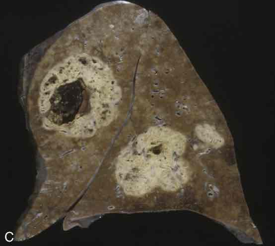
ANCA-associated small vessel vasculitis. (B and C) Granulomatosis with polyangiitis. (C) Lung from a patient with granulomatosis with polyangiitis, demonstrating large nodular cavitating lesions.
Thromboangiitis obliterans (Buerger disease). The lumen is occluded by thrombus containing a sterile abscess (arrow), and the vessel wall is infiltrated with leukocytes.
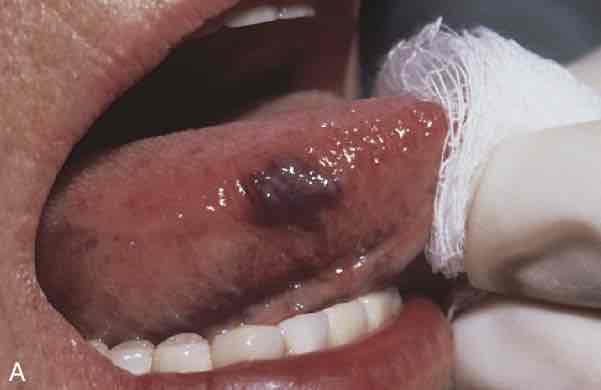
Hemangiomas. (A) Hemangioma of the tongue.
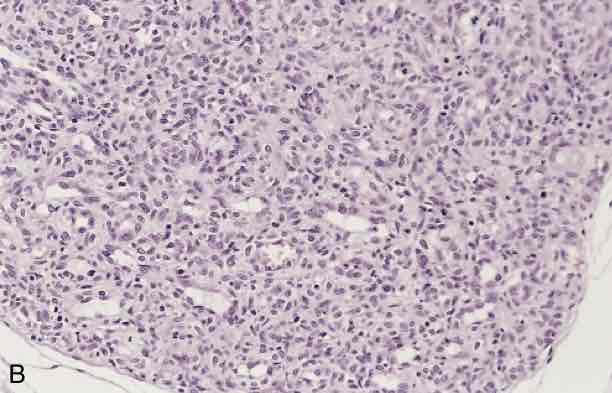
Hemangiomas. (B) Histologic appearance of infantile capillary hemangioma.
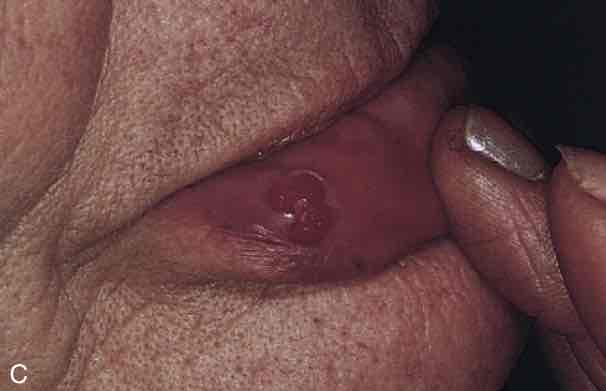
Hemangiomas. (C) Pyogenic granuloma of the lip.
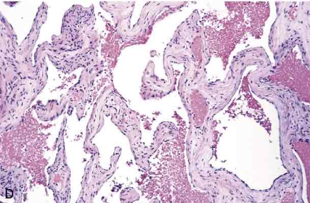
Hemangiomas. (D) Histologic appearance of cavernous hemangioma.
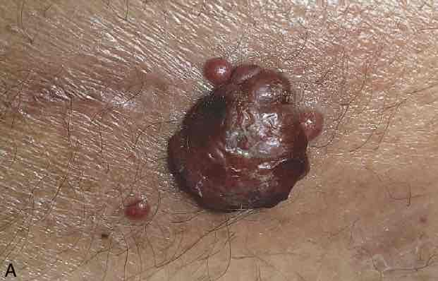
Bacillary angiomatosis. (A) Characteristic cutaneous lesion.
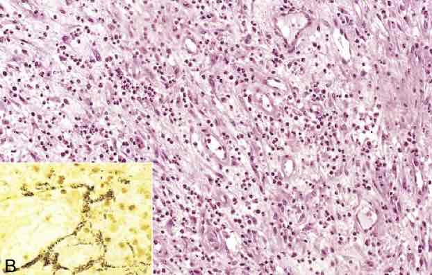
Bacillary angiomatosis.(B) Histologic features are those of acute inflammation and capillary proliferation. Inset, Modified silver (Warthin-Starry) stain demonstrates clusters of tangled bacilli (black).
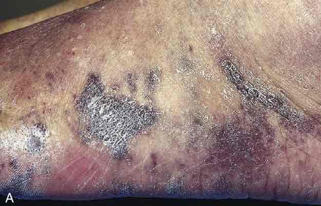
Kaposi sarcoma. (A) Characteristic coalescent cutaneous red-purple macules and plaques.
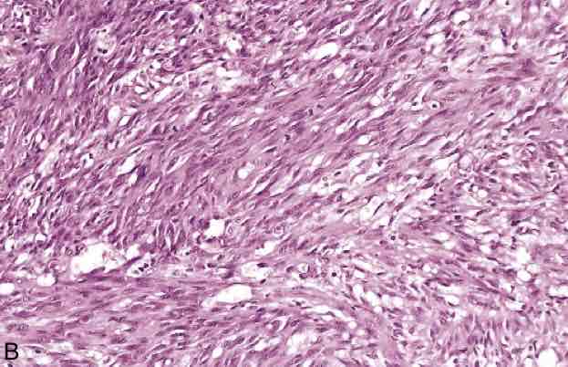
Kaposi sarcoma. (B) Histologic view of the nodular stage, demonstrating sheets of plump, proliferating spindle cells and slitlike vascular spaces
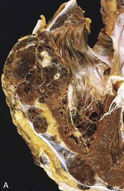
Angiosarcoma. (A) Angiosarcoma of the right ventricle.
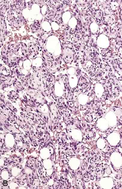
Angiosarcoma. (B) Moderately differentiated angiosarcoma with dense clumps of atypical cells lining distinct vascular lumina.
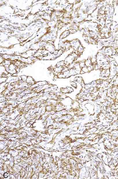
Angiosarcoma. (C) Immunohistochemical staining of angiosarcoma for the endothelial cell marker CD31.