Anatomy Lecture: Cellular Level of Organization (Ch.3)
 Call Kai
 Learn
Learn Practice Test
 Spaced Repetition
 Match
Match Flashcards
 Knowt Play
1/27

Earn XP
Description and Tags
Review of cell membrane features
Name | Mastery | Learn | Test | Matching | Spaced | Call with Kai | Chat |
|---|
No analytics yet
Send a link to your students to track their progress
28 Terms
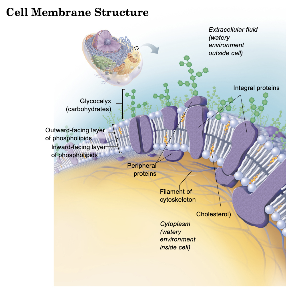
What are the 4 Functions of the Cell/Plasma Membrane?
Physical Barrier
Regulates Transport —>Influx, efflux [selective permeability]
Communication —> variety of receptors
Structural Support —> Anchoring point for cytoskeleton; junctions between cells or cell-extracellular matrix
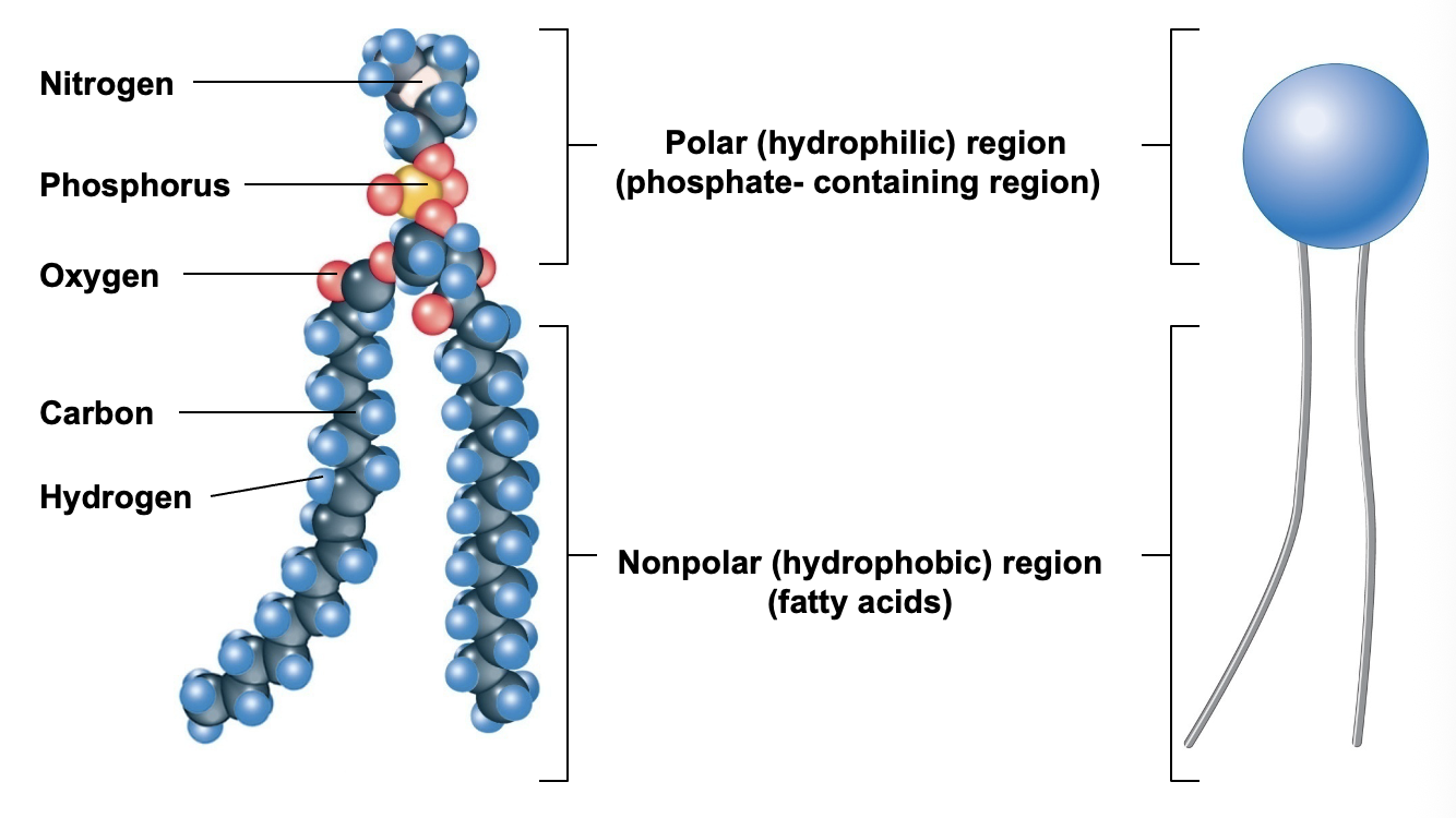
What are properties of a phospholipid?
Major membrane component
Amphipathic properties
Hydrophilic (polar) head
Hydrophobic (non polar) tail
What are the 2 major fluid compartments and what do they contain?
Intracellular fluid —> Water within the cell (2/3 of all body water)
Extracellular fluid —> Everything outside the cells (1/3 of body water)
Interstitial Fluid
Plasma (blood)
Cerebrospinal Fluid
Contain:
Different ionic compositions
Same Osmotic Concentrations
Exchange between compartments
Interstitial Fluid is within which fluid compartments and what is it?
Within the extracellular fluid compartment
Its the water sitting between the cells

Plasma (blood) Fluid is within which fluid compartments and what is it?
Within the extracellular fluid compartment
Its the liquid part of the blood

Cerebrospinal Fluid is within which fluid compartments and what is it?
Within the extracellular fluid compartment
Its the fluid surrounding the brain and spinal cord

In relation to the fluid compartments in the body, what does different ionic compositions refer to?
It means that the two sides (intracellular & extracellular) are chemically opposite.
Inside cells, potassium dominates.
Outside, sodium and chloride dominate.
Cells spend energy constantly pumping sodium out and potassium in to keep it that way, and those gradients are what let nerves and muscles fire

In relation to the fluid compartments in the body, what does same osmotic concentration refer to?
It means that even though the ions differ, the total number of dissolved particles is the same everywhere, around 290 mOsm/kg.
Water crosses cell membranes so easily that any imbalance gets evened out almost immediately by water shifting to the more concentrated side.
In relation to the fluid compartments in the body, what does exchange between compartments refer to?
It means that the compartments aren't sealed off.
Water and solutes move between them constantly: across cell membranes by osmosis and transport proteins, and across capillary walls by pressure.
This is why a change in one compartment shows up in the others. It's also why fluid can pool where it shouldn't and cause swelling.
What are the 3 Cell-Cell Junctions?
Tight Junctions
Desmosomes
Gap Junctions
What is the function of Tight Junctions?
Prevents passage between cells (permeability barrier)

What is the function of Desmosomes?
Anchoring Junction
Prevents cells from being torn apart ('“zipper”)

What is the function of Gap Junctions?
Allows for communication between adjacent cells (intracellular communication)
Interlocking membrane proteins

Function of Cell-Matrix Junctions?
Anchoring of cells to underlying tissues —> attaches to the matrix beneath it
Hemidesmosome —> name means "half a desmosome"
cytoskeleton fibers link to matrix proteins
it passes from the matrix, through the integrin, into the cell's internal skeleton. That's why an epithelium can resist shearing forces instead of tearing.

What are the 2 types of Membrane Transport Mechanisms?
Passive
Active
What is Passive Transport and provide examples of Passive Transport?
Passive transport does not use ATP. Things move down their gradient, from high to low concentration.
Diffusion
small, nonpolar molecules (O₂, CO₂, steroids) slip straight through the lipid bilayer
Facilitated Diffusion —> Protein channels and carriers
molecule needs help crossing → Channels are pores that open and let ions through fast → Carriers bind the molecule and change shape to pass it across (slower, and they can saturate). Ex: Glucose
Osmosis
water moves toward the side with more solute, usually through aquaporins

What is Active Transport and provide examples of Active Transport?
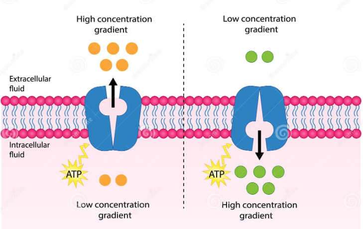
Active Transport requires the use of ATP and moves things against their gradient
Primary
pump itself hydrolyzes ATP. Ex: Na⁺/K⁺-ATPase
Secondary → Antiporters, Symporters
no direct ATP use, it borrows the gradient a primary pump already built. Sodium rushing back into the cell drags another molecule along. Symporters carry both in the same direction (Na⁺ plus glucose in the gut) → antiporters swap them in opposite directions (Na⁺ in, Ca²⁺ out)
Vesicular —> for cargo too large for any protein: the membrane itself wraps around it
Endocystosis → brings material in
Exocytosis → a vesicle fuses with the membrane and dumps contents out
Transcytosis → take something in one side of the cell and release it out the other, ferrying it across an entire cell layer
What are the 4 Membrane Receptors?
Endocytosis
Chemical (ligand)-gated ion channel
Enzymes
Linked to G proteins (2nd messengers)

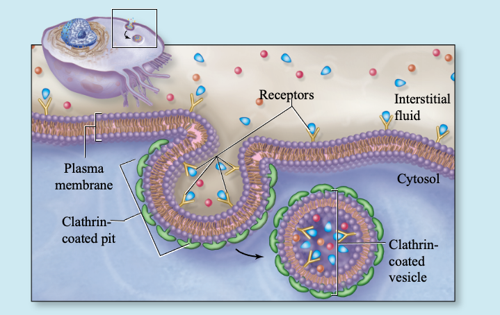
What is Receptor-Mediated Endocytosis?
Receptor grabs its ligand and the membrane pulls the whole complex inside as a vesicle
What is Chemical (ligand) - Gated Ion Channels?
Binding pops it open and ions flood through

What are Enzyme Receptors?
Receptor spans the membrane with a binding site outside and an enzyme domain inside. Ligand binding switches the enzyme on, and it starts modifying proteins in the cytoplasm

What are G-Protein Coupled Membrane Receptors?
Binding activates a G protein sitting on the inner surface, which in turn triggers an enzyme that produces a second messenger (cAMP, IP₃, calcium). That messenger then spreads through the cell and activates many enzymes at once

3 Types of Ion Channels:
Leakage (passive) Channels —> always open
Chemical (ligand)-gated Channels —> Requires chemical binding to open/close
Voltage-gated Channels —> Requires membrane voltage change to open/close
When ions move, the membrane potential changes and serves as a signal to change cell function

What type of cell-cell junctions allows communication between cells?
Gap Junctions
What type of cell-cell junction might be found in a tissue that has force applied to it (like the external surface of the skin) and why?
Desmosomes. This is because they are the strongest lateral anchoring junction and so stretching or shearing force gets distributed rather than concentrated on individual membranes

What would be the advantage of receptor-mediated endocytosis over pinocytosis?
Selectivity and concentration.
Pinocytosis is nonspecific the cell just sips extracellular fluid and takes in whatever happens to be dissolved in it, so a rare molecule gets brought in only in trace amounts.
Receptor-mediated endocytosis uses receptors that bind one specific ligand, so the target gets concentrated on the membrane surface before the vesicle forms.
The cell can take up large amounts of a substance present at very low outside concentrations, and control uptake by adjusting how many receptors it displays.
Why are membrane channels required for ions to enter/exit cells?
Because the interior of the membrane is hydrophobic. Ions are charged and stay surrounded by a shell of water molecules, so crossing the lipid core would mean stripping off that hydration shell and forcing a charged particle into a fatty, nonpolar environment
Channels provide a water-lined pore that lets the ion pass, avoiding contact with the lipid.
Channels also add selectivity and control: a given channel admits only certain ions and can be gated open or closed
What effect does movement of a cation or anion have on the membrane potential? (Note: You should be able to provide the specific electrical term for a membrane potential that is more positive or negative then it was at rest/before the ion movement occurred.)
The terms are depolarization (membrane becomes more positive than resting) and hyperpolarization (more negative than resting). Returning toward resting after depolarization is repolarization.
The effect depends on charge and direction:
Cation into the cell (Na⁺, Ca²⁺) adds positive charge inside → depolarization
Cation out of the cell (K⁺ leaving) removes positive charge → hyperpolarization
Anion into the cell (Cl⁻ entering) adds negative charge → hyperpolarization
Anion out of the cell removes negative charge → depolarization
So the rule is just: whatever makes the inside less negative depolarizes, whatever makes it more negative hyperpolarizes.