1.1 The Cardiovascular System
1/70
Name | Mastery | Learn | Test | Matching | Spaced |
|---|
No study sessions yet.
71 Terms
Heart
Deliver o2
Gather waste products
Transport heat
Atria - thin muscular walls
Ventricles - thick walls
Left - oxygenated
Right - deoxygenated
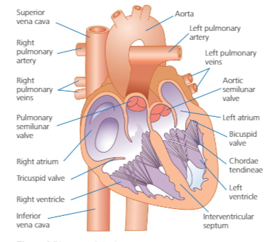
Blood vessels of the heart
Vena cava - brings deoxygented blood back to the right atrium
Pulmonary artery - leaves right ventricle with deoxygenated blood to lungs
Aorta - o2 to body
Cardiac Conduction System
Group of specialised cells located in the wall of the heart.
Cardiac Conduction Pathway
.The hearts myogenic beats send impulses to SAN
2.The impulse spreads like a way through heart 3.
The impulses from SAN spread through atria walls. 4.
This causes atrium to contract, forcing blood into ventricles 5.
The impulses then passes through atrioventricular node (AVN) 6.
AVN delays transmission 7.
Allows atria to fully contracts before ventricular systole
8.The impulse passes through Bundle of his
9.Impulse then arrives at purkinje fibres
10.Ventricular systole
CCC Pathway
San
Atrial walls
Avn
Bundles of his
Purkinje fibres
Ventricular systole
Factors affecting the change in rate of the conduction system
neural control mechanism
Role of AVN
Receive impulse from SAN
Delay transmission of impulse'
Allow atria wall to fully contract
Send impulse to Bundle of His
Myogenic
Heart generates its own impulses
Diastole
Relax (filling of chambers)
Systole
Contract (emptying of chambers)
Neural control mechanism
Sympathetic nervous system
Parasympathetic nervous system
2 systems coordinated by CCC located in the medulla oblongata
Send impulse to SAN
Sympathetic Nervous System
stimulates hearts to beat faster
Parasympathetic Nervous System
returns heart to resting level
Nervous System Pathway
1.Receptors detect change
2.Send impulses to cardiac control centre in medulla oblongata
3.Send impulse to SAN to increase/decrease (sympathetic/para)
4.Atrial systole
5.AVN
6.Bundle of his
7.Purkinje fibres
8.Ventricular systole
Chemoreceptors
Detect increase in carbon dioxide/ acidity levels causing heart rate to increase
Baroreceptors
Detect decrease in blood pressure causing heart rate to increase
Proprioreceptors
Detect increase in movements causing heart rate to increase
Hormonal control mechanism
Release Adrenaline
Adrenalin
Stress hormone released by sympathetic nervous system and cardiac nerve during exercise, causing increase in heart rate.
Prior to exercise - anticipatory rise
Send to SA node
Increase CO
More blood to working muscles
Stroke Volume
Volume of blood pumped out of the heart ventricles in each contraction
What is stroke volume dependent on?
1.Venous return - increases, SV increases
2.Elasticity of cardiac fibres - the more stretched, greater ejection fraction
Stroke volume in response to exercise
Increases with intensity
Only up to 40-60% max effort (plateaus)
Increase HR near max effort results in shorter diastolic phase
Ventricles don’t have time to fill with blood - can’t pump as much out
Heart rate
number of times the heart beats per minute (72)
Trained perfromer has a greater HR range - resting HR is lower and max higher
Cardiac hypertrophy
thickening of muscular wall of heart so it becomes bigger and stronger
Bradycardia
decrease resting heart rate below 60 per min
Cardiac output
Volume of blood pumped out of the heart ventricles per minute'
HR X SV = CO
Cardiac Output in response to exercise
Increase as intensity increases until plateaus
Increase max CO - transport more blood to working muscles + o2
Less blood to organs
Venous return
Volume of blood returning to the heart via veins
Starling's law
1.Increased venous return
2.Greater diastolic filling
3.This causes cardiac muscles to stretch and strengthen
4.This leads to greater force of contraction
5.Increase ejection fraction
Elasticity of cardiac fibres
how much the cardiac tissues stretch during diastole
Ejection fraction
the percentage of blood pumped out the left ventricle per beat
Heart Disease
CHD
Cause (Atherosclerosis) - coronary arteries become blocked/narrow by gradual build up of fatty deposits (caused by: high blood pressure, high cholesterol, lack of exercise, smoking)
Atheroma = fatty deposits in inner lining of artery
Angina = chest pains that occur when blood supply through coronary arteries to heart muscles restricted - blood clot
Exercise keep heart healthy - pump more blood, bigger + stronger, increase SV, flexible blood vessels, blood flow
High blood pressure
Force exerted by blood against the blood vessel wall
Extra strain on arteries
Increase: heart failure/attack, stroke
Aerobic exercise - reduce
Lower systolic + diastolic pressure
Cholesterol
HDL - transport excess in blood back to liver where it’s broken down (lower heart disease)
LDL - transport cholesterol in blood to tissues (increase heart disease)
Stroke
Blood supply to brain cut off
Brain needs constant supply of o2 - maintain function (energy)
Brain cells die
Injury, death, disability'
Ischaemic - blood clot stops blood
Haemorrhagic - weakened blood vessel bursts
Skeletal muscle pump
1.Change in shape of muscles when they contract/relax
2.Presses on veins causing a pumping effect
3.Squeezes blood to heart
AO2 - Increased use of muscles during specific body parts in sport which presses veins pushing blood back to heart
AO3 - Avoid fatigue + lactic acid build up
AO3 - Starling’s law + venous return
Respiratory pump
1.During breathing, muscles relax and contract
2.Pressure changes occur in thoracic + abdominal cavities
3.Pressure changes compress veins + assist blood return to heart
AO2 - Increased breathing rate during exercise causes increased effect of respiratory pump returning more blood to heart
AO3 - More CO2 exchanged for O2
Pocket valves
Prevent back flow by forcing blood in one direction back to heart
Smooth muscle
found in veins and contacts to push blood back to heart
AO2 - suction pump of the heart increase as heart beats harder/faster during exercise
AO3 - more blood to working muscles supplying O2 for resynthesis of ATP
Gravity
Causes blood in upper body to return back to heart
Venous return relationship with blood pressure
1, Increase - increases systolic pressure
Decrease - decreases systolic pressure
Transportation of o2 during exercise
Haemoglobin - found in red blood cells'
Combines o2 = oxyhaemoglobin
PP lungs high = full saturation OH
PP muscles low - offloads o2 to oxides glycogen + fats
Myoglobin - muscle haemoglobin
higher affinity for o2 then haemo
addition o2 store in muscles
Released to working muscles - extend aerobic respiration
Oxyhaemo diss curve - relationship between blood transport + release of o2
Bohr shift - ODC shifts to right
lower pH due to increase CO2
Product of aerobic respiration
Increased dissociation of o2 to muscles
Greater quantity of o2 to mitochondria
Mitochondria - cell respiration
higher aerobic intensity
Myoglobin
oxygen stored in muscles (high affinity for oxygen)
Affinity
How easily a substance combines with another
Haemoglobin role during exercise
1.Transport oxygen in blood to muscles
2.Less saturation of haemoglobin during exercise
3.Increase in oxygen release
4.Muscle receive more oxygen
Myoglobin role during exercise
1.Stores oxygen in muscles
2.Has higher affinity for oxygen
3.Oxygen dissociates from myoglobin at lower P02
Bohr Shift
movement of the s-shaped oxygen dissociation curve shifts to the right
Bohr Shift Explained
1.Oxygen becomes more easily dissociated from haemoglobin in blood capillaries to the muscles.
2.Reduction of affinity of haemoglobin for oxygen
3.Muscles require more oxygen in exercise - leads to dissociation of oxygen from haemoglobin
Factors responsible for Bohr shift
1.Increase In Blood Temperature - when blood is in muscles, temp increases causing oxygen to dissociate from haemoglobin more readily
Increase in Partial Pressure Of C02 - C02 levels rising during exercise leads to oxygen dissociating quicker
3.Decrease in pH - more C02 will lower pH in blood, causing oxygen to dissociate quicker
Redistribution Of Blood
During exercise , skeletal muscles require more oxygen so more blood needs to be redirected to them as they require more oxygen
What control blood flow?
Vasomotor centre which control vasodilation and vasoconstriction
Blood flow and brain
Blood flow to the brain must remain constant to ensure brain function is maintained as the brain needs oxygen for energy.
Vasoconstriction
Narrowing of blood vessel diameter to decrease blood flow to capillaries
Vasodilation
Widening of blood vessel diameter to increase blood flow to capillaries
Redistribution Of Blood During Exercise
1.Vasodilation - more oxygen is needed at working muscles so vasodilation will occur in the arterioles supplying the muscles, increasing blood flow and bringing in oxygen that's needed.
2.Vasoconstriction - occurs in arterioles supplying non-essential organs (eg liver + intestines)
What happens when sympathetic stimulation increases?
1.Vasoconstriction occurs
2.Blood flow reduces
3.This means blood can be redistributed to muscles in exercise
What happens when sympathetic stimulation decreases?
1.Vasodilation occurs
2.Increases blood flow to that body part
Pre-capillary sphincters
1.Tiny rings located at opening of capillaries
2.Contract - restrict blood flow
3,Relax - increased blood flow
4.During exercise - relax to supply skeletal muscles
Why Is redistribution of blood important?
1.Increase supply of oxygen to working muscles
2.Remove waste products
3.Increase blood to skin - regulate body temp + remove heat (sweat)
4.Direct more blood flow to heart
Aterio-venous difference (A-V02 Diff)
The difference in volume of oxygen between arteries and veins
A-V02 Diff at rest
Low - little oxygen required at muscles
A-V02 Diff at exercise
High - lots of oxygen required at muscles
Increase of A-VO2 Effects
1.This increase will affect gaseous exchange at alveoli - more oxygen is taken in and more carbon dioxide is removed.
2.Training increases A-VO2 diff - trained performers can extract more oxygen from blood
A-VO2 Diff at start of exercise
1.Increase in difference
2.More oxygen is extracted by working muscles + stored in myoglobin
3.Venous blood therefore has less oxygen returning to heart
How training increases max A-VO2 Diff and how body systems adapt to allow this
Increased o2 content in arterial blood due to more haemoglobin carrying capacity
Increased gas exchange at muscle due to more myoglobin which has a greater affinity for o2 than haemoglobin so can store more o2 in muscle
Increased gas exchange at muscle due to more efficient mitochondria allowing for more o2 to be used in muscle so less return in venous blood
Cardiovascular drift
Increase in heart rate in order to try and maintain cardiac output due to decrease in stroke volume (cardiac output increases during prolonged exercise to cool body)
When does cardiovascular drift occur
After 10 minutes
Warm environment
Steady state exercise
What causes cardiovascular drift
Caused by reduction of fluid in blood plasma due to an increase in sweating - makes blood more viscous
2.This means venous return increases - reduces atrial filling and SV
Vasodilation occurs at skin to cool it down
Anticipatory rise
increase in heart rate before exercise due to adrenalin
Bradycardia
decrease in resting heart rate
How does increased CO2 increase Breathing Rate
Increased blood acidity
Detected by chemorecpetors
Impulse sent to respiratory centre
Increased impulses to respiratory muscles to contract faster