Ch 06 Anatomic Descriptiors and Fundamentals (General Anatomy)
 Call Kai
 Learn
Learn Practice Test
 Spaced Repetition
 Match
Match Flashcards
 Knowt Play
1/74

There's no tags or description
Looks like no tags are added yet.
Name | Mastery | Learn | Test | Matching | Spaced | Call with Kai | Chat |
|---|
No analytics yet
Send a link to your students to track their progress
75 Terms
anatomical position
a reference posture of the human body, in which the anterior view of the human body is shown standing with legs slightly apart, feet forward, palms facing forward.
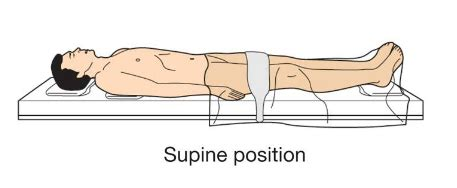
supine
lying or resting on the back with the face, palm, etc., upwards
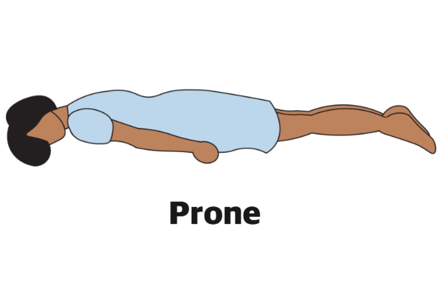
prone
Lying with the front or face downward.
Superior
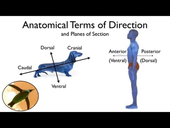
Superior (or cranial, or rostral) means to the top or towards the head-end of the body.
Both superior (to the top) and inferior (to the bottom) describe location of structures along the vertical axis of the body. For example, the arms are superiorly situated as compared to the legs, which are inferiorly situated.
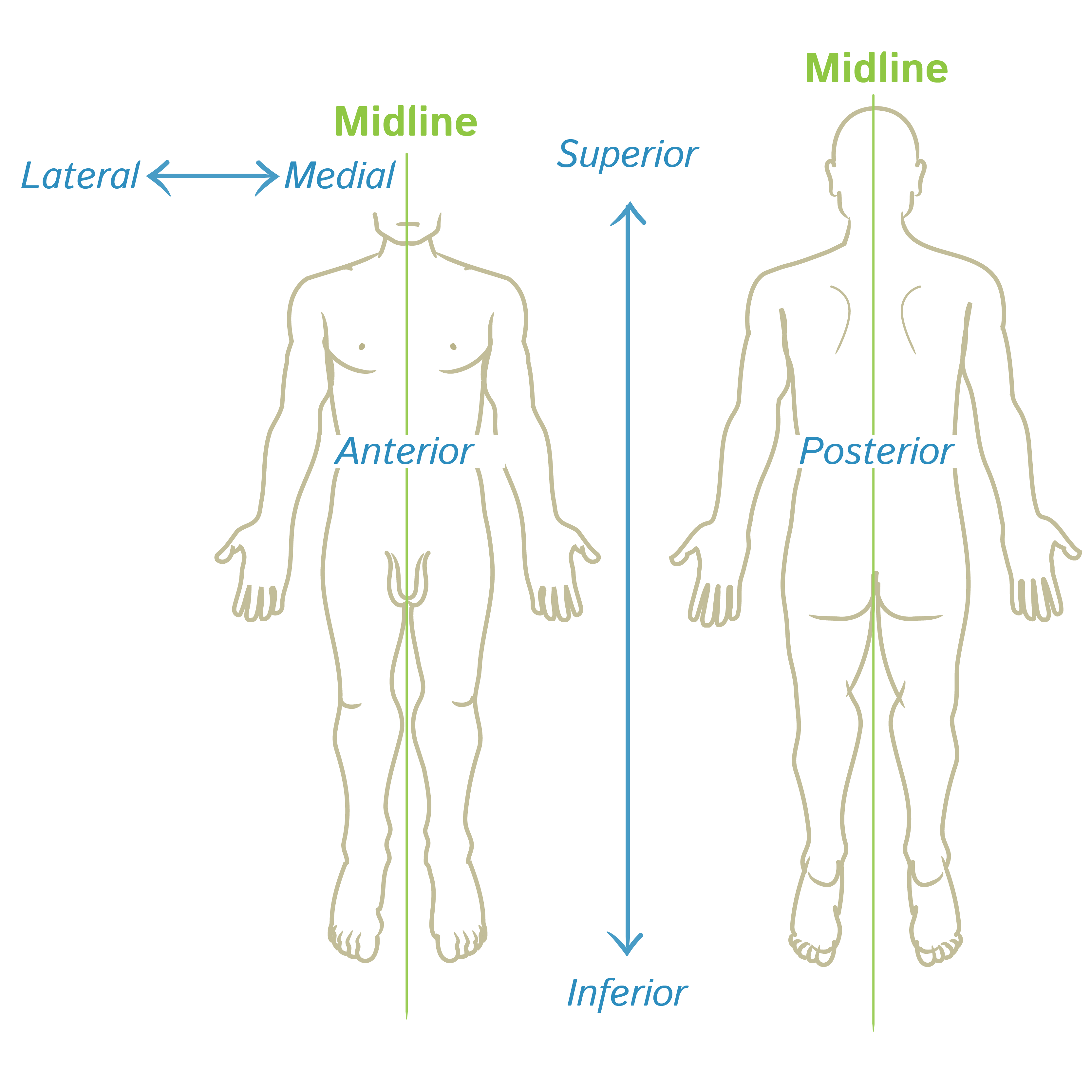
Inferior
Inferior (or caudal) means towards the bottom or away from the head-end of the body.
Both inferior (towards the bottom) and superior (to the top) describe location of structures along the vertical axis of the body. For example, the feet are inferiorly situated as compared to the hands, which are superiorly situated.
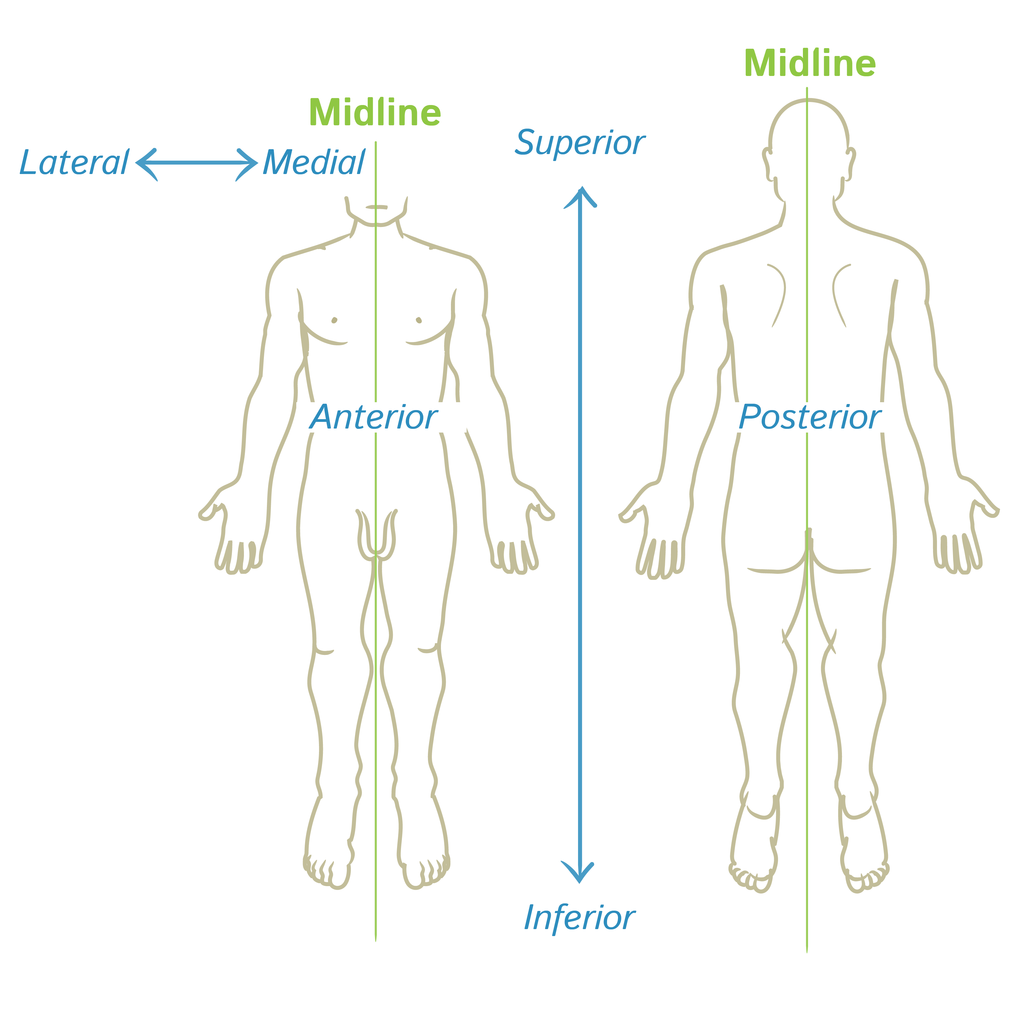
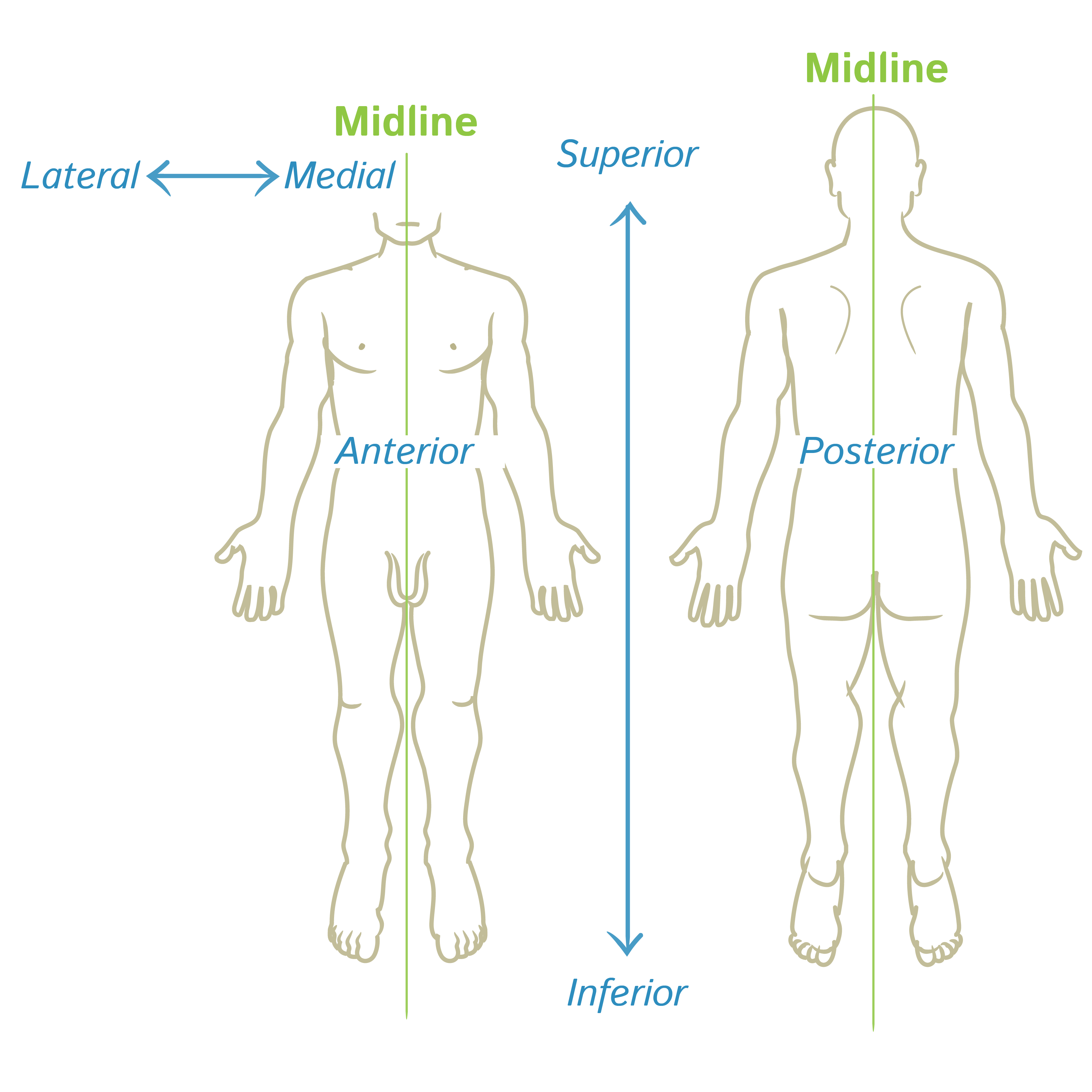
Anterior
Anterior (or ventral) means towards the front.
Both anterior and posterior are directional terms which describe the location of the body parts to the front or to the back. For instance, sternum is located anteriorly, while the vertebral column is a posteriorly situated structure.
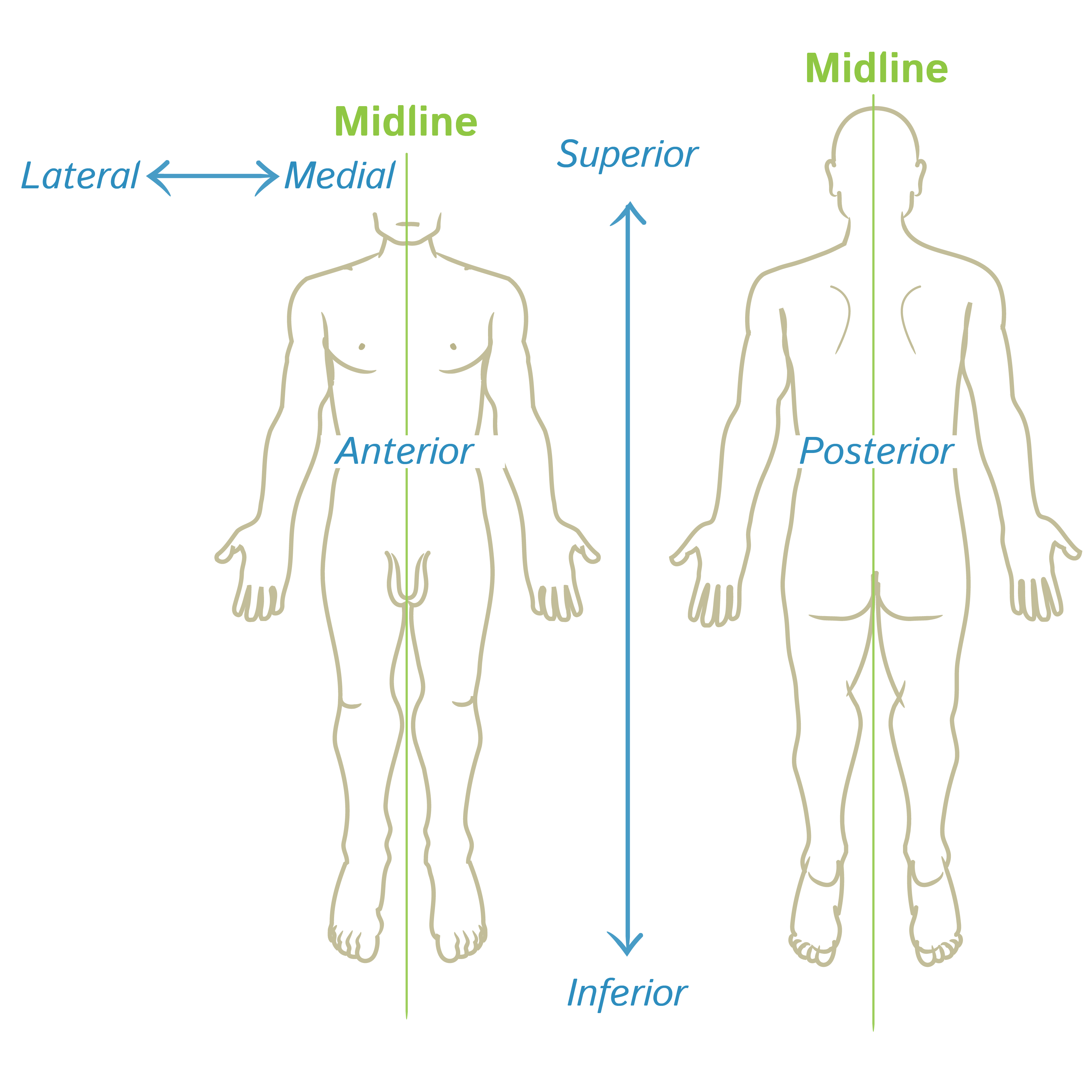
Posterior
Posterior (or dorsal) means towards the back.
Both posterior and anterior are directional terms which describe the location of the body parts to the back or to the front. For instance, the vertebral column is located posteriorly, while the sternum is a anteriorly situated structure.
Medial
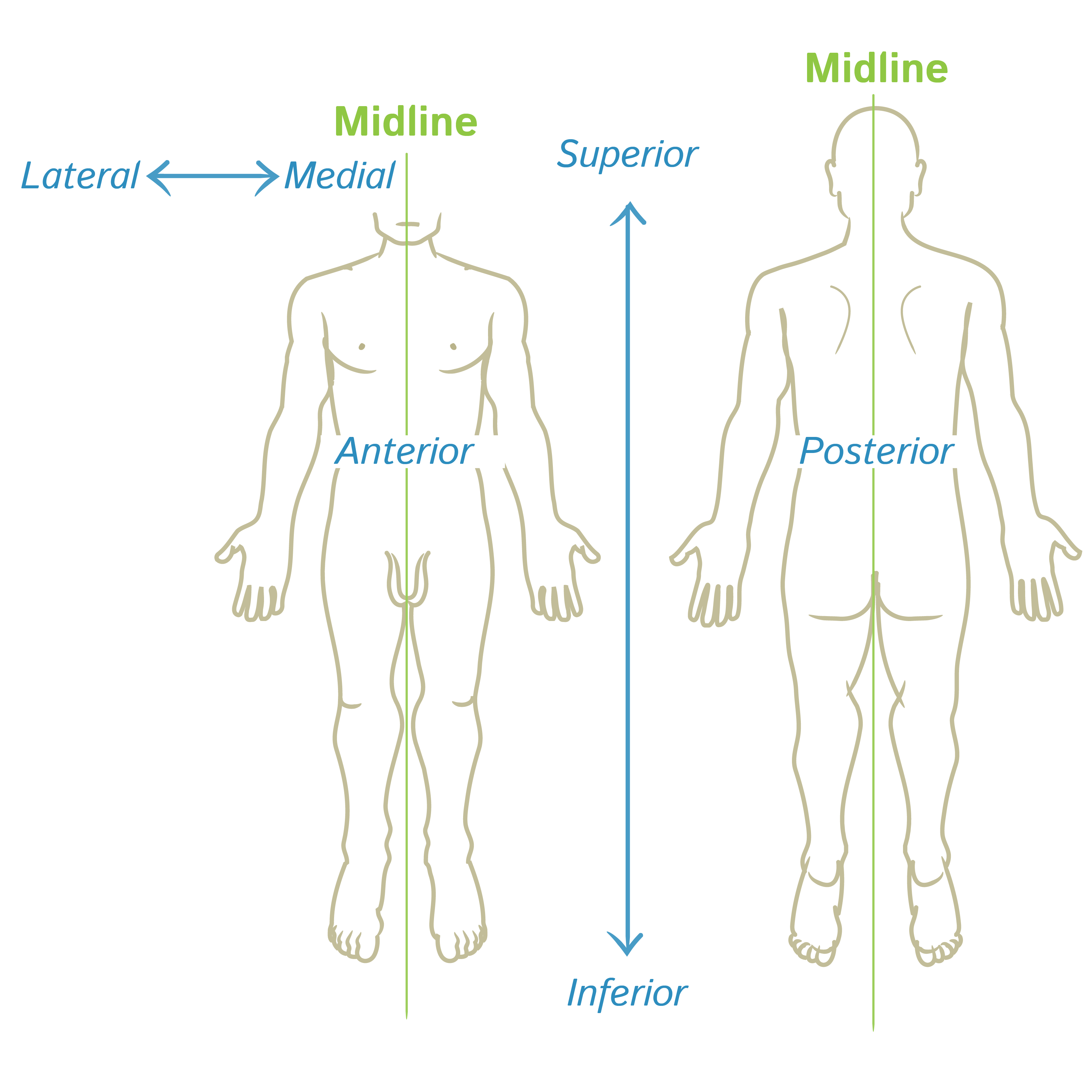
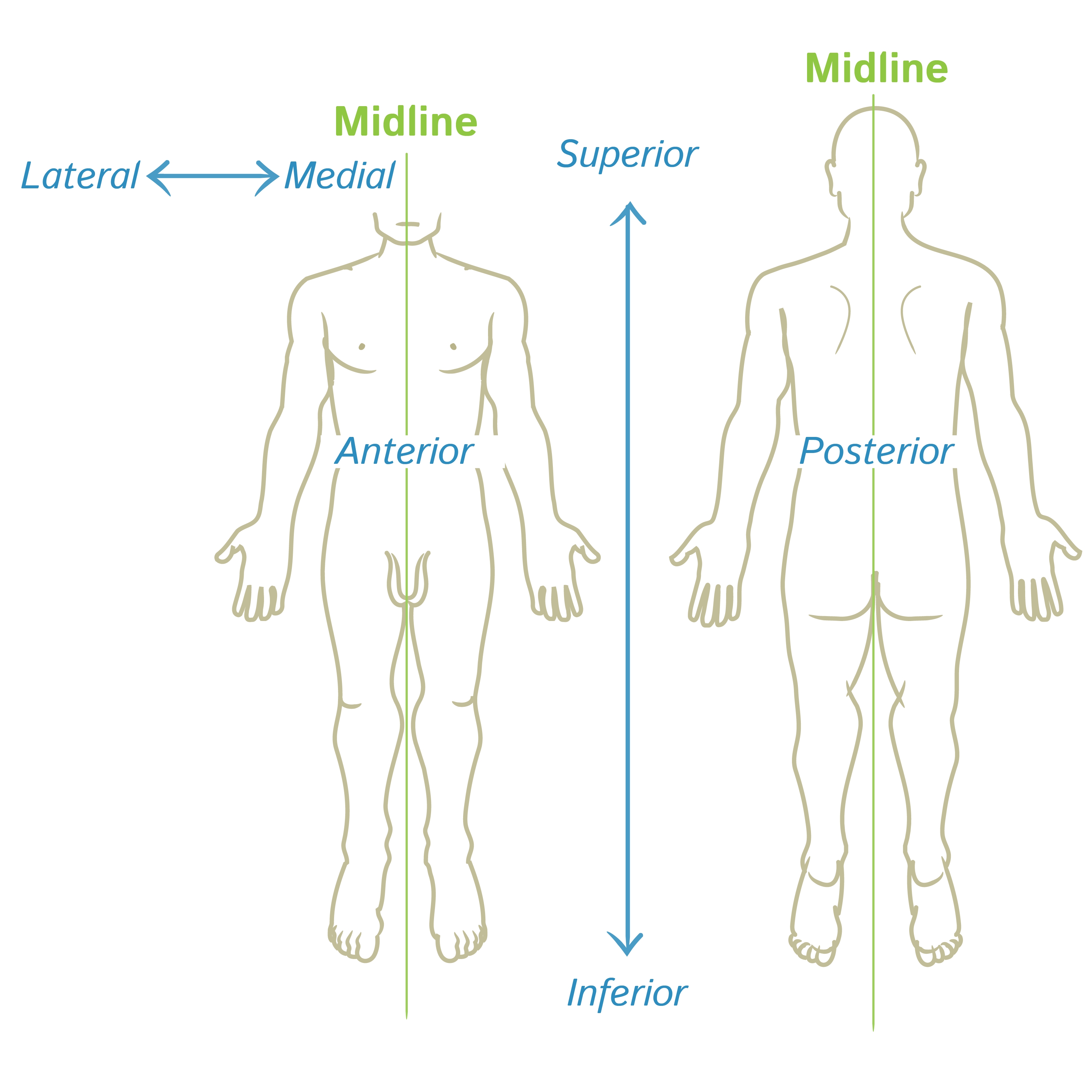
Medial is a term used to describe the location of a body part ‘towards’ the midline or the middle of the body. For instance, the eyes are medial compared to the ears, meaning thereby that the eyes are closer to the midline as compared to the ears.
Lateral
Lateral is a term used to describe the location of a body part ‘away’ from the midline or the middle of the body. For instance, the little toe is located on the lateral side of the foot, or the ears are lateral to the eyes, meaning thereby that the ears are farther from the midline as compared to the eyes.
Proximal
The term proximal is used to describe location of a structure or a body part closer to the trunk or near its origin. This is contrary to the term ‘distal’ which refers to part of the body being farthest from the trunk or from its origin.
For example, the quadriceps muscles in the anterior thigh are attached at their origin to the hip bone proximally, while they insert into the upper end of tibia distally. Similarly, the femur articulates with the hip bon proximally (closer to the trunk or axial body) while it articulates with the tibia distally (away from the trunk or axial body).
Distal
The term distal is used to describe location of a structure or a body part farthest from the trunk or away from its origin. This is contrary to the term ‘proximal’ which refers to part of the body being closer to the trunk or near its origin.
For example, the quadriceps muscles in the anterior thigh insert into the upper end of tibia distally, while they are attached at their origin to the hip bone proximally. Similarly, the femur articulates with the tibia distally (away from the trunk or axial body), while it articulates with the hip bon proximally (closer to the trunk or axial body).
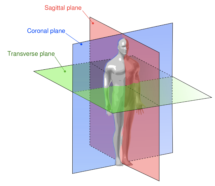
Sagittal planes
Sagittal planes refer to vertically-oriented, antero-posterior planes which, when pass through the body in its anatomical position, divide it into right and left parts.
The sagittal planes lie at right angles to the vertically-oriented coronal planes, while they perpendicularly intersect the transverse (a.k.a. horizontal) planes.
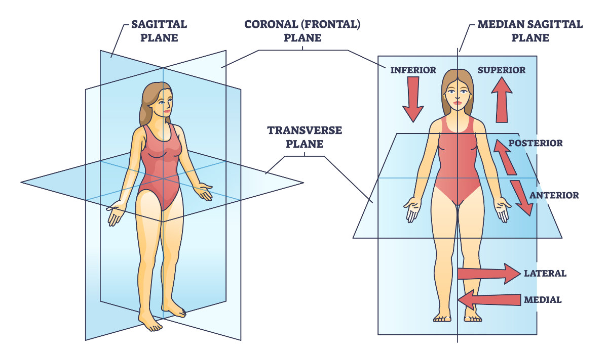
Transverse plane
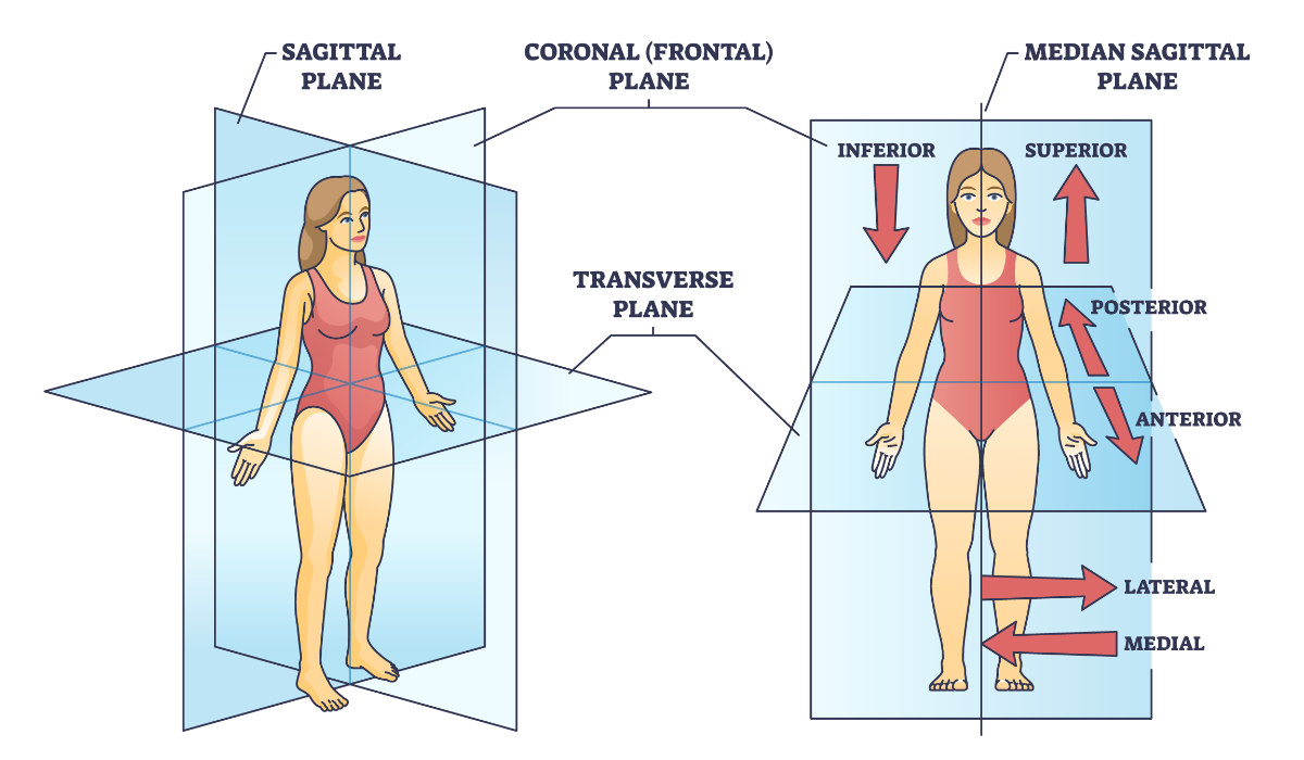
Transverse (a.k.a. axial) plane refers to a horizontally-oriented plane which, when passes through the body in its anatomical position, divides it into upper and lower parts.
The transverse plane is intersected at right angles by the vertically-oriented coronal (side-to-side) and sagittal (antero-posterior) planes.
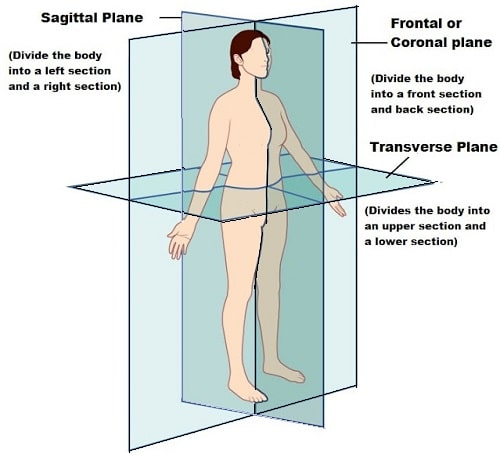
Frontal plane
the frontal plane, in anatomy, is an imaginary plane that divides the body into anterior (ventral) and posterior (dorsal) halves along the longitudinal (left-right) axis.
Deep
The terms deep and superficial are used to describe locations of body structures with reference to the ‘surface’ of the body. Deep means away from the body surface (usually more internally placed), while ‘superficial’ means closer to the surface of the body.
For instance, the brain and spinal cord are deeper structures which are being protected within the bony skull and vertebral column, that are relatively more superficial. Another example could be that of the fascial wrappings of the body. The outer superficial fascia containing cutaneous vessels and nerves is closer to the body surface as compared to the deep fascia which is more internally placed.
Ventral
Ventral (or anterior) means towards the front.
Both ventral (or anterior) and dorsal (or posterior) are directional terms which describe the location of the body parts to the front or to the back. For instance, sternum is located ventrally, while the vertebral column is a dorsally situated structure.
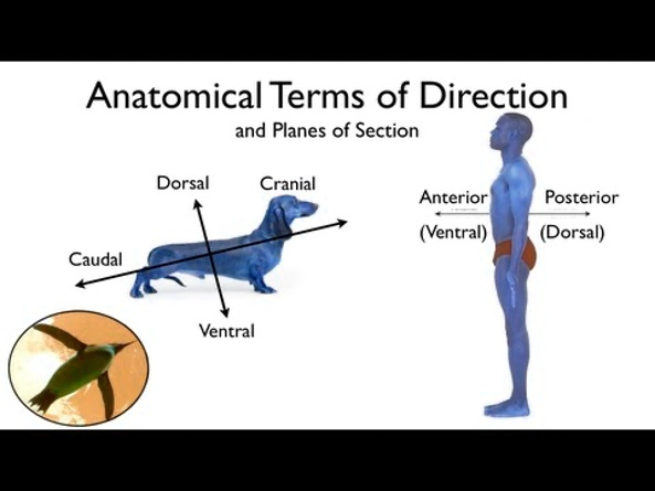
Dorsal
Dorsal (or posterior) means towards the back.
Both dorsal (or posterior) and ventral (or anterior) are directional terms which describe the location of the body parts to the back or to the front. For instance, the vertebral column is located dorsally, while the sternum is a ventrally situated structure. Similarly, shoulder blades are situated on the dorsal aspect of the rib cage.
thoracic cavity
The space within the walls of the chest, bounded below by the diaphragm and above by the neck, and containing the heart and the lungs.
abdominal cavity
The space bounded by the abdominal walls, diaphragm, and pelvis and containing most of the organs of digestion, the spleen, the kidneys, and the adrenal glands.
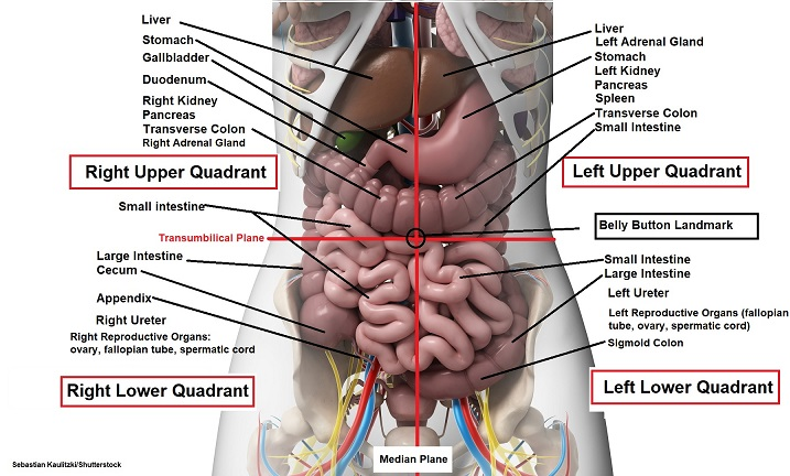
Right Upper Quadrant: Liver, stomach, gallbladder, duodenum, right kidney, pancreas, and the right adrenal gland.
Left Upper Quadrant: Liver, stomach, pancreas, left kidney, spleen, and the left adrenal gland.
Right Lower Quadrant: appendix, reproductive organs, right ureter.
Left Lower Quadrant: left ureter, reproductive organs
NOTE: All four quadrants contain portions of the small and large intestines.
pelvic cavity
The space bounded by the bones of the pelvis and pelvic girdle.
abdominopelvic cavity
the space between the diaphragm and the groin. There is no structurally distinct separation between the abdomen and pelvic regions.
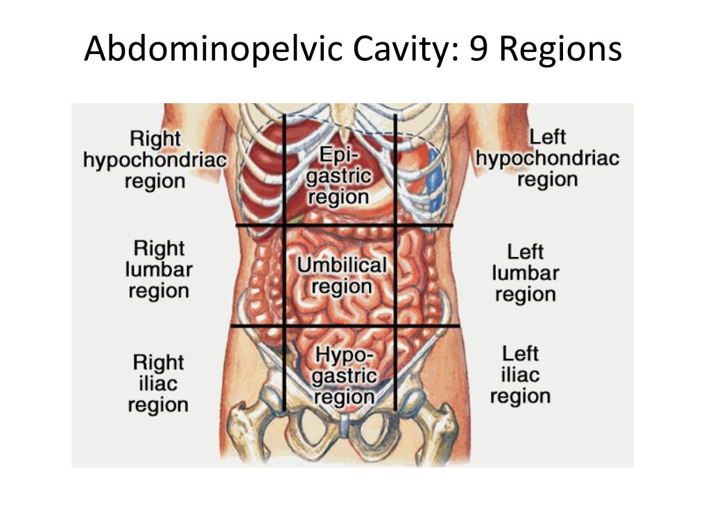
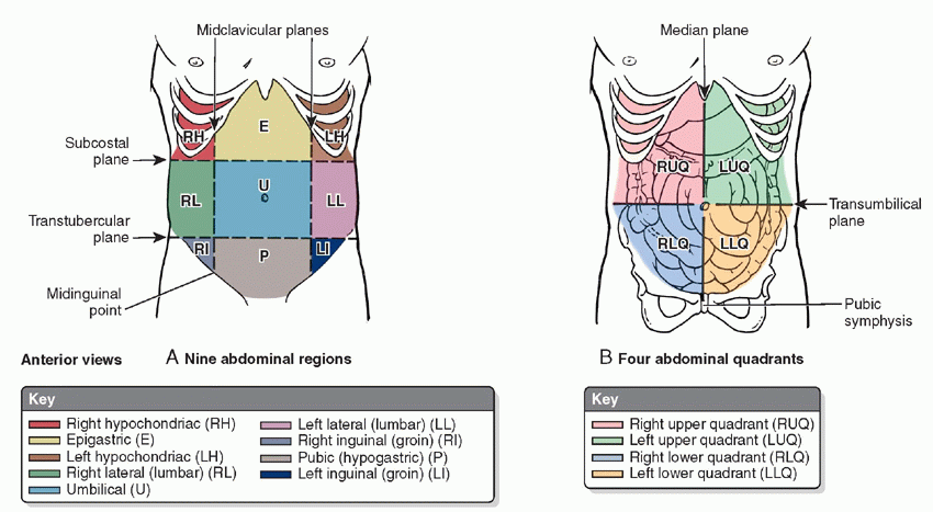
Hypochondriac Regions (Left and Right): the prefix “hypo” means below or under. The word “chondriac” means cartilage, which is referring to the cartilage of the ribs. When we put them together, this is the abdominal region that is under the ribs. Boom. Easy. So you have a right and left hypochondriac region.
Lumbar Regions (Left and Right): The word lumbar refers to the vertebrae in your lower back, which are the bones nearest to the lumbar region. That’s where this region gets its name.
Iliac Region (Left and Right): The top of the hip bone has what is called the iliac crest, and that’s the bone closest to the iliac region.
Epigastric Region: The prefix “epi” means above, or over, and “gastric” means stomach or belly. Therefore, this is the region over and above the belly.
Umbilical Region: This is easy to remember because the umbilical region contains your navel, which is also called the umbilicus. The navel is the landmark for the four quadrant point of intersection, and it can also help you remember that the umbilical region makes up the middle of the nine abdominal regions.
Hypogastric Region: We’ve already learned that hypo means “below,” and gastric refers to the stomach or belly. So when we put the two together, we know that the hypogastric region is the region below the belly.
Cervical
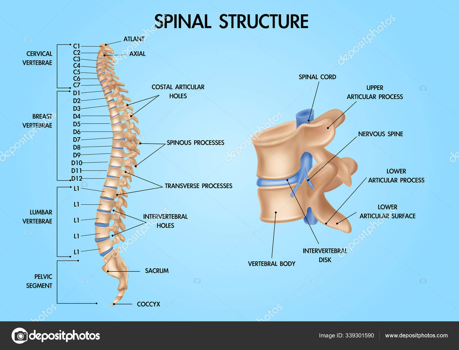
Relating to the top part of the spine that is composed of the seven vertebrae of the neck and the disks that separate them.
Occipital
pertaining to the back of the head.
lumbar
pertaining to the part of the body between the thorax and the pelvis.
Vertical plane
A vertical axis or plane goes up and down. A few examples of vertical planes include the sagittal and coronal planes.
Horizontal plane
Horizontal (a.k.a. transverse or axial) plane divides the body or any of its parts into upper and lower parts.
The horizontal plane is intersected at right angles by the vertically-oriented coronal (side-to-side) and sagittal (antero-posterior) planes.
Median plane
Median plane refers to an antero-posterior sagittal plane that passes through the midline or the middle of the body and divides it into right and left equal halves. It is a.k.a. the median sagittal plane.
The median (or median sagittal) plane lies at right angle to the vertically-oriented coronal plane, while it perpendicularly intersect the transverse (or horizontal) plane.
Coronal plane
Coronal plane (a.k.a. frontal plane) pertains to a vertical, side-to-side plane that, when it traverses the body in its anatomical position, separates it into anterior and posterior portions.
The coronal plane is perpendicular to the vertical sagittal plane and intersects the transverse (a.k.a. horizontal) plane at a right angle.
Intermediate
Intermediate means something in between two structures. For instance, the sternum is a midline structures, while upper end of shoulder is laterally placed. Here the clavicle is intermediate between the sternum and the shoulder-tip.
Frontal
Frontal means to the front and adjacent to the forehead or the frontal bone. For instance, the lobe of the brain which is situated in the front (near the forehead) is called the frontal lobe, as compared to the posteriorly situated lobe which is called the occipital lobe.
Occipital
Occipital means to the back of the head or adjacent to the occipital bone. For instance, the lobe of the brain which is situated in the back of the cranial cavity (adjacent to the occipital bone) is called the occipital lobe, as compared to the anteriorly situated lobe which is called the frontal lobe.
Cranial
Cranial (or rostral) means towards the head-end of the body. It is commonly used interchangeably with the term ‘superior’, when the body is in its anatomical position.
Both cranial (towards the head-end) and caudal (away from the head-end / towards the tail) describe location of structures along the long axis of the body. For example, the arms are cranially situated as compared to the legs, which are caudally located.
Caudal
Caudal means towards the tail or away from the head-end of the body. It is commonly used interchangeably with the term ‘inferior’, when the body is in its anatomical position.
Both caudal (away from the head-end / towards the tail) and cranial (towards the head-end) describe location of structures along the long axis of the body. For example, the legs are caudally situated compared to the arms, which are cranially (or rostrally) located.
Rostral
Rostral (or cranial) means towards the head-end of the body. It is commonly used interchangeably with the term ‘superior’, when the body is in its anatomical position.
Both rostral or cranial (towards the head-end) and caudal (away from the head-end / towards the tail) describe location of structures along the long axis of the body. For example, the arms are rostrally situated as compared to the legs, which are caudally located.
Apical
Apical (or the apex) refers to the tip of a rounded or pyramid-shaped structure. It usually faces opposite the base of that specific body part.
For instance, the apical part of the lung lies at the level of the inlet of thoracic cavity, and it faces opposite the base of the lung which lies on the diaphragm. Similarly, apex of the heart lies at the level of left fifth intercostal space and it faces opposite the base which is formed by atria and lies at the back of the heart.
Basal
Basal refers to the base of a rounded or pyramid-shaped structure. It usually faces opposite the tip of that specific body part.
For instance, the basal part of the lung rests on the diaphragm and it faces opposite the apex of the lung which lies at the level of the thoracic inlet.
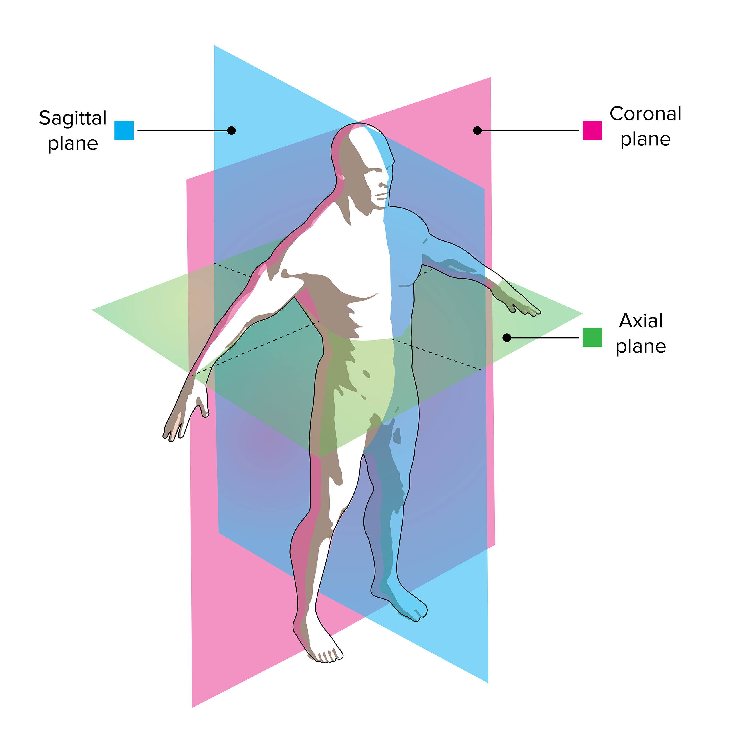
Longitudinal planes
Longitudinal planes refer to the vertically-oriented planes which are aligned along the long axis of the body. These could be sagittal or coronal.
The sagittal plane refers to a vertically-oriented, antero-posterior plane which, when passes through the body in its anatomical position, divides it into right and left parts.
The coronal plane refers to a vertically-oriented, side-to-side plane which, when passes through the boy in its anatomical position, divides it into anterior and posterior parts.
Both sagittal and coronal longitudinal planes perpendicularly intersect the transverse (or horizontal) plane.
Axial plane
Axial (a.k.a. transverse) plane refers to a horizontally-oriented plane which, when passes through the body in its anatomical position, divides it into upper and lower parts.
The axial plane is intersected at right angles by the vertically-oriented coronal (side-to-side) and sagittal (antero-posterior) planes.
External
External means to the outside or away from the center of the an organ or cavity.
For instance, if we take the example of the muscles of anterior abdominal wall, we will find three main muscles situated from outside to inside. The outermost muscle is called external oblique as it is externally situated compared to the other muscles, i.e. the internal oblique and transversus abdominis muscles.
Internal
Internal means to the inside or closer to the center of the an organ or cavity.
For instance, heart, lungs are examples of internal body organs situated within the thoracic cavity. Liver and stomach are examples of internal organs situated within the abdominal cavity.
Similarly, if we take the example of the muscles of anterior abdominal wall, we will find three main muscles situated from outside to inside. The outermost muscle is called external oblique as it is externally situated compared to the internal oblique muscle, which is more internally situated.
Luminal
Luminal refers to being within the lumen of a membrane-bound cavity or a tubular structure. For instance, various blood cells, such as the red and white blood cells and platelets are found within the lumen of the blood vessels that are lined by endothelium.
The opposite of luminal would be extraluminal that refers to structures present outside the lumen of the membrane-bound space.
Superficial
The terms superficial and deep are used to describe locations of body structures with reference to the ‘surface’ of the body. Superficial means closer to the surface of the body, as compared to ‘deep’ which means away from the body surface (usually more internally placed).
For instance, the bony skull and vertebral column are superficial to the brain and spinal cord which are deeper structures. Another example could be that of the fascial wrappings of the body. The outer superficial fascia containing cutaneous vessels and nerves is closer to the body surface as compared to the deep fascia which is more internally placed.
Central
The term central describes the location of a structure closer to the center of the body or closer to the trunk. This is contrary to the term ‘peripheral’ which refers to the structure being away from the center of the body or trunk region.
For example, our nervous system can be divided into two sub-parts, namely the ‘central’ and the peripheral nervous systems. The central nervous system comprises of brain and spinal cord which reside within the axial skeleton, close to the body center. The brain is protected inside the bony skull and the spinal cord resides within the vertebral column. On the contrary, the peripheral nervous system comprises of cranial and spinal nerves, which innervate structures away from the body center, such as, the appendicular muscles.
Peripheral
The term peripheral describes the location of a structure away from the center of the body or the trunk region. This is contrary to the term ‘central’ which refers to the structure being closer to the body center or the trunk region.
For example, our nervous system can be divided into two sub-parts, namely the ‘central’ and the ‘peripheral’ nervous systems. The peripheral nervous system comprises of cranial and spinal nerves, which innervate structures away from the body center, such as, the appendicular muscles. On the contrary, the central nervous system comprises of brain and spinal cord which reside within the axial skeleton, close to the body center. The brain is protected inside the bony skull and the spinal cord resides within the vertebral column.
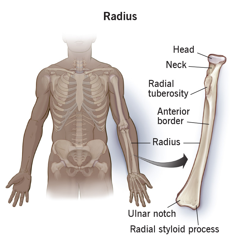
Radial
Radial means along the side of the radius or the radial bone.
Radius is a long bone which is situated in the lateral half of the forearm, when the upper limb has been positioned in its normal anatomical orientation (i.e. the elbow extended, forearm supinated and palm facing forwards).
Ulnar
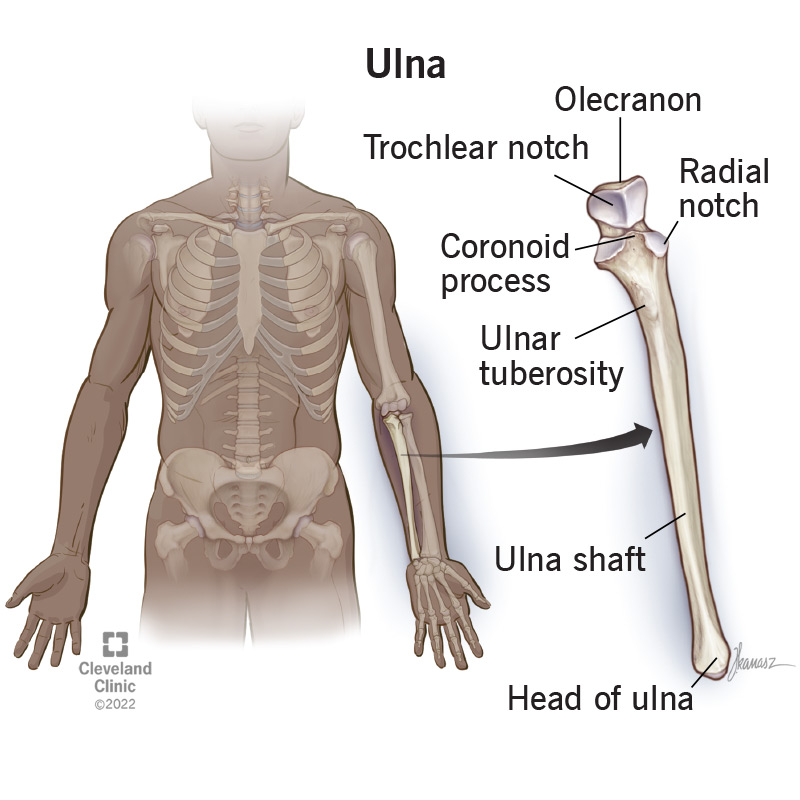
Ulnar means along the side of the ulna or the ulnar bone.
Ulna is a long bone which is situated in the medial half of the forearm, when the upper limb has been positioned in its normal anatomical orientation (i.e. the elbow extended, forearm supinated and palm facing forwards).
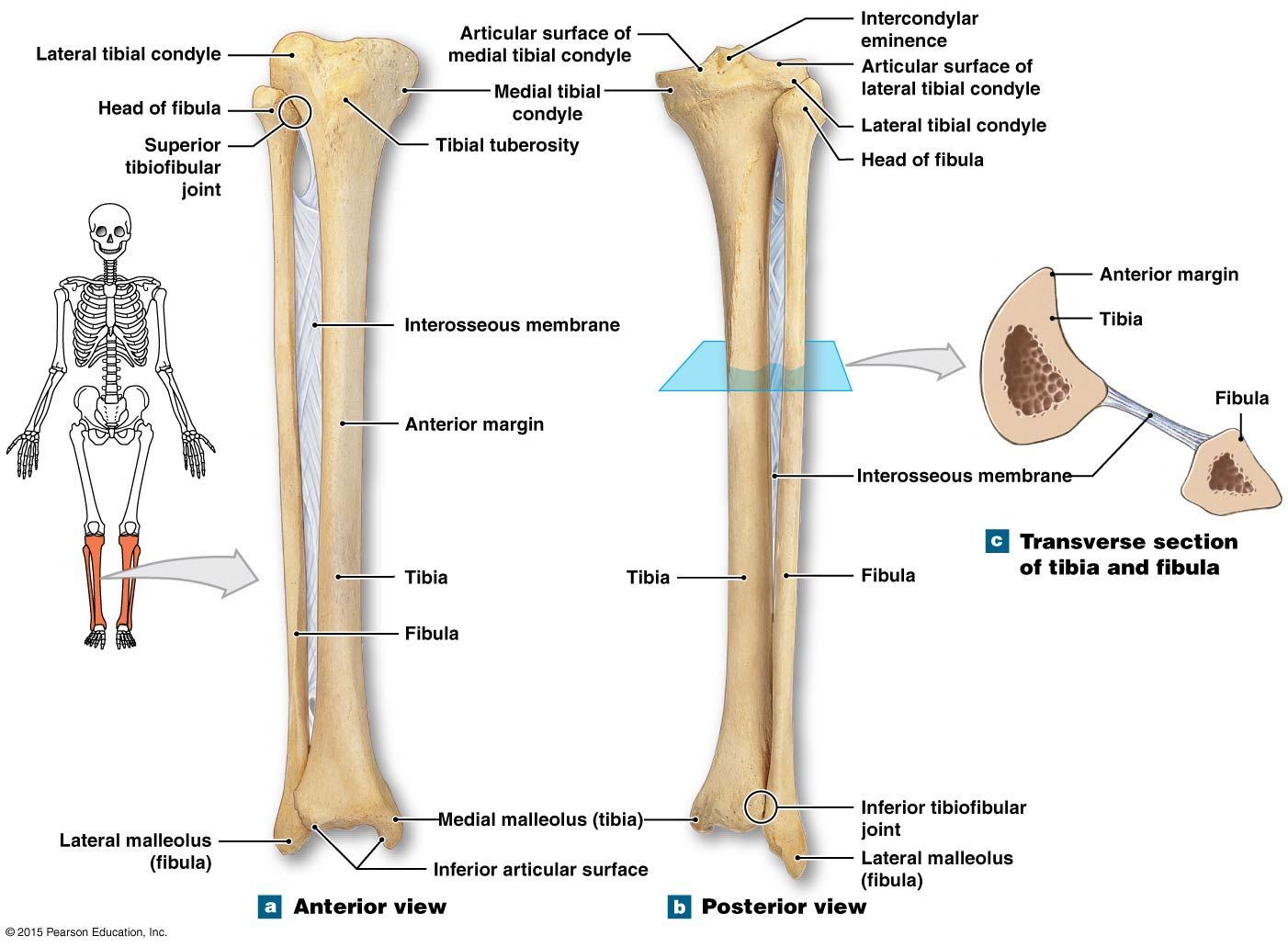
Fibular
Fibular (a.k.a. peroneal) means along the side of the fibula or the fibular bone.
Fibula is a long bone which is situated in the lateral aspect of the leg, when the lower limb has been positioned in its normal anatomical orientation (i.e. the hip and knee extended).
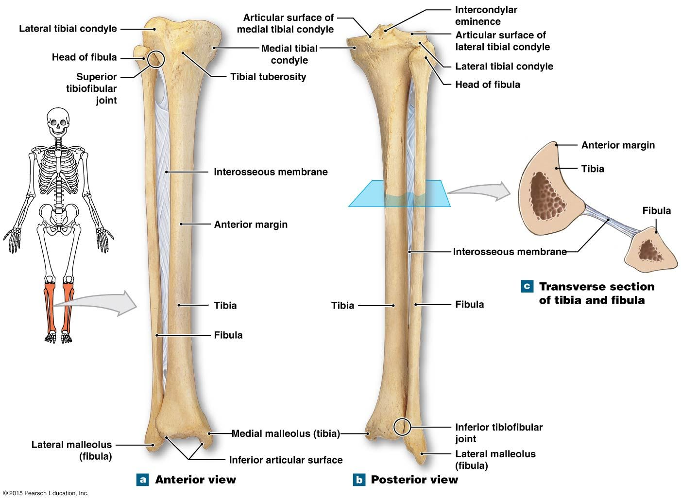
Tibial
Tibial means along the side of the tibia or the tibial bone.
Tibia is a long bone which is situated on the medial side in the leg, when the lower limb has been positioned in its normal anatomical orientation (i.e. the hip and knee extended).
Palmar
Palmar refers to the palm or the anterior surface of the hand. It is also known as the flexor or the ventral surface of hand, when kept in the normal anatomical position, (i.e. the arm externally rotated, elbow extended, forearm supinated and wrist and fingers extended with the thumb facing laterally).
For instance, palmar aponeurosis and palmaris longus muscle, both are examples of palmar structures and are situated on the ventral surface of the hand.
Plantar
Plantar refers to the sole of the foot. It is also known as the flexor or the inferior surface of the foot, when kept in the normal anatomical position, (i.e. the hip, knee and ankle extended, with sole facing downwards and/or backwards)
For instance, plantar aponeurosis is an example of a plantar structure and is situated in the sole of the foot.
Flexor
Flexor surface refers to the body surface which is on the inner side of a joint. For example, the ventral body surface situated on the front of the elbow or that which is located behind the knee joint.
The flexor surfaces cover the flexor muscles, which overlay the ventral aspects of joints. Upon contraction, the flexor muscles bend the joint and bring the flexor surfaces in close apposition with each other, thereby reducing the angle between the bones.
In case of hands and feet, the flexor surfaces are specifically known as ‘palmar’ and ‘plantar’, respectively.
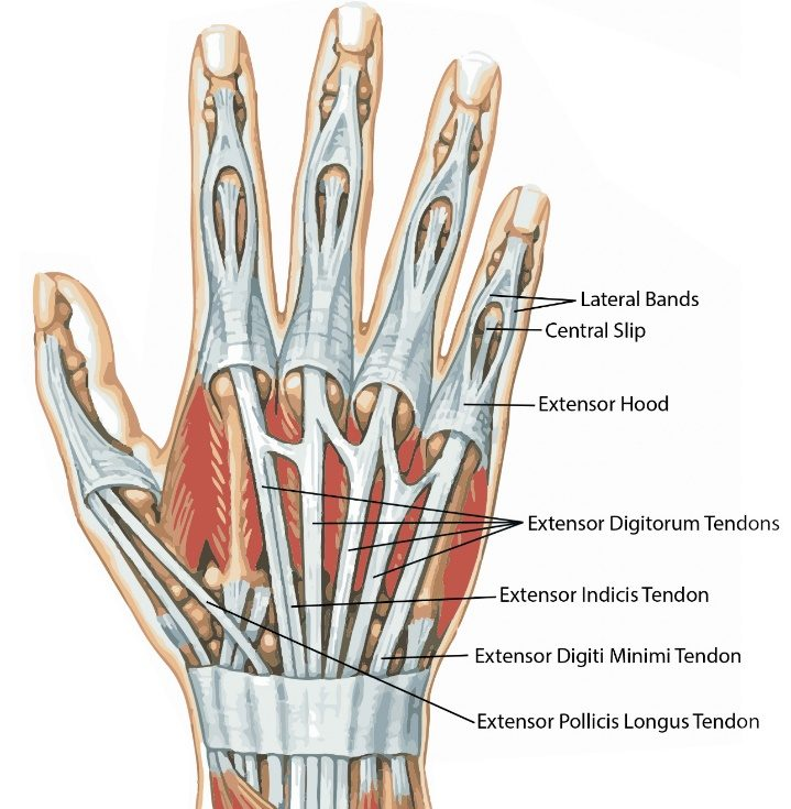
Extensor
Extensor surface refers to the body surface which is on the outer aspect of a joint. For example, the dorsal body surface situated on the back of the elbow or that which is located in front of the knee joint.
The extensor surfaces cover the extensor muscles, which overlay the dorsal aspects of joints. Upon contraction, the extensor muscles straighten up the joint and pull the flexor surfaces away from each other, thereby increasing the angle between the bones.
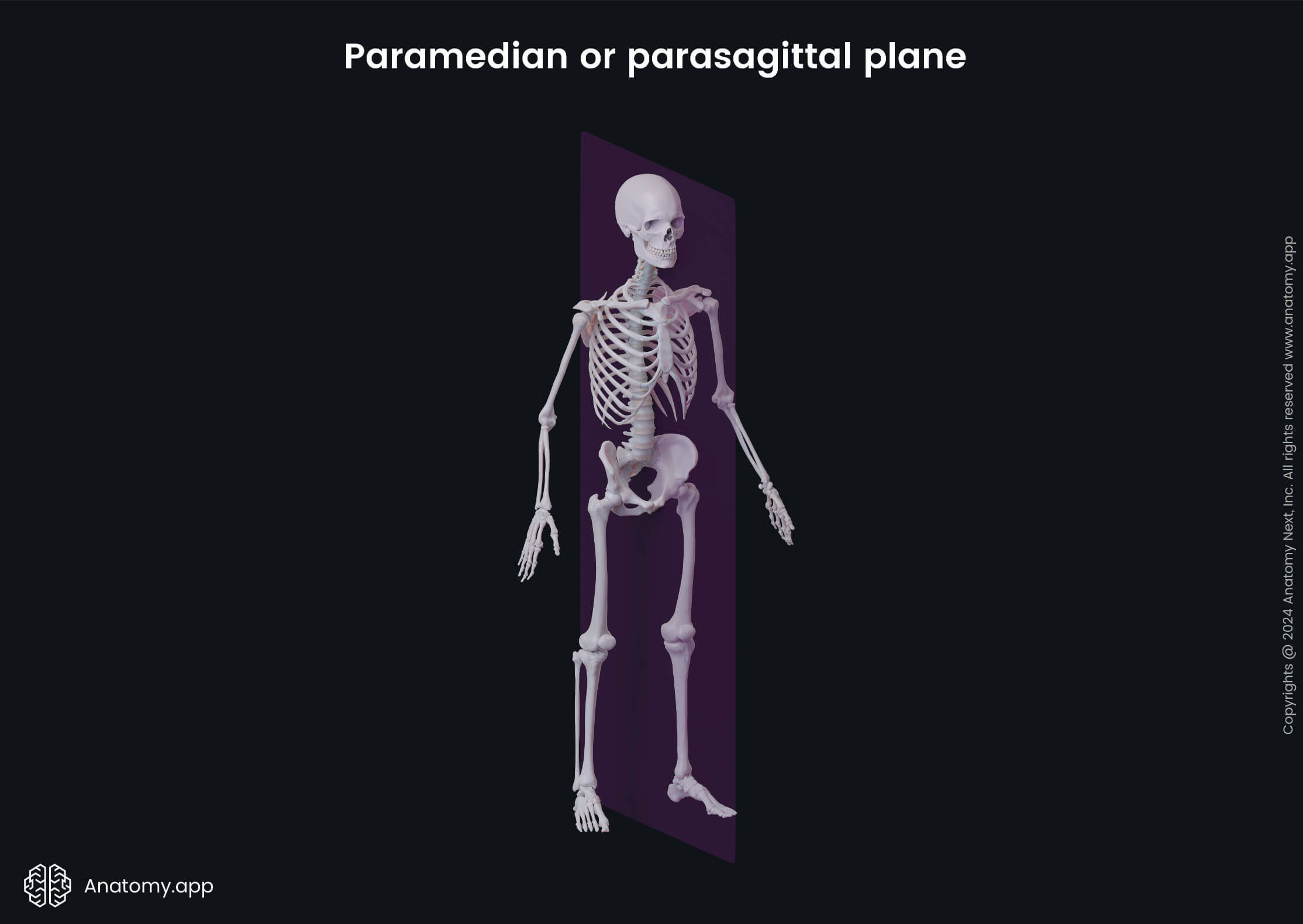
Paramedian plane
Paramedian plane refers to an antero-posterior sagittal plane that lies lateral to–but in close proximity of–the median sagittal plane.
Like all other sagittal planes, the paramedian plane also lies at right angle to the vertically-oriented coronal plane, while it perpendicularly intersects the transverse (or horizontal) plane.
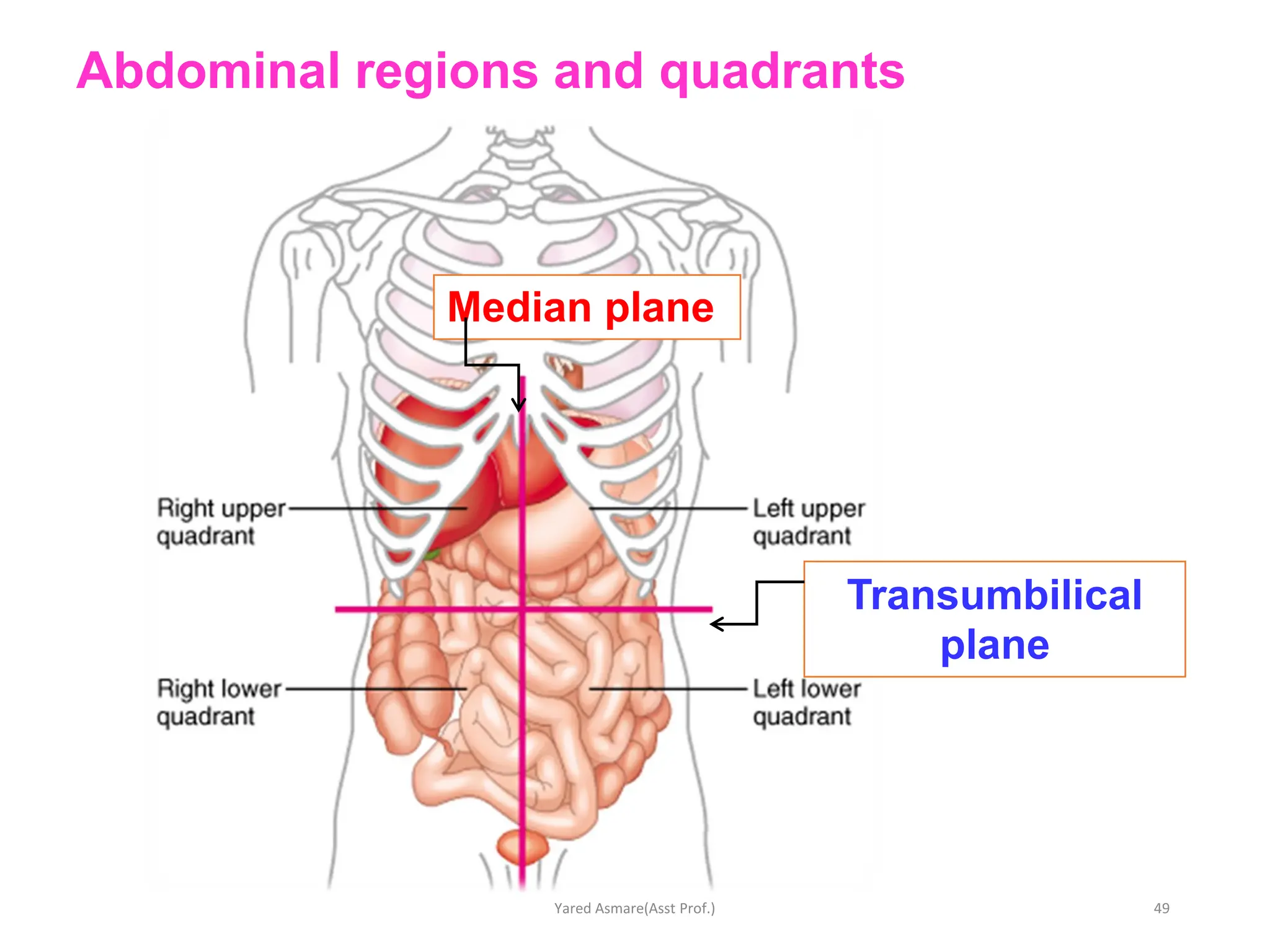
Transumbilical plane
The transumbilical plane or umbilical plane, is an example of a transverse (or horizontal or axial) reference plane in the human body. Anteriorly, it passes through the umbilicus, while posteriorly it is situated at the level of the intervertebral disc between the third (L3) and fourth (L4) lumbar vertebrae.
The transumbilical plane perpendicularly intersects the median sagittal plane in the midline, thus dividing the abdomen into four quadrants, namely the right upper, right lower, left upper and left lower quadrants. This division of abdomen into four quadrants–along with the conventional method of dividing abdomen into nine quadrants–makes it convenient for the clinicians to describe the location of viscera and the pathological lesions associated with them.
Transpyloric plane
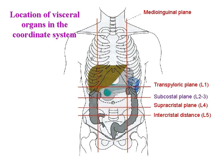
Transpyloric plane refers to a horizontal (or transverse) plane which lies at level of the costal margin, i.e., connecting the 9th costal cartilages of right and left sides with each other. The level of the transpyloric plane can also be marked by drawing a horizontal line midway between the jugular notch and the pubic symphysis or by marking a level midway between the lower end of body of sternum (above) and umbilicus (below). Posteriorly, the position of transpyloric plane equates to the lower border of the first lumbar (L1) vertebrae.
(pylorus = The passage at the lower end of the stomach that opens into the duodenum)
Subcostal plane
The subcostal plane refers to a horizontal (or transverse) plane which lies just below the level of the costal margin, i.e., connecting the 10th costal cartilages of right and left sides with each other. Posteriorly, the position of subcostal plane equates to the body of the third lumbar (L3) vertebrae.
Abduction
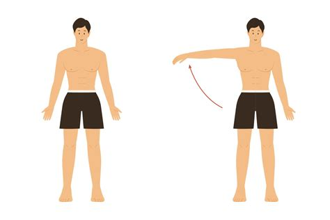
Abduction is defined as the movement of a limb or appendage, away from midline.
For instance, when the hand is placed in the normal anatomical position, the outward movement of the index finger away from the midline (i.e. middle finger) and the movement of ring and little fingers away from the midline (or middle finger), are both examples of abduction movements of the fingers. Similarly, swinging the arms upwards from the sides of the body, towards the shoulder or higher is also an example of abduction.
The opposite of abduction is adduction, where the limb or the appendage moves towards the midline.
Adduction
Adduction is defined as the movement of a limb or appendage, towards the midline.
For instance, when the hand is placed in a normal anatomical position, the inward movement of the index finger towards the midline (i.e. middle finger) and the movement of ring and little fingers towards the midline (or middle finger), are both examples of adduction movements of the fingers. Similarly, bringing the arms downwards and inwards towards the sides of the body is also an example of adduction.
The opposite of adduction is abduction, where the limb or the appendage moves away from the midline.
Lateral rotation
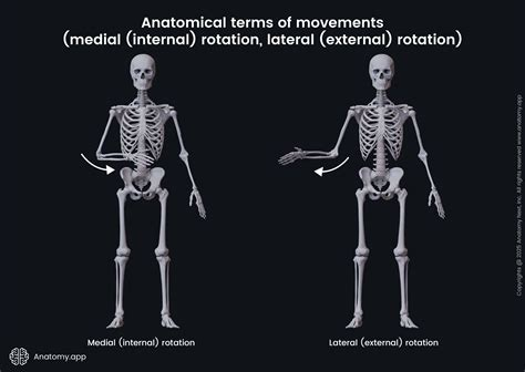
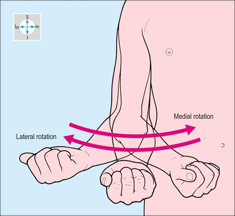
Lateral rotation is defined as the external rotation of the limb away from the midline. For instance, the lateral rotation at the hip joint can be brought about by externally rotating a fully extended lower limb, such that the big toe points outwards. The rotational movement occurs along the long axis of the lower limb. Similarly, when the arms are placed on the side of the chest and elbows flexed (as if holding a tray), then lateral rotation at the shoulder joint can be brought about by bringing the forearm and hand outwards. Here, the rotational movement occurs along the long axis of the humerus.
Medial rotation
Medial rotation is defined as the internal rotation of the limb towards the midline. For instance, the medial rotation at the hip joint can be brought about by internally rotating a fully extended lower limb, such that the big toe points inwards. The rotational movement occurs along the long axis of the lower limb. Similarly, when the arms are placed on the side of the chest and elbows flexed (as if holding a tray), then medial rotation at the shoulder joint can be brought about by bringing the forearm and hand inwards. The rotation movement occurs along the long axis of the humerus.
Circumduction
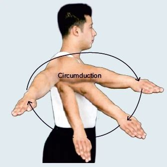
Circumduction is a movement which can be brought about at the shoulder or the hip joints (both being ball and socket variety of joints). For example, circumduction at the shoulder joint can be brought about by moving the outstretched arm in a circle and is brought about by a combination of movements including flexion, extension, adduction and abduction.
Flexion
Flexion is defined as a movement which occurs across a joint and leads to a decrease in the angle between two body parts or surfaces. For instance, the flexion at the knee joint brings the posterior surface of the leg closer to the hamstring region in the thigh, thus decreasing the angle between the femur (above) and tibia and fibula (below).
The opposite of flexion is extension which increases the angle between two body parts or surfaces. Both flexion and extension usually happen along a sagittal plane.
Extension
Extension is defined as a movement which occurs across a joint and leads to an increase in the angle between two body parts or surfaces. For instance, the extension at the knee joint moves apart the posterior surface of the leg from the hamstring region in the thigh, thus increasing the angle between the femur (above) and tibia and fibula (below).
The opposite of extension is flexion which decreases the angle between two body parts or surfaces. Both flexion and extension usually happen along a sagittal plane.
Pronation
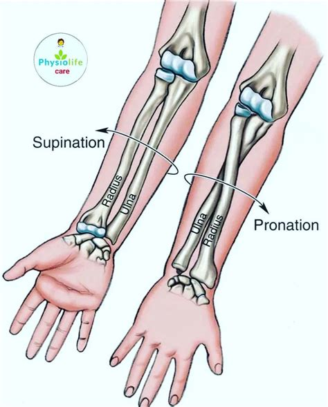
Pronation and supination of the hand are rotational movements which occur at the superior radioulnar joint.
These rotational movements are facilitated due to the presence of an annular ligament which holds the upper end of the radius in close apposition with the ulna. Since the upper radial end is freely mobile within the annular ligament, it allows the radius to rotate medially and laterally around the long axis of the ulna, bringing about pronation and supination movements.
For instance, if we place the forearm on a flat surface and flip the hand such that the palm faces downwards, this is pronation. Here, the distal end of radial bone has rotated medially over the ulna. On the contrary, if we bring the hand back to its original position with palm facing upwards, this is supination movement. In this case, the distal radius has rotated laterally over the ulna.
Two major pronators of the forearm are pronator teres and pronator quadratus muscles.
Supination
Supination and pronation of the hand are rotational movements which occur at the superior radioulnar joint.
These rotational movements are facilitated due to the presence of an annular ligament which holds the upper end of the radius in close apposition with the ulna. Since the upper radial end is freely mobile within the annular ligament, it allows the radius to rotate medially and laterally around the long axis of the ulna, bringing about pronation and supination movements.
For instance, if we place the forearm on a flat surface and flip the hand such that the palm faces downwards, this is pronation. Here, the distal end of radial bone has rotated medially over the ulna. However, if we bring the hand back to its original position with palm facing upwards, this is supination movement. In this case, the distal radius has rotated laterally over the ulna.
Two major supinators of the forearm are supinator muscle and the long head of biceps muscle.
Opposition
Opposition of the thumb is defined as the positioning of the thumb pad (or tip of the thumb) against the distal pad (or tip) of any of the fingers. The opposite of opposition is reposition, where the thumb moves away from the tip of the other finger.
Opposition and reposition movements occur at the first carpometacarpal joint, i.e. the joint between trapezium and first metacarpal bone. The opposition movement is helpful in creating a pincer grip of the hand for holding objects.
Reposition
Reposition is defined as the opposite of opposition, where the thumb moves away from the tip of the other finger, for instance, to release a grip against an object. On the contrary, opposition of the thumb is defined as the positioning of the thumb pad (or tip of the thumb) against the distal pad (or tip) of any of the fingers.
Opposition and reposition movements occur at the first carpometacarpal joint, i.e. the joint between trapezium and first metacarpal bone.
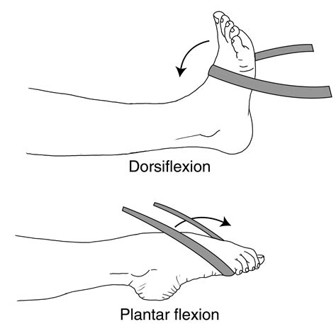
Plantar flexion
Plantar flexion occurs at the ankle joint and involves the sole of the foot moving downwards towards the floor. It is in fact, the flexion movement at the ankle joint. Daily life activities involving plantar flexion at the ankle joint include pressing the peddle of the car for escalating or breaking, standing on tippy toes (such as, in ballet dancing) and jumping off the floor.
Plantar flexion is brought about by the combined action of flexor muscles in the back of the leg (gastrocnemius, soleus, plantaris, flexor hallucis longus, flexor digitorum longus and flexor hallucis longus) and by the peroneal compartment muscles, namely the peroneus longus and brevis muscles.
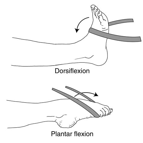
Dorsiflexion
Dorsiflexion occurs at the ankle joint and involves the sole of the foot moving upwards away from the floor. It is in fact, the extension movement at the ankle joint.
Dorsiflexion is brought about by the combined action of extensor muscles in the front of the leg (extensor hallucis longus, extensor digitorum longus, extensor hallucis longus and peroneus tertius).
The opposite of dorsiflexion is plantar flexion, which also occurs at the ankle joint but it involves the foot pointing downwards towards the floor.
Elevation
Elevation refers to raising or lifting up a body part. For instance, moving shoulder girdle superiorly or moving the forearm upwards by flexing at the elbow joint, when arms hang on the side of the body in the normal anatomical position.
Depression
Depression refers to lowering a body part. For instance, moving shoulder girdle inferiorly or lowering the forearm by extending the elbow joint, when arms hang on the side of the body in the normal anatomical position.
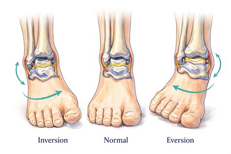
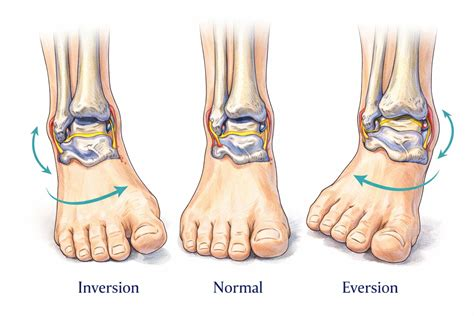
Eversion
Eversion of the foot is a movement which involves lifting the lateral border of the foot such that the sole of the foot faces laterally or outwards. The movement occurs at the intertarsal and tarsometatarsal joints and NOT on the ankle joint.
The opposite of eversion is inversion, where the sole of the foot faces inwards or medially.
Inversion
Inversion of the foot is a movement which involves tilting of the foot such that the sole of the foot faces medially or inwards. The movement occurs at the intertarsal and tarsometatarsal joints and NOT at the ankle joint.
The opposite of inversion is eversion, where the sole of the foot faces outwards or laterally.
Protraction
Protraction means to protrude or stick out a part of the body. For instance, when throwing a punch with abducted arms, a muscle originating from the chest wall–called serratus anterior–contracts and drags the scapula forwards over the rib cage, causing protraction of scapula. Similarly, sticking the mandible or the lower jaw bone outwards is called protraction of mandible.
The opposite of protraction is retraction which refers to bringing the protracted scapula or the mandible back into their original positions.
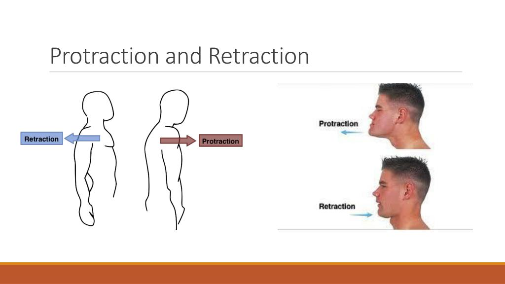
Retraction
Retraction is the opposite of protraction movement, which refers to bringing the protracted scapula or the mandible back into their original positions.
Protraction means to protrude or stick out a part of the body. For instance, when throwing a punch with abducted arms, a muscle originating from the chest wall–called serratus anterior–contracts and drags the scapula forwards over the rib cage, causing protraction of scapula. Similarly, sticking the mandible or the lower jaw bone outwards is called protraction of mandible. As mentioned above, bringing them back to their original positions is called retraction.