Balance Wk1 bootcamp LG1
 Call Kai
 Learn
Learn Practice Test
 Spaced Repetition
 Match
Match Flashcards
 Knowt Play
1/130

There's no tags or description
Looks like no tags are added yet.
Name | Mastery | Learn | Test | Matching | Spaced | Call with Kai | Chat |
|---|
No analytics yet
Send a link to your students to track their progress
131 Terms
Which structures are located in the renal cortex?
Answer: The glomerulus, proximal convoluted tubule, distal convoluted tubule, and cortical collecting duct.
Extra Information:
These are the cortical structures identified on the Bootcamp slide.
🧠 “Cortex Girls Prefer Drinking Coffee”
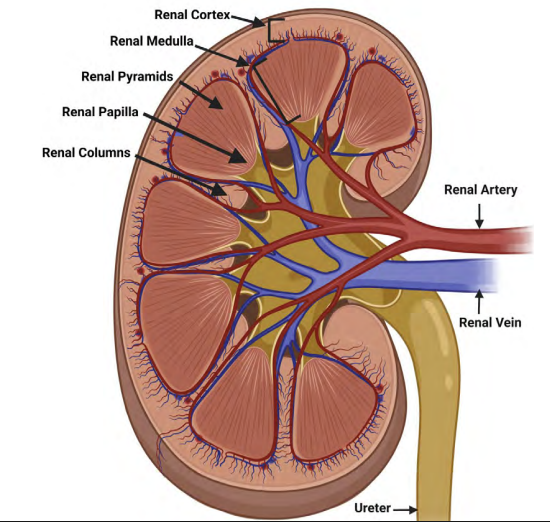
Which structures are located in the renal medulla?
Answer: Renal pyramids, renal columns, the Loop of Henle, and the medullary collecting duct.
Extra Information:
These are the medullary structures identified on the Bootcamp slide.
🧠 “Please Come Learn Medulla”

Trace the renal arterial supply from the renal artery to the glomerulus.
Answer: Renal artery → segmental artery → interlobar artery → arcuate artery → interlobular artery → afferent arteriole → glomerulus.
Extra Information:
This is the complete preglomerular arterial pathway shown on the slide.
Recall the sequence in order from the largest vessel to the glomerulus.
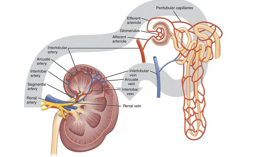
Which vessel immediately delivers blood into the glomerulus?
Answer: The afferent arteriole.
Extra Information:
The afferent arteriole comes after the interlobular artery in the arterial pathway.

Which vessel carries blood away from the glomerulus?
Answer: The efferent arteriole.
Extra Information:
Blood then enters the peritubular capillaries or vasa recta.

Trace blood flow from the glomerulus back toward the renal vein.
Answer: Glomerulus → efferent arteriole → peritubular capillaries or vasa recta → venous outflow pathway → renal vein.
Extra Information:
This is the postglomerular blood-flow pathway shown on the slide.
What structures make up the juxtaglomerular apparatus on this Bootcamp slide?
Answer: The macula densa and juxtaglomerular cells.
Extra Information:
The macula densa is associated with the distal convoluted tubule.
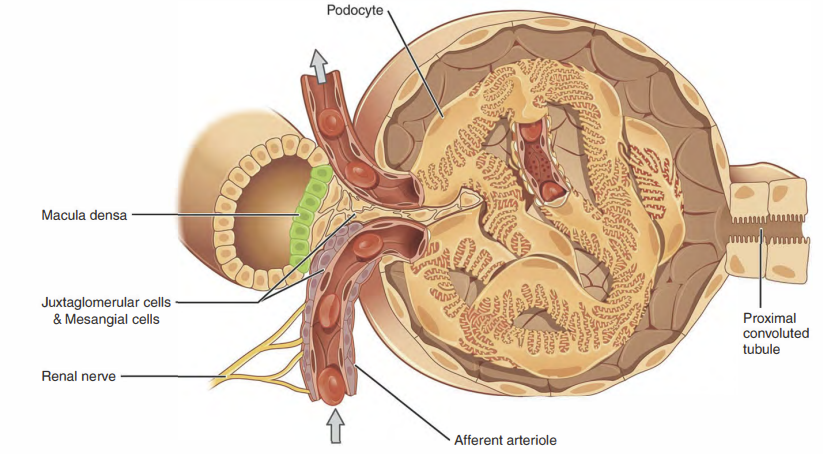
The macula densa is associated with the ______.
Answer: Distal convoluted tubule.
Extra Information:
The macula densa is identified as part of the juxtaglomerular apparatus.
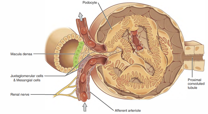
Juxtaglomerular cells are located in the ______ and secrete ______.
Answer: Afferent arteriole and renin.
Extra Information:
Juxtaglomerular cells are part of the juxtaglomerular apparatus.

What is the function of mesangial cells identified on this slide?
Answer: Mesangial cells remove debris.
Extra Information:
This is the specific mesangial cell function emphasized on the Bootcamp slide.
How does the length of the left renal vein compare with the right renal vein?
Answer: The left renal vein is longer than the right renal vein.
Extra Information:
This anatomical relationship is marked as high yield on the slide.

Which gonadal vein drains into the left renal vein?
Answer: The left gonadal vein.
Extra Information:
The slide contrasts this relationship with the right gonadal vein.

What is nutcracker syndrome?
Answer: Compression of the left renal vein by the superior mesenteric artery.
Extra Information:
This relationship is emphasized as a clinically important consequence of left renal vein anatomy.

What clinical presentation is associated with nutcracker syndrome?
Answer: Abdominal pain, hematuria (blood in urine), and varicocele.
Extra Information:
The underlying abnormality is compression of the left renal vein by the superior mesenteric artery.

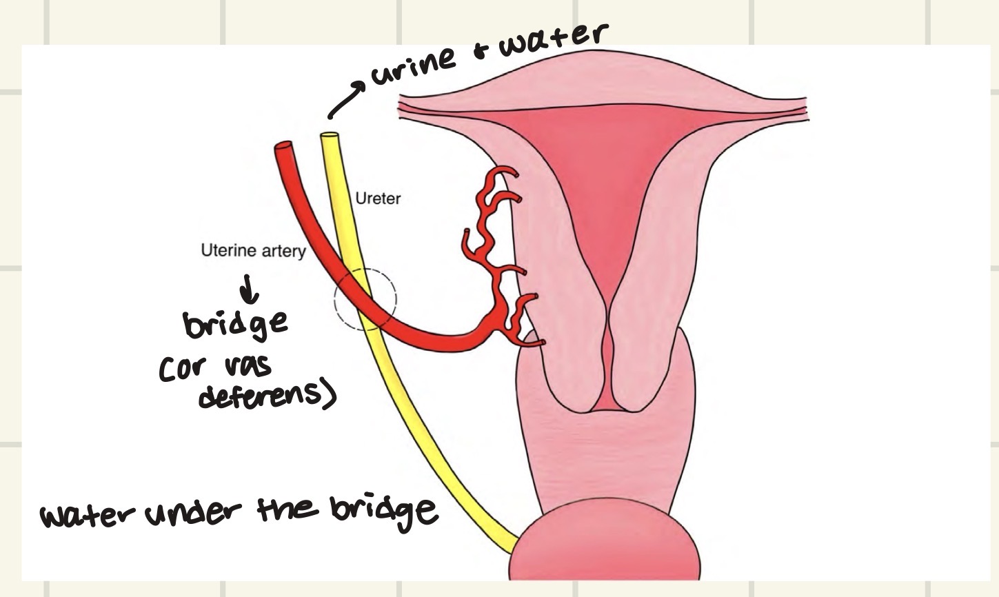
What does “water under the bridge” describe in ureter anatomy?
Answer: The ureter passes under the vas deferens or uterine artery.
Extra Information:
“Water” represents the ureter.
The slide uses this phrase to remember the anatomical relationship.
The ureter passes over the ______ artery.
Answer: Common iliac artery.
Extra Information:
The slide emphasizes both the structures the ureter passes under and the vessel it passes over.

Why can the ureter be damaged during gynecologic procedures?
Answer: The ureter is in close proximity to the uterine artery.
Extra Information:
The ureter passes under the uterine artery.
This close anatomical relationship creates a risk of ureteral injury.

Where are the ureteral orifices located within the bladder?
Answer: The trigone.
Extra Information:
The ureteral orifice is one of the bladder structures highlighted on the slide.
The trigone of the bladder is a smooth, triangular area located at the base of the urinary bladder
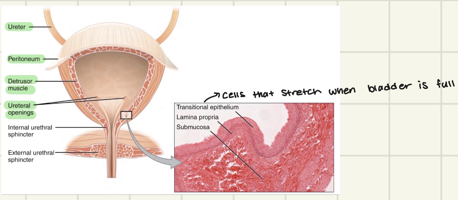
Which urethral sphincter is involuntary versus voluntary?
Answer: The internal urethral sphincter is involuntary, while the external urethral sphincter is voluntary.
Extra Information:
Distinguishing the two sphincters by voluntary control is emphasized on the slide.

The detrusor muscle of the bladder is under ______ control.
Answer: Autonomic control.
Extra Information:
The detrusor muscle is the bladder muscle specifically identified on the Bootcamp slide.

What is renal secretion?
Answer: Movement of a substance from the circulation into the nephron.
Extra Information:
The direction is blood → nephron.

What is renal reabsorption?
Answer: Movement of a substance from the nephron into the circulation.
Extra Information:
The direction is nephron → blood.
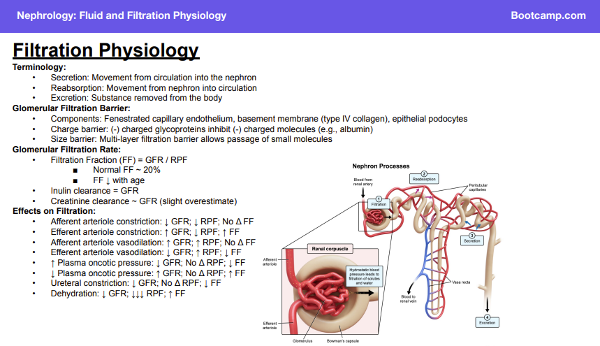
What is renal excretion?
Answer: Removal of a substance from the body.
Extra Information:
Excretion represents substance leaving the body.

What are the three components of the glomerular filtration barrier?
Answer: Fenestrated capillary endothelium → basement membrane → epithelial podocytes.
Extra Information:
The basement membrane contains type IV collagen.
Together, these layers form a size-selective filtration barrier.
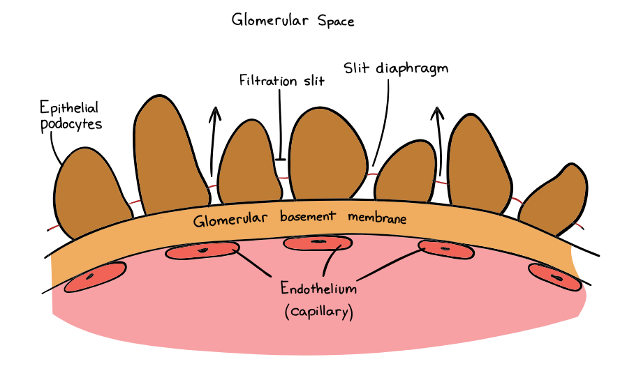
What type of collagen is found in the glomerular basement membrane?
Answer: Type IV collagen.
Extra Information:
The basement membrane is one component of the glomerular filtration barrier.

How does the charge barrier of the glomerulus restrict filtration?
Answer: Negatively charged glycoproteins inhibit the filtration of negatively charged molecules.
Extra Information:
Albumin is an example of a negatively charged molecule restricted by this barrier.

Why can small molecules pass through the glomerular filtration barrier more readily than large molecules?
Answer: The multilayer filtration barrier acts as a size barrier that permits passage of small molecules.
Extra Information:
Glomerular filtration depends partly on molecular size.

What is the equation for filtration fraction?
Answer: Filtration fraction = glomerular filtration rate / renal plasma flow.
Extra Information:
Filtration fraction represents the fraction of renal plasma flow that undergoes filtration.

What is the normal filtration fraction?
Answer: Approximately 20%.
Extra Information:
Filtration fraction decreases with age.
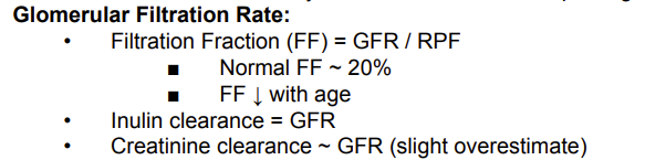
How does filtration fraction change with age?
Answer: Filtration fraction decreases with age.
Extra Information:
Normal filtration fraction is approximately 20%.

Which substance has a clearance equal to glomerular filtration rate?
Answer: Inulin.
Extra Information:
Inulin clearance = glomerular filtration rate.
Filtration = movement of a substance from the glomerular capillaries → Bowman’s space/tubule.
Clearance = the volume of plasma completely cleared of a substance per unit time (usually mL/min).
Think:
Filtration = what crosses into the nephron
Clearance = how effectively the kidney removes that substance from plasma

How does creatinine clearance compare with glomerular filtration rate?
Answer: Creatinine clearance approximates glomerular filtration rate but slightly overestimates it.
Extra Information:
The slide distinguishes creatinine from inulin, whose clearance equals glomerular filtration rate.
creatinine: produced by the body
Creatinine clearance slightly OVERestimates true GFR.
💡 Memory trick: “Creatinine cheats a little” → it sneaks some extra into the tubule through secretion.
Creatine phosphate → helps make ATP → creatinine is produced as a waste product

What happens to glomerular filtration rate, renal plasma flow, and filtration fraction with afferent arteriole constriction?
Answer: ↓ glomerular filtration rate, ↓ renal plasma flow, and no change in filtration fraction.
Extra Information:
Both glomerular filtration rate and renal plasma flow decrease.
Because they decrease together, the slide indicates no change in filtration fraction.

What happens to glomerular filtration rate, renal plasma flow, and filtration fraction with afferent arteriole vasodilation?
Answer: ↑ glomerular filtration rate, ↑ renal plasma flow, and no change in filtration fraction.
Extra Information:
Both glomerular filtration rate and renal plasma flow increase.
The slide indicates no change in filtration fraction.

What happens to glomerular filtration rate, renal plasma flow, and filtration fraction with efferent arteriole constriction?
Answer: ↑ glomerular filtration rate, ↓ renal plasma flow, and ↑ filtration fraction.
Extra Information:
Glomerular filtration rate and renal plasma flow move in opposite directions.
The resulting filtration fraction increases.
1. More gets filtered
Blood has trouble exiting → pressure builds up in the glomerulus → ↑ glomerular hydrostatic pressure → ↑ GFR
2. But blood/plasma can't flow through as easily
The efferent arteriole is narrow → ↑ resistance → ↓ RPF

What happens to glomerular filtration rate, renal plasma flow, and filtration fraction with efferent arteriole vasodilation?
Answer: ↓ glomerular filtration rate, ↑ renal plasma flow, and ↓ filtration fraction.
Extra Information:
Glomerular filtration rate decreases while renal plasma flow increases.
The resulting filtration fraction decreases.
Efferent dilation →
↓ resistance → ↑ RPF (plasma flows through more easily)
↓ backup pressure in glomerulus → ↓ glomerular hydrostatic pressure
↓ pressure pushing fluid into Bowman space → ↓ GFR
Therefore → ↓ FF

What happens to glomerular filtration rate, renal plasma flow, and filtration fraction when plasma oncotic pressure increases?
Answer: ↓ glomerular filtration rate, no change in renal plasma flow, and ↓ filtration fraction.
Extra Information:
The change in plasma oncotic pressure affects filtration without changing renal plasma flow on this slide.
oncotic pressure pulls fluid in
So you have opposing forces:
Glomerular hydrostatic pressure: pushes fluid OUT → favors filtration
Glomerular oncotic pressure: pulls fluid IN → opposes filtration
Therefore:
↑ oncotic pressure → ↑ pulling fluid into/keeping fluid in capillary → ↓ filtration → ↓ GFR
But importantly, this isn't narrowing an arteriole, so you're not increasing vascular resistance.
➡ RPF stays about the same.
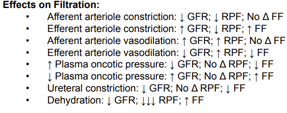
What happens to glomerular filtration rate, renal plasma flow, and filtration fraction when plasma oncotic pressure decreases?
Answer: ↑ glomerular filtration rate, no change in renal plasma flow, and ↑ filtration fraction.
Extra Information:
Renal plasma flow remains unchanged while glomerular filtration rate increases.
↓ Glomerular oncotic pressure = fewer proteins pulling water INTO the blood
So:
↓ oncotic pull inward → more fluid can be pushed OUT into Bowman space → ↑ GFR
But you haven't changed the diameter of the arterioles, so:
RPF stays about the same.

What happens to glomerular filtration rate, renal plasma flow, and filtration fraction with ureteral constriction?
Answer: ↓ glomerular filtration rate, no change in renal plasma flow, and ↓ filtration fraction.
Extra Information:
Renal plasma flow remains unchanged while glomerular filtration rate decreases.
Blood vessel constriction = changes FLOW
Urinary obstruction = changes BACKPRESSURE against filtration
That's why ureteral obstruction can lower GFR without directly lowering RPF. With prolonged obstruction, secondary changes can eventually reduce renal blood flow too.
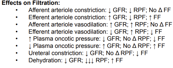
What happens to glomerular filtration rate, renal plasma flow, and filtration fraction during dehydration?
Answer: ↓ glomerular filtration rate, ↓↓↓ renal plasma flow, and ↑ filtration fraction.
Extra Information:
Renal plasma flow decreases much more substantially than glomerular filtration rate.
Because renal plasma flow falls disproportionately, filtration fraction increases.
Dehydration → ↓ blood/plasma volume → ↓ blood reaching the kidneys → ↓ renal plasma flow (RPF)
Then there's another effect:
Dehydration also causes ↑ plasma protein concentration because there's less water in the blood.
So:
Dehydration
→ ↓ plasma volume → ↓ RPF
→ ↑ plasma oncotic pressure → pulls/holds more water in glomerular capillaries → ↓ GFR

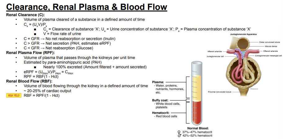
What is renal clearance?
Answer: The volume of plasma cleared of a substance in a defined amount of time.
Extra Information:
Clearance describes how effectively the kidneys remove a particular substance from plasma.
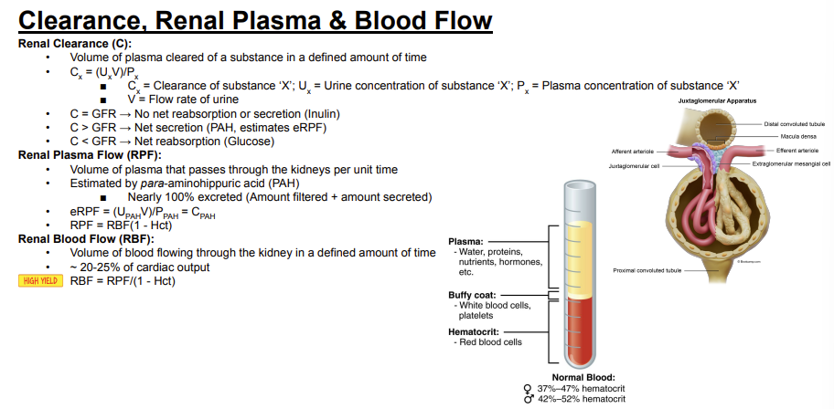
What is the equation for renal clearance of substance X?
Answer: Cx = (Ux × V) / Px.
Extra Information:
Cx is the clearance of substance X.
The equation uses urine concentration, urine flow rate, and plasma concentration.

In the renal clearance equation, what do Ux, Px, and V represent?
Answer: Ux = urine concentration of substance X, Px = plasma concentration of substance X, and V = urine flow rate.
Extra Information:
These variables are used in Cx = (Ux × V) / Px.

What does it mean if the clearance of a substance equals the glomerular filtration rate?
Answer: There is no net reabsorption or secretion of the substance.
Extra Information:
Inulin is the example given on the slide.
Inulin clearance therefore equals glomerular filtration rate.

What does it mean if the clearance of a substance is greater than the glomerular filtration rate?
Answer: The substance undergoes net secretion.
Extra Information:
Para-aminohippuric acid is the example given on the slide.
Its clearance is used to estimate renal plasma flow.
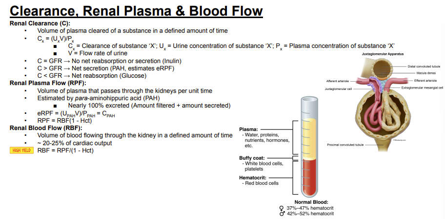
What does it mean if the clearance of a substance is less than the glomerular filtration rate?
Answer: The substance undergoes net reabsorption.
Extra Information:
Glucose is the example given on the slide.

How does inulin clearance compare with glomerular filtration rate, and what does this indicate?
Answer: Inulin clearance equals glomerular filtration rate, indicating no net reabsorption or secretion.
Extra Information:
Inulin represents the C = glomerular filtration rate relationship on the slide.

How does para-aminohippuric acid clearance compare with glomerular filtration rate, and what does this indicate?
Answer: Para-aminohippuric acid clearance is greater than glomerular filtration rate, indicating net secretion.
Extra Information:
Para-aminohippuric acid clearance is used to estimate renal plasma flow.
Renal Plasma Flow = how much plasma flows through the kidney.
Imagine 100 people walk into a store every minute.
RPF = 100 people/min
ERPF
We can't easily stand there and count exactly how much plasma flows through the kidney, so we use PAH to estimate it.
PAH is special because the kidney removes almost all of it from the plasma in one trip.
So:
PAH in blood → kidney → almost all PAH removed into urine
Therefore, by measuring how quickly the kidney clears PAH, we can estimate:
“How much plasma must have passed through the kidney?”
That's effective renal plasma flow (ERPF).
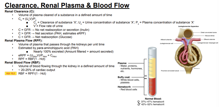
How does glucose clearance compare with glomerular filtration rate, and what does this indicate?
Answer: Glucose clearance is less than glomerular filtration rate, indicating net reabsorption.
Extra Information:
Glucose represents the C < glomerular filtration rate relationship on the slide.
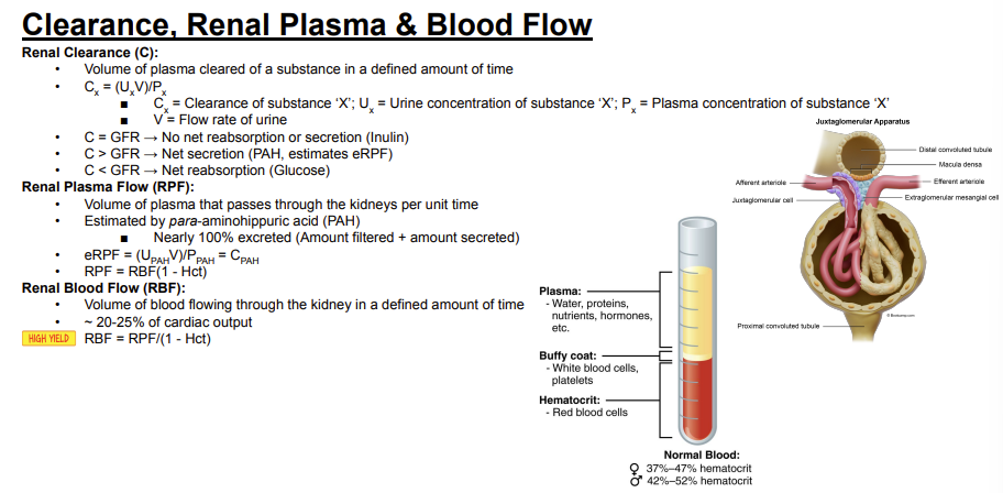
What is renal plasma flow?
Answer: The volume of plasma that passes through the kidneys per unit time.
Extra Information:
Renal plasma flow specifically refers to the plasma component of blood.

Which substance is used to estimate renal plasma flow?
Answer: Para-aminohippuric acid.
Extra Information:
Para-aminohippuric acid is nearly 100% excreted.
Its excretion consists of the amount filtered plus the amount secreted.
Plasma carries PAH into kidney → kidney removes almost all the PAH → measure how much PAH was cleared → estimate how much plasma must have flowed through the kidney.
Think:
“If I know how much PAH the kidney cleared, I can work backward to estimate how much plasma brought that PAH to the kidney.”

Why can para-aminohippuric acid be used to estimate renal plasma flow?
Answer: Para-aminohippuric acid is nearly 100% excreted through filtration plus secretion.
Extra Information:
Its clearance therefore estimates renal plasma flow.

What is the equation for estimated renal plasma flow using para-aminohippuric acid?
Answer: Estimated renal plasma flow = (UPAH × V) / PPAH = CPAH.
Extra Information:
CPAH represents para-aminohippuric acid clearance.

What is the relationship between renal plasma flow, renal blood flow, and hematocrit?
Answer: Renal plasma flow = renal blood flow × (1 − hematocrit).
Extra Information:
The factor (1 − hematocrit) represents the non-red-blood-cell portion of blood.

What is renal blood flow?
Answer: The volume of blood flowing through the kidney in a defined amount of time.
Extra Information:
Unlike renal plasma flow, renal blood flow refers to the entire blood volume.

Approximately what percentage of cardiac output goes to renal blood flow?
Answer: Approximately 20–25% of cardiac output.
Extra Information:
This value is marked as high yield on the Bootcamp slide.
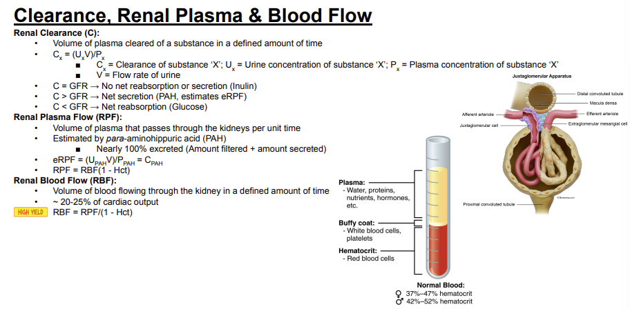
How can renal blood flow be calculated from renal plasma flow and hematocrit?
Answer: Renal blood flow = renal plasma flow / (1 − hematocrit).
Extra Information:
This equation converts plasma flow into total blood flow by accounting for hematocrit.
What is the normal blood glucose range shown on the Bootcamp slide?
Answer: Approximately 60–120 mg/dL.
Extra Information:
This range is listed under normoglycemic conditions.
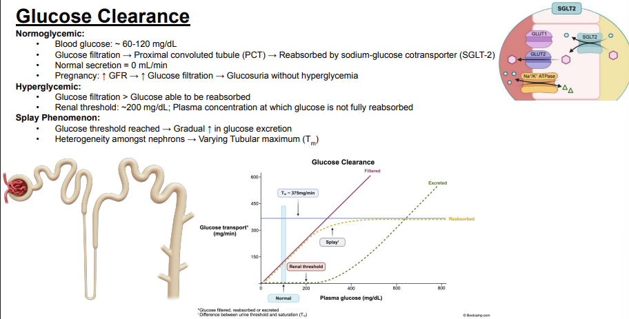
Trace the handling of filtered glucose under normal conditions.
Answer: Glucose is filtered → enters the proximal convoluted tubule → is reabsorbed by the sodium-glucose cotransporter 2.
Extra Information:
Under normoglycemic conditions, filtered glucose is reabsorbed.

Where is filtered glucose reabsorbed according to the slide?
Answer: The proximal convoluted tubule.
Extra Information:
Glucose reabsorption occurs after glucose is filtered at the glomerulus.

Which transporter reabsorbs glucose in the proximal convoluted tubule according to the slide?
Answer: Sodium-glucose cotransporter 2.
Extra Information:
Sodium-glucose cotransporter 2 is abbreviated SGLT-2.

What is the normal secretion rate of glucose?
Answer: 0 mL/min.
Extra Information:
The slide lists no normal glucose secretion.
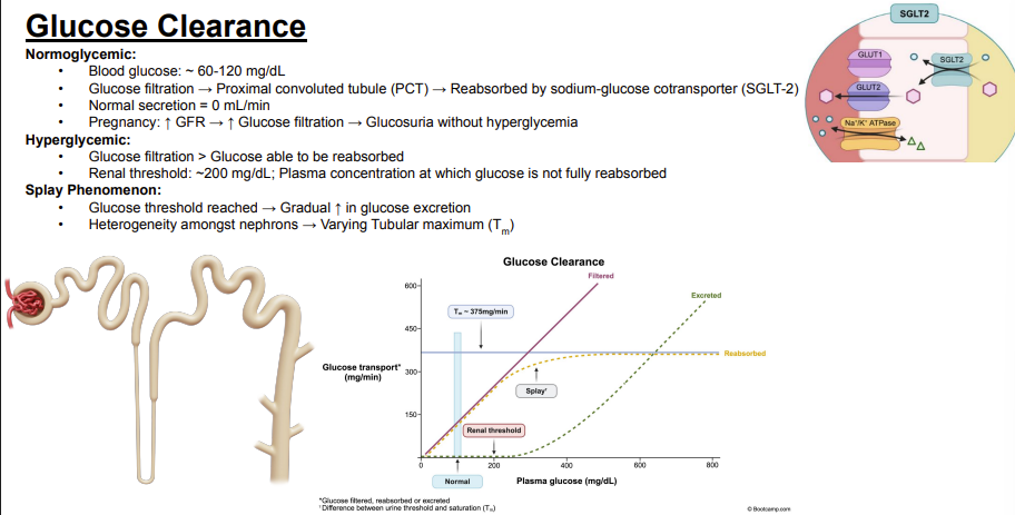
Why can pregnancy cause glucosuria without hyperglycemia?
Answer: Pregnancy increases glomerular filtration rate → increases glucose filtration → causes glucosuria without hyperglycemia.
Extra Information:
The increased filtered glucose load is driven by the increased glomerular filtration rate.
↑ GFR → ↑ amount of glucose filtered → proximal tubule may not reabsorb all of the increased filtered glucose → some glucose remains in tubule → glucose in urine (glycosuria)

What happens to renal glucose handling during hyperglycemia?
Answer: Glucose filtration can exceed the amount of glucose that can be reabsorbed.
Extra Information:
Once reabsorptive capacity is exceeded, glucose remains available for excretion.

What is the renal threshold for glucose?
Answer: Approximately 200 mg/dL.
Extra Information:
This is a plasma glucose concentration.

What does the renal threshold for glucose represent?
Answer: The plasma glucose concentration at which glucose is no longer fully reabsorbed.
Extra Information:
The slide gives a renal threshold of approximately 200 mg/dL.

What is the splay phenomenon?
Answer: After the glucose threshold is reached, glucose excretion increases gradually rather than abruptly.
Extra Information:
The graph shows the transition from complete reabsorption toward increasing glucose excretion.

What causes the splay phenomenon?
Answer: Heterogeneity among nephrons causes different nephrons to have varying tubular maximums.
Extra Information:
The nephrons therefore do not all reach their reabsorptive limits at exactly the same plasma glucose concentration.
Below Tm → all glucose can be reabsorbed → no glucose in urine
Reach/exceed Tm → transporters are saturated → glucose spills into urine
🧠 Tm = Transporters MAXED out.

What does the tubular maximum for glucose represent on the glucose clearance graph?
Answer: The maximum rate at which glucose can be reabsorbed.
Extra Information:
The graph shows reabsorption approaching a plateau as the tubular maximum is reached.
The slide labels a tubular maximum of approximately 375 mg/min.
↑ filtered glucose → transporters start reaching capacity → glucose starts appearing in urine → eventually all transporters max out (Tm) → any additional filtered glucose MUST be excreted.

Approximately how much filtered glucose and amino acids are normally reabsorbed by the proximal convoluted tubule?
Answer: Approximately 100%.
Extra Information:
The proximal convoluted tubule normally reabsorbs essentially all filtered glucose and amino acids.

Which transporter is responsible for glucose reabsorption in the proximal convoluted tubule?
Answer: The sodium-glucose cotransporter.
Extra Information:
Sodium and glucose are transported together across the apical side.

At approximately what blood glucose concentration does proximal convoluted tubule glucose transport become saturated according to this slide?
Answer: Approximately 350 mg/dL.
Extra Information:
Above this level, the sodium-glucose cotransporter cannot continue increasing glucose reabsorption proportionally.

Which drugs block proximal convoluted tubule sodium-glucose cotransport?
Answer: Sodium-glucose cotransporter 2 inhibitors.
Extra Information:
These drugs inhibit the sodium-glucose cotransporter responsible for glucose reabsorption.

In what form is bicarbonate reabsorbed across the apical side of the proximal convoluted tubule?
Answer: Carbon dioxide and water.
Extra Information:
The slide contrasts this with basolateral transport as bicarbonate and sodium.
The apical side of a nephron cell faces the inside lumen containing urine, while the basal (basolateral) side faces the outer interstitial fluid and blood vessels
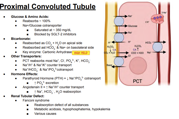
In what form is bicarbonate transported across the basolateral side of the proximal convoluted tubule?
Answer: Bicarbonate and sodium.
Extra Information:
On the apical side, bicarbonate is handled as carbon dioxide and water.
Apical side: HCO₃⁻ can't easily cross by itself because it's charged.
So HCO₃⁻ gets converted → CO₂ + H₂O
CO₂ diffuses into the cell
Inside the cell → CO₂ gets converted back into HCO₃⁻
Basolateral side: a Na⁺/HCO₃⁻ cotransporter carries bicarbonate out of the cell toward the blood

What key enzyme is involved in proximal convoluted tubule bicarbonate reabsorption?
Answer: Carbonic anhydrase.
Extra Information:
The slide marks carbonic anhydrase as high yield.
It participates in the interconversion involving carbon dioxide, water, and bicarbonate.

Trace bicarbonate handling across a proximal convoluted tubule cell.
Answer: Bicarbonate is reabsorbed as carbon dioxide and water on the apical side → converted through a carbonic anhydrase-dependent process → transported as bicarbonate with sodium on the basolateral side.
Extra Information:
Carbonic anhydrase is the key enzyme emphasized for this process.

Which major electrolytes and solutes are mostly reabsorbed by the proximal convoluted tubule according to the slide?
Answer: Sodium, chloride, phosphate, potassium, and bicarbonate.
Extra Information:
The proximal convoluted tubule reabsorbs most of each of these filtered substances.

Which countertransporters are identified in the proximal convoluted tubule?
Answer: Sodium/hydrogen and sodium/potassium countertransport.
Extra Information:
These are listed among the other proximal convoluted tubule transporters on the slide.

Which cotransporters are identified in the proximal convoluted tubule?
Answer: Sodium/bicarbonate and sodium/phosphate cotransport.
Extra Information:
Sodium and the accompanying solute move together through these transport processes.

What effect does parathyroid hormone have on sodium/phosphate cotransport in the proximal convoluted tubule?
Answer: Parathyroid hormone decreases sodium/phosphate cotransport.
Extra Information:
The resulting effect is increased phosphate excretion.
phosphate trashing hormone

How does parathyroid hormone affect renal phosphate excretion?
Answer: Parathyroid hormone increases phosphate excretion.
Extra Information:
It does this by decreasing sodium/phosphate cotransport in the proximal convoluted tubule.
phosphate trashing hormone

What effect does angiotensin II have on sodium/hydrogen countertransport in the proximal convoluted tubule?
Answer: Angiotensin II increases sodium/hydrogen countertransport.
Extra Information:
This promotes increased reabsorption within the proximal convoluted tubule.

How does angiotensin II affect sodium, bicarbonate, and water reabsorption in the proximal convoluted tubule?
Answer: Angiotensin II increases sodium, bicarbonate, and water reabsorption.
Extra Information:
This occurs with increased sodium/hydrogen countertransport.
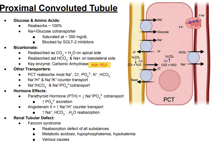
What proximal convoluted tubule disorder causes a generalized reabsorption defect?
Answer: Fanconi syndrome.
Extra Information:
The slide describes it as a reabsorption defect of all substances.
It can have various causes.

What renal tubular defect occurs in Fanconi syndrome?
Answer: A reabsorption defect of all substances in the proximal convoluted tubule.
Extra Information:
Fanconi syndrome therefore represents generalized proximal tubular dysfunction.

What metabolic abnormalities are associated with Fanconi syndrome on this slide?
Answer: Metabolic acidosis, hypophosphatemia, and hypokalemia.
Extra Information:
These abnormalities result from the generalized proximal convoluted tubule reabsorption defect.

What does the thin descending Loop of Henle passively reabsorb?
Answer: Water.
Extra Information:
Water reabsorption is passive.
It is driven by medullary hypertonicity.

What drives passive water reabsorption from the thin descending Loop of Henle?
Answer: Medullary hypertonicity.
Extra Information:
The hypertonic medulla draws water out of the descending limb.
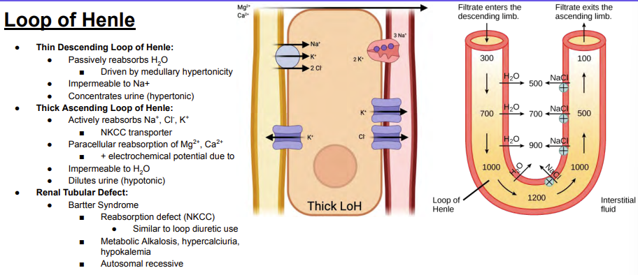
What is the thin descending Loop of Henle impermeable to according to the slide?
Answer: Sodium.
Extra Information:
In contrast, the thin descending limb is permeable to water.
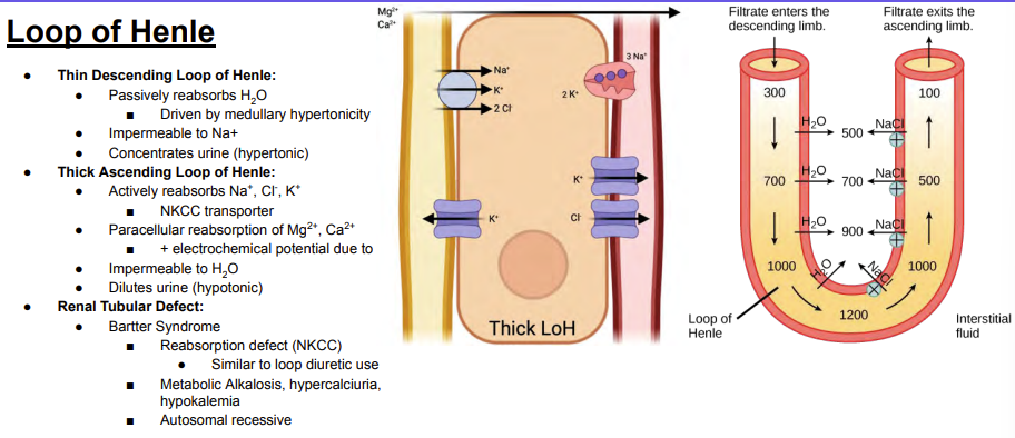
How does the thin descending Loop of Henle affect tubular fluid concentration?
Answer: It concentrates the tubular fluid, making it hypertonic.
Extra Information:
Water leaves the descending limb as the filtrate travels deeper into the hypertonic medulla.

What happens to water and tubular fluid concentration as filtrate travels down the thin descending Loop of Henle?
Answer: Water is passively reabsorbed → water leaves the tubule → the tubular fluid becomes progressively more concentrated.
Extra Information:
This process is driven by increasing medullary hypertonicity.
The slide's diagram shows increasing osmolarity deeper in the medulla.
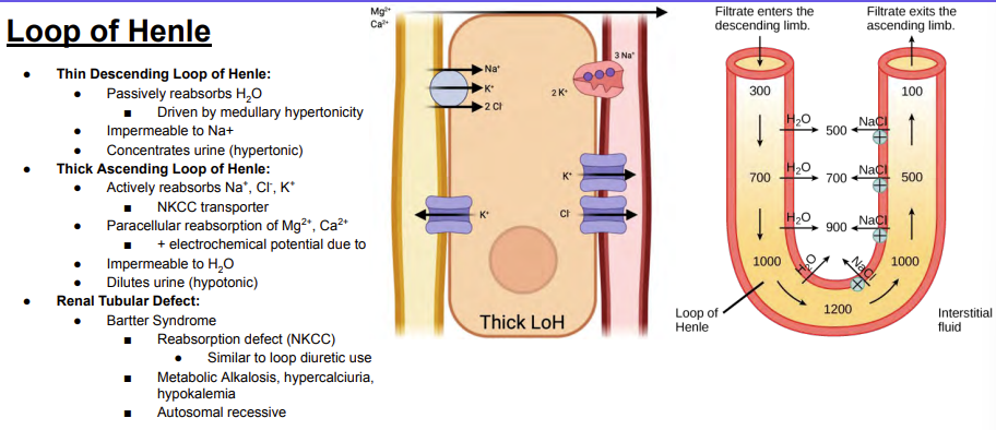
Which ions are actively reabsorbed by the thick ascending Loop of Henle?
Answer: Sodium, chloride, and potassium.
Extra Information:
These ions are reabsorbed through the NKCC transporter.
NKCC stands for:
N = Sodium (Na⁺)
K = Potassium (K⁺)
C = Chloride (Cl⁻)
C = Chloride (Cl⁻)
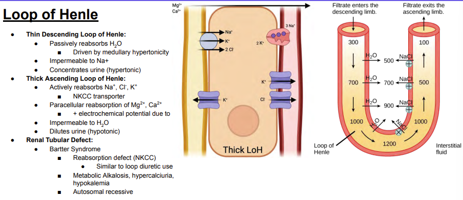
Which transporter actively reabsorbs sodium, potassium, and chloride in the thick ascending Loop of Henle?
Answer: The NKCC transporter.
Extra Information:
NKCC is the transporter affected in Bartter syndrome on this slide.

Which ions undergo paracellular reabsorption in the thick ascending Loop of Henle?
Answer: Magnesium and calcium.
Extra Information:
Their paracellular reabsorption is driven by a positive electrochemical potential.

What drives paracellular magnesium and calcium reabsorption in the thick ascending Loop of Henle?
Answer: A positive electrochemical potential due to potassium.
Extra Information:
This promotes paracellular reabsorption of magnesium and calcium.
Thick ascending limb: K⁺ recycling creates a positive luminal potential that drives paracellular reabsorption of Ca²⁺ and Mg²⁺.

What is the thick ascending Loop of Henle impermeable to?
Answer: Water.
Extra Information:
Despite active solute reabsorption, water does not follow.

Why does the thick ascending Loop of Henle dilute the tubular fluid?
Answer: It actively reabsorbs sodium, chloride, and potassium while remaining impermeable to water.
Extra Information:
Solute leaves without water following.
The resulting tubular fluid becomes hypotonic.

Compare the major functions of the thin descending and thick ascending limbs of the Loop of Henle.
Answer: The thin descending limb reabsorbs water and concentrates tubular fluid, while the thick ascending limb reabsorbs solutes without water and dilutes tubular fluid.
Extra Information:
The descending limb is impermeable to sodium.
The thick ascending limb is impermeable to water.

What transporter is defective in Bartter syndrome according to the slide?
Answer: The NKCC transporter in the thick ascending Loop of Henle.
Extra Information:
The defect causes impaired reabsorption in the thick ascending limb.
The slide compares Bartter syndrome with loop diuretic use.
