Synovial Joints
1/6
There's no tags or description
Looks like no tags are added yet.
Name | Mastery | Learn | Test | Matching | Spaced |
|---|
No study sessions yet.
7 Terms
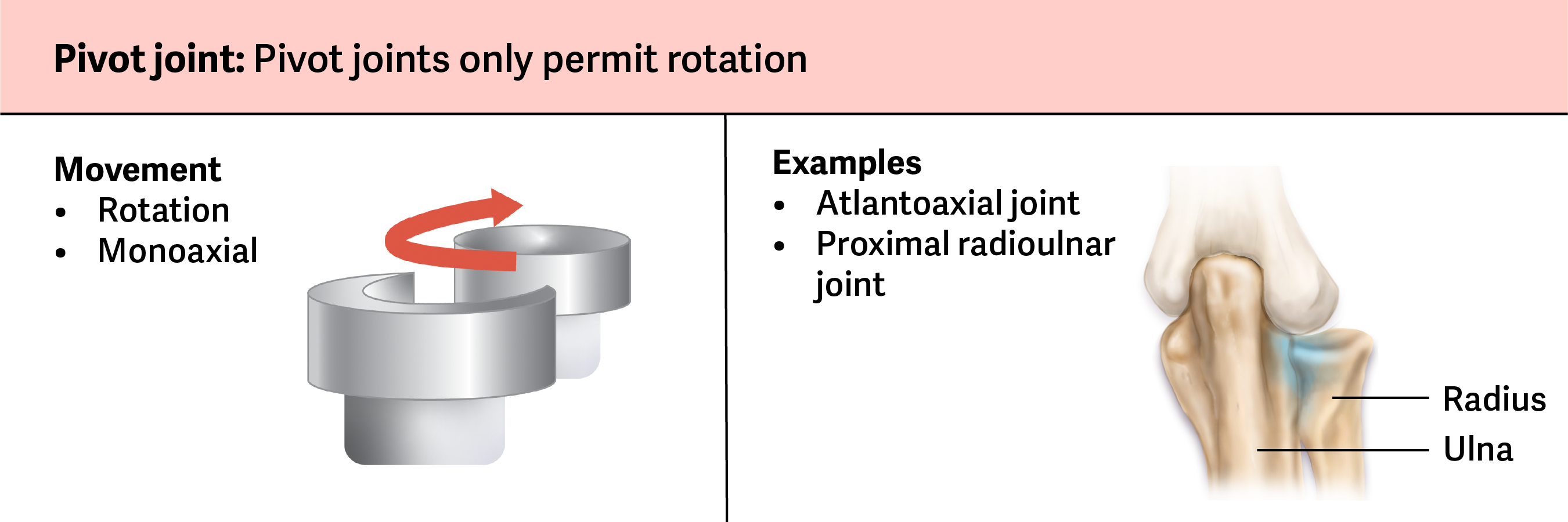
Pivot Joint
At a pivot joint, a rounded portion of a bone is enclosed within a ring formed partially by the articulation with another bone and partially by a ligament (Figure 4.22). The bone rotates within this ring. Because the rotation is around a single axis, pivot joints are functionally classified as a uniaxial diarthrosis type of joint.
Examples of Pivot Joints
The atlantoaxial joint, found between the C1 (atlas) and C2 (axis) vertebrae. The upward projecting dens of the axis articulates with the inner aspect of the atlas, where it is held in place by a ligament. Rotation at this joint allows turning of the head from side to side.
The proximal radioulnar joint. The head of the radius is largely encircled by a ligament that holds it in place as it articulates with the radial notch of the ulna. Rotation of the radius allows for supination and pronation.
Examples of Hinge Joints
The elbow joint, with the articulation between the trochlea of the humerus and the trochlear notch of the ulna.
The knee, ankle, and interphalangeal joints between the phalanx bones of the fingers and toes.

Examples of Condyloid Joints
The knuckle (metacarpophalangeal) joints of the hand between the distal end of a metacarpal bone and the proximal phalanx bone are condyloid joints. This allows for bending and straightening of the fingers in addition to abduction or adduction of spreading the fingers apart and bringing them together.
The radiocarpal joint of the wrist between the shallow depression at the distal end of the radius bone and the rounded scaphoid. The first axis of movement is flexion and extension at the wrist, while the second axis of motion allows for abduction and adduction of the wrist.

Examples of Saddle Joints
The first carpometacarpal joint between the trapezium (a carpal bone) and the first metacarpal bone at the base of the thumb.
This joint provides the thumb the ability to move away from the palm of the hand along two planes. The thumb can move within the same plane as the palm of the hand, or it can move out anteriorly, perpendicular to the palm. This movement of the first carpometacarpal joint is what gives "opposable" thumbs.
The sternoclavicular joint between the sternum and clavicle.

Examples of Plane Joints
Intercarpal joints between the carpal bones of the wrist and intertarsal joints between the tarsal bones of the foot.
Acromioclavicular joint between the clavicle and acromion of the scapula.
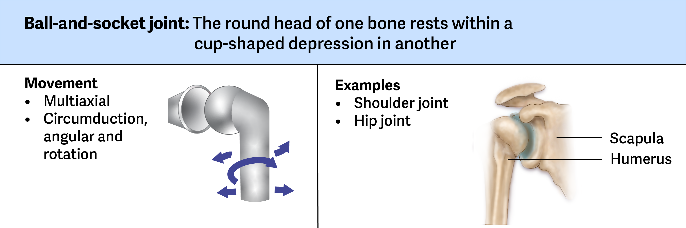
Examples of Ball and Socket Joints
At the hip joint, the head of the femur articulates with the acetabulum of the pelvis.
The deep socket of the acetabulum and the strong supporting ligaments of the hip joint serve to constrain movements of the femur, balancing the need for stability and weight-bearing ability with increased movements at the hip.
At the glenohumeral joint, the head of the humerus articulates with the glenoid cavity of the scapula.
The humerus has a smoothly rounded head that fits into the socket of the scapula at the glenoid cavity. The shallow socket formed by the glenoid cavity allows the shoulder joint to have an extensive range of motion. The glenoid cavity is very shallow and much smaller than the head of the humerus. The head of the humerus needs to be held to the shallow glenoid cavity by the rotator cuff muscles and other ligaments. The structure of the shoulder permits movement of the arm in almost any direction but provides little stability.