Cell Structure
1/109
There's no tags or description
Looks like no tags are added yet.
Name | Mastery | Learn | Test | Matching | Spaced | Call with Kai |
|---|
No analytics yet
Send a link to your students to track their progress
110 Terms
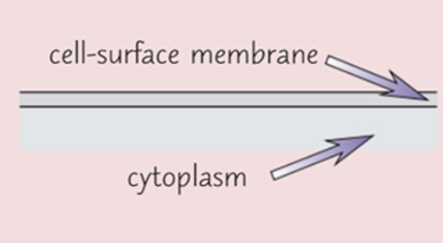
What is the function of the cell surface membrane?
Controls the movement of substances into and out of the cell
What is the function of the nucleus?
Controls the cells activities by controlling the transcription of DNA.
List 5 structures in a nucleus
- chromosomes
- one or more nucleoli
- nuclear envelope
- nuclear pores
- nucleoplasm
Describe a nuclear envelope
double membrane with pores that surrounds the nucleus
What is the function of a nuclear envelope?
separates the nucleus from the cytoplasm
Describe the function of nuclear pores
allows the passage of large molecules (e.g. RNA) out
How many pores are there typically in each nucleus?
3000, each 40-100nm in diameter
Describe the nucleoplasm
The granular, jelly-like material that makes up the bulk of the nucleus
What do the chromosomes in the nucleus consist of?
Protein bound, linear DNA
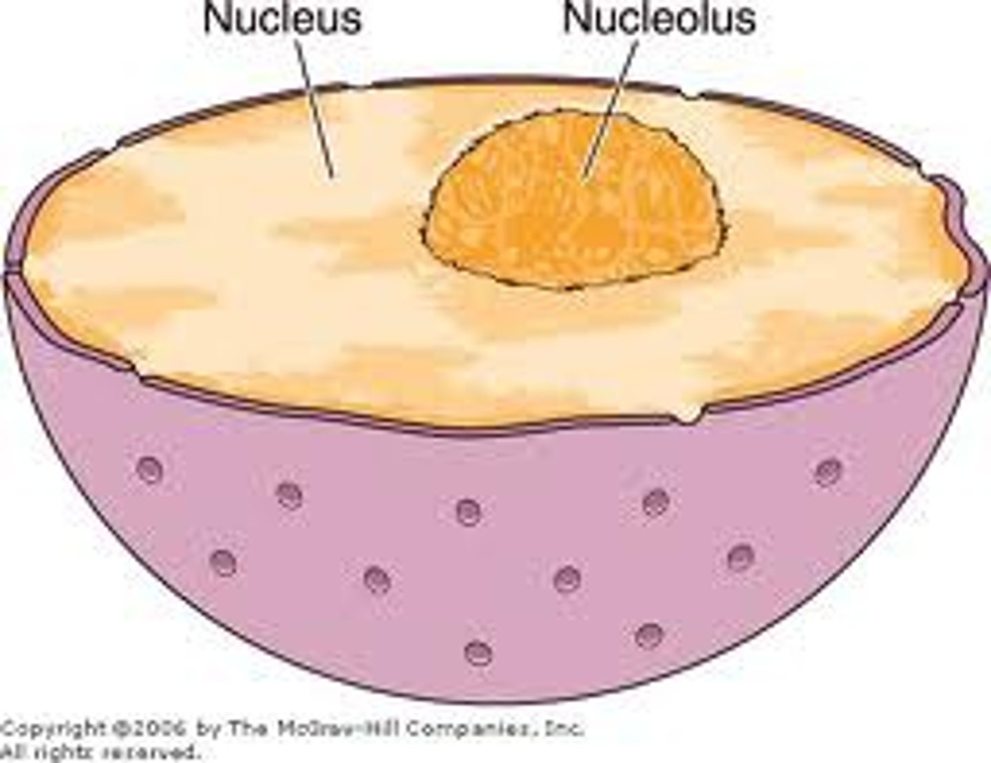
What is the nucleolus?
A small spherical region within the nucleoplasm
What is the function of the nucleolus?
Makes ribosomes
How does the nucleus control cell activity?
By controlling the transcription of DNA
What is the function of the mitochondria?
Site of aerobic respiration, producing ATP.
Describe the structure of the mitochondria
Double membranes - an outer membrane and a cristae or inner membrane
Intermembrane space - the space between the membranes
Matrix - the innermost part of the mitochondria
What are the cristae in the mitochondria?
folds in the inner membrane of mitochondria
What is the function of cristae in the mitochondria?
provides large surface area needed for attachment of enzymes and other proteins involved in respiration
What is the matrix of the mitochondria?
fluid inside cristae
What is the function of the matrix in the mitochondria?
Contains enzymes used for respiration
What is the function of chloroplast?
Site of photosynthesis
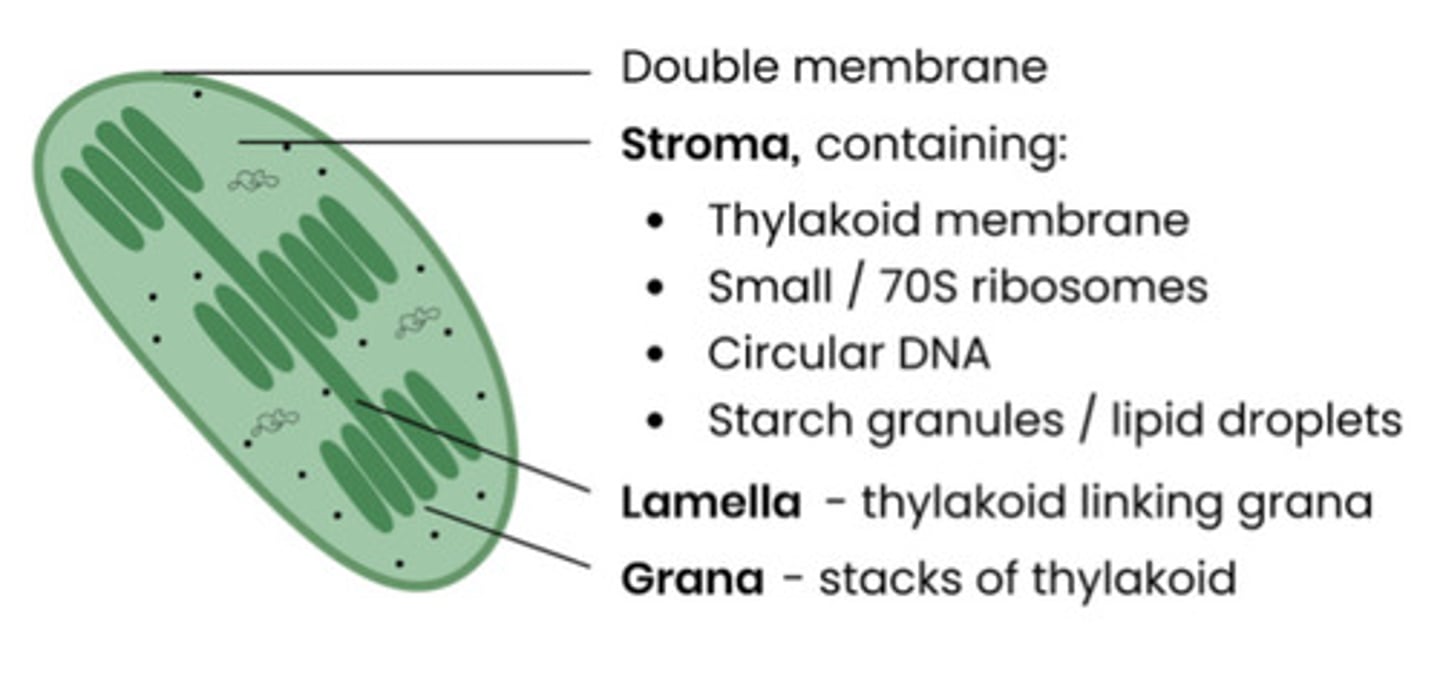
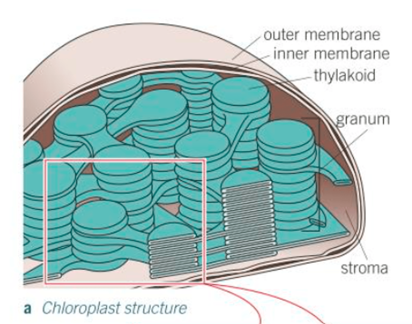
Describe the structure of the chloroplasts
stacks of thylakoids called granum surrounded by stroma with a double membrane
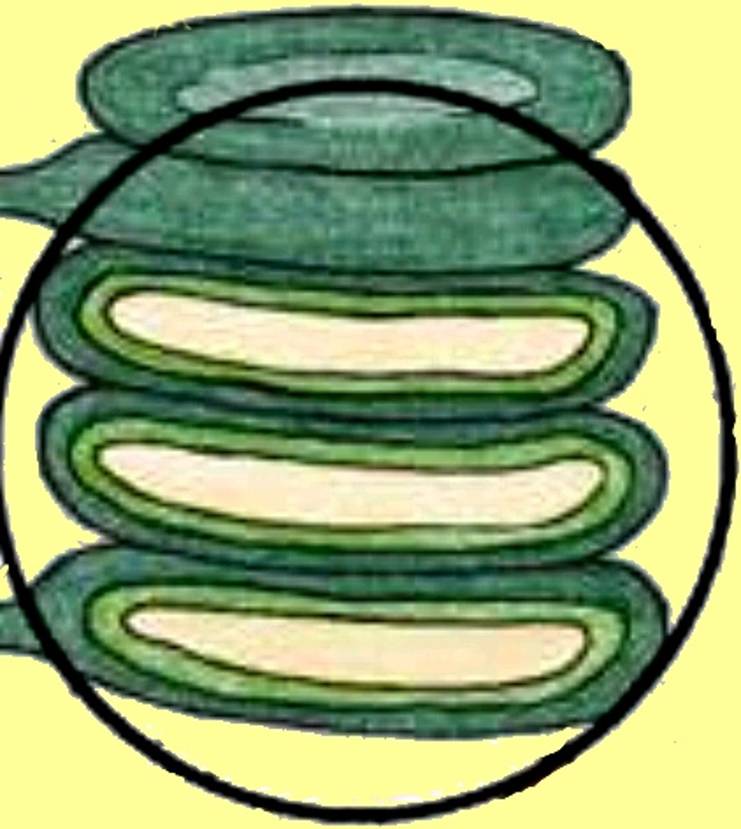
What are thylakoids?
saclike photosynthetic membranes that stack to form grana
What is the function of thylakoids?
Helps plants capture and convert light into energy. (first stage of photosynthesis)
What are grana linked by?
Lamellae - thin, flat pieces of thylakoid membrane.
What is the stroma?
fluid portion of the chloroplast; outside of the thylakoids
What is the function of the stroma?
Where 2nd stage of photosynthesis (synthesis of sugars) takes place
List the adaptations chlorophyll has for its function
- The granal membranes
- Fluid of stroma
- Contains both DNA and ribosomes
How do granal membranes help chloroplasts adapt for function?
- Provide large surface area for attachment of chlorophyll, electron carriers and enzymes that carry out the first stage of photosynthesis (chemicals are attached to membrane in order)
How do fluid of stroma help chloroplasts adapt for their function?
Possesses all the enzymes needed to make sugars in the second stage of photosynthesis
How does chloroplasts containing DNA and ribosomes help adapt it for it's function?
So they can quickly and easily manufacture some of the proteins needed for photosynthesis
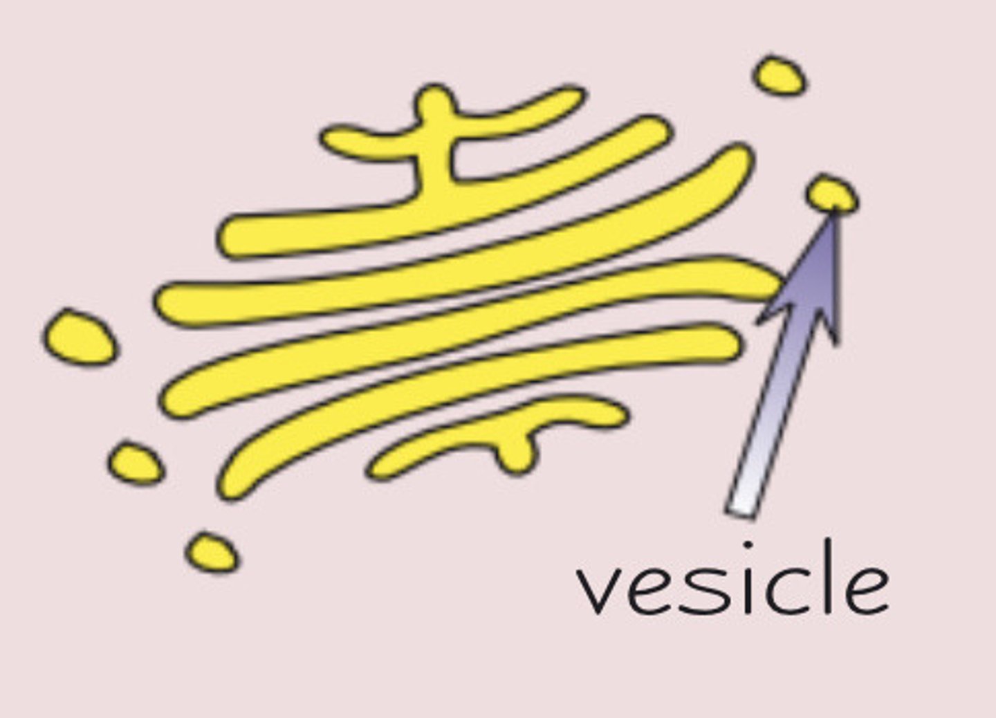
What is the function of Golgi apparatus?
Processes and packages new lipids and proteins and makes lysosomes
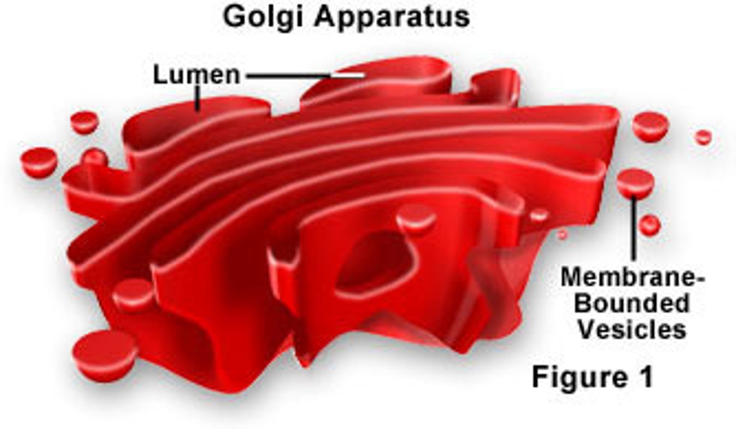
What is the function of the Golgi Vesicle?
Stores lipids and proteins made by the Golgi apparatus and transports them out of the cell.
In what type of cells are golgi bodies often found?
secretory cells e.g. epithelial cells in intestine
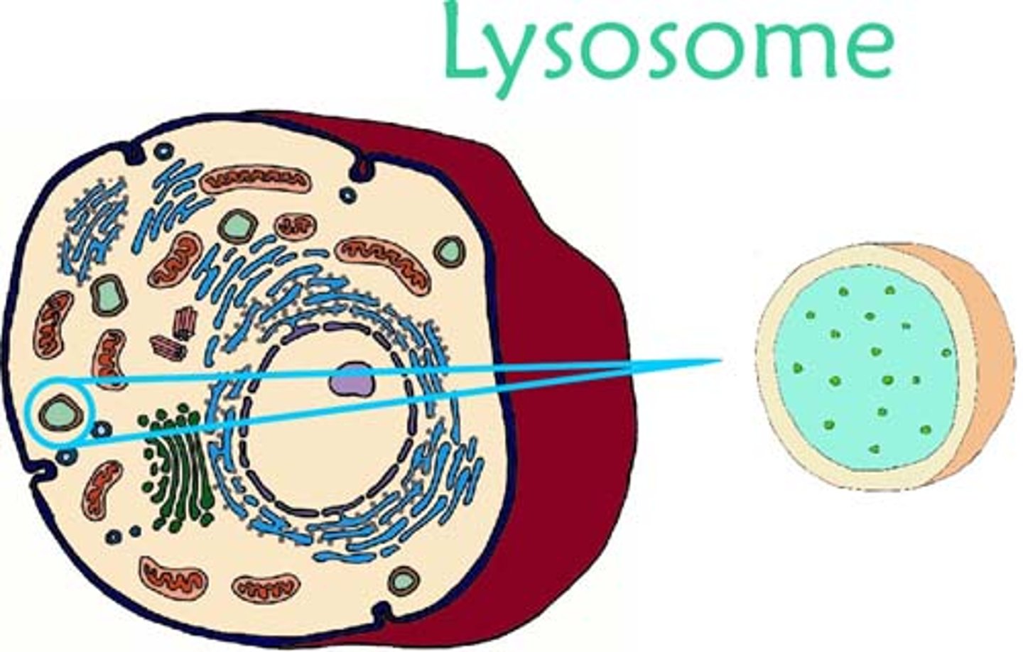
What is the function of the lysosome?
Digests cellular waste through the release of lysozymes.
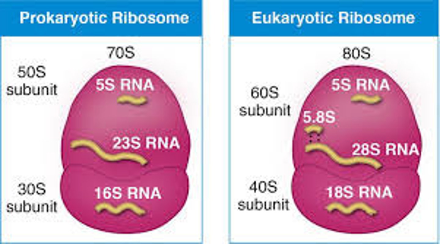
What is the function of the ribosome?
protein synthesis
What are the two types of ribosomes?
· 80s-eukaryotic - 25nm diameter
· 70s-prokaryotic - slightly smaller
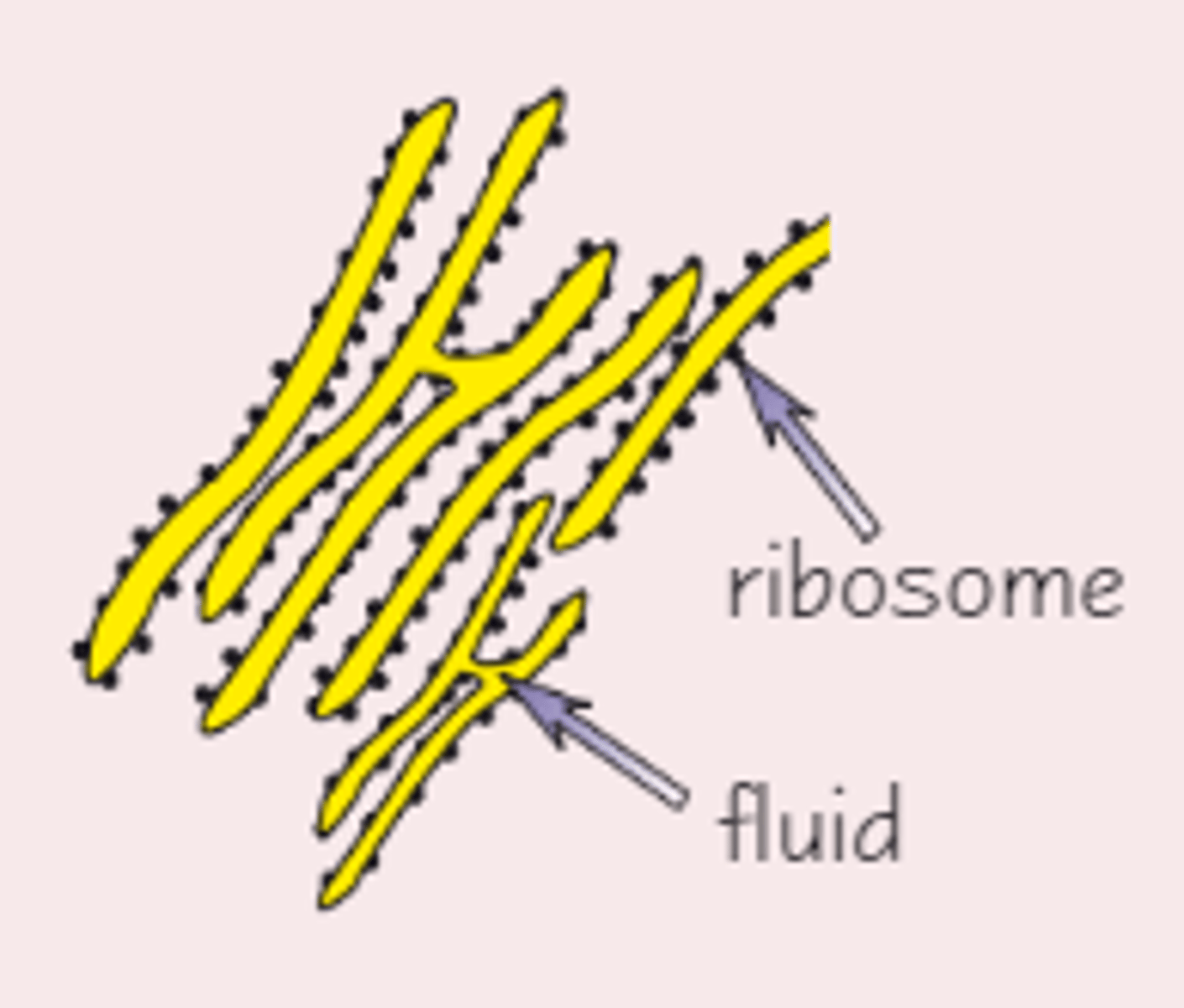
What is the function of Rough Endoplasmic Reticulum(RER)?
Folds and processes proteins made at the ribosomes
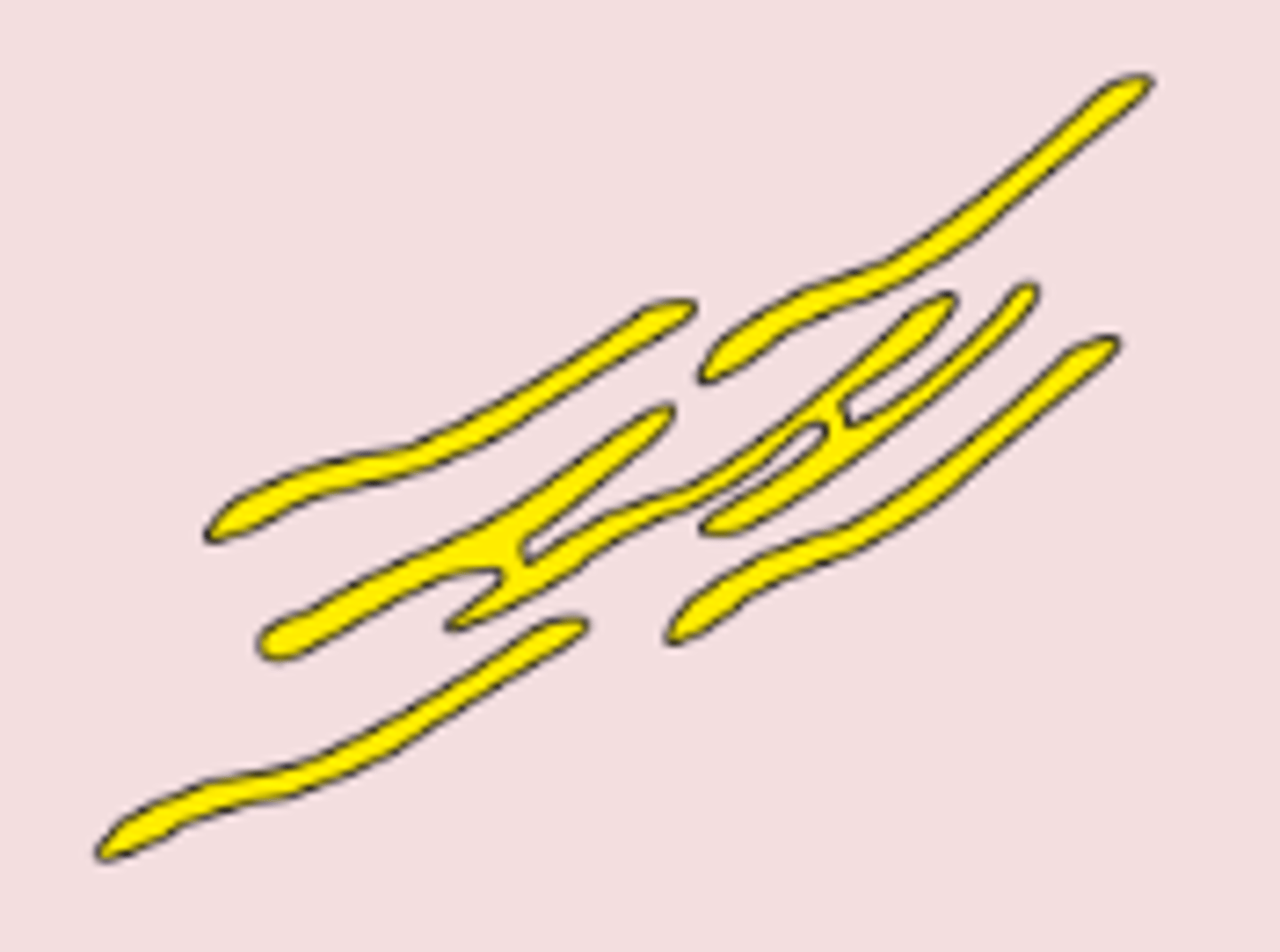
What is the function of Smooth Endoplasmic Reticulum(SER)?
Synthesises and processes lipids
Describe the structure of the cell wall in plants and algae.
- consists of microfibrils of cellulose, embedded in a matrix
- thin layer, called middle lamella, marks the boundary between cells and cements cells together
Describe the cell wall of fungi
External rigid cell wall made of chitin
What is the function of the cell wall? (3)
- stops cell bursting by osmosis
- gives mechanical strength to plant as a whole
- allows water to pass along it (contributes to the movement of water throughout the plant)
What is the function of the cell vacuole?
- Helps maintain pressure inside the cell, keeps it rigid which stops the plants from wilting.
- It is involved in the isolation of unwanted chemicals inside the cell.
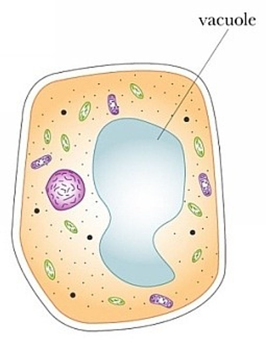
What is the single membrane around the cell vacuole called?
Tonoplast
What does the cell vacuole contain?
Cell sap - a weak solution of sugar and salts.
What's the difference between algal cells and plant cells?
Algal cells contain chloroplasts with different shapes to those found in plant cells.
How do prokaryotic cells differ from eukaryotic cells? (6 ways)
- much smaller
- have cytoplasm that lacks membrane-bound organelles
- have smaller ribosomes (70S)
- have no nucleus
- have a cell wall that contains murein, a glycoprotein
- have one or more plasmids - a capsule surrounding the cell - one or more flagella.
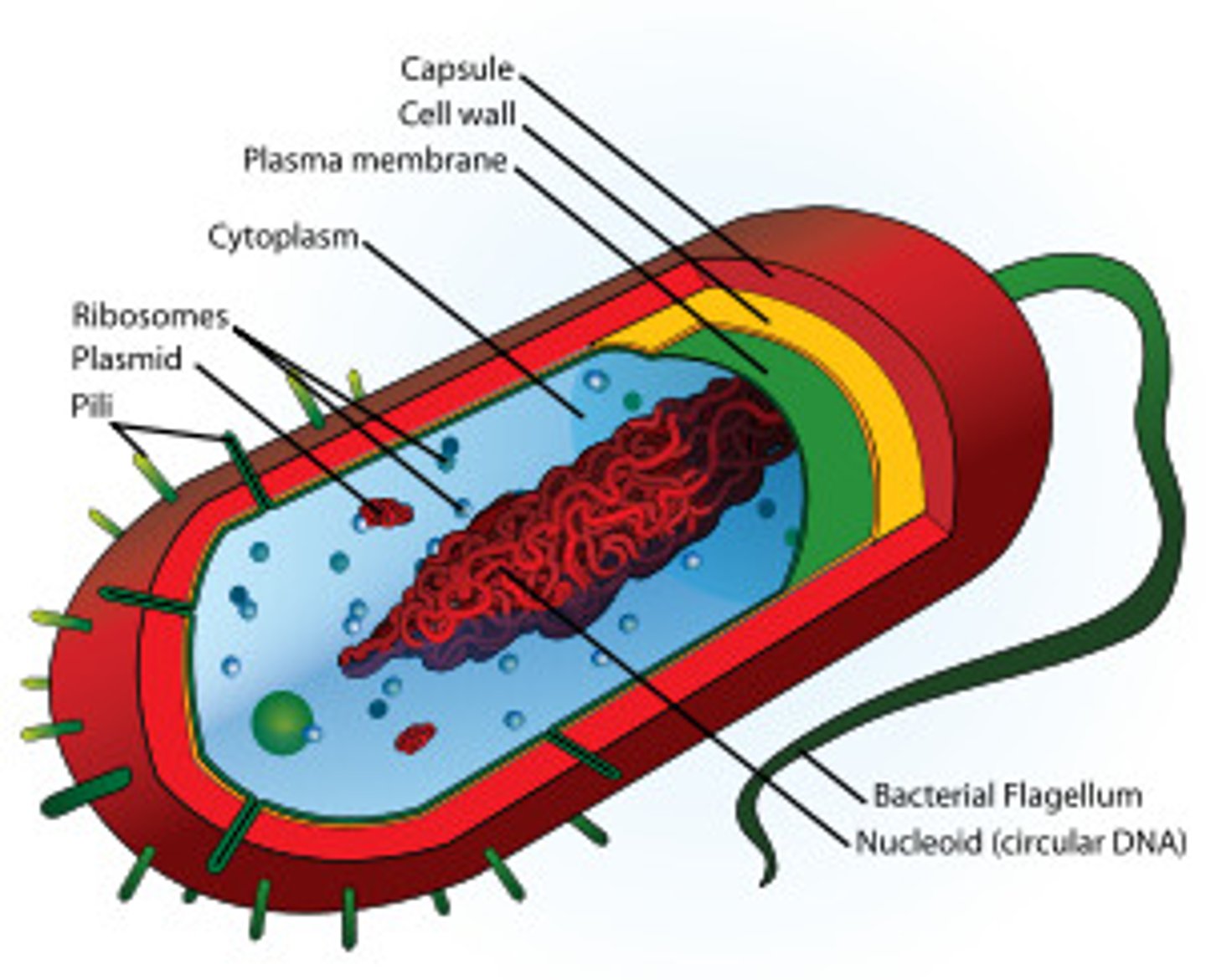
List the structures in a bacterial cell (8)
- Cell Wall
- Slime Capsule
- Plasmid
- Flagellum
- Pili
- Ribosomes
- Mesosomes
- Circular DNA
Describe the bacterial cell wall
the cell's rigid outer covering made of peptidoglycan
What is the bacterial cell wall made of?
Murein (a glycoprotein) - A polymer of polysaccharides and polypeptides
What is the function of the bacterial cell wall?
Provides the cell with strength and support.
Describe the slime capsule
protective slimy layer of polysaccharide which helps the cell to retain moisture and adhere to surfaces.
How does the slime capsule aid the bacterial cell?
- stop the cell drying out
- stop cells sticking together
- protect the cell against the action of a host's digestive enzymes
- gives extra protection to the cell
Describe the DNA in prokaryotic cells.
- Nucleoid - single circular piece of DNA with no nuclear membrane
- Plasmids - separate circles of DNA carrying only a few genes
What are plasmids?
small rings of DNA
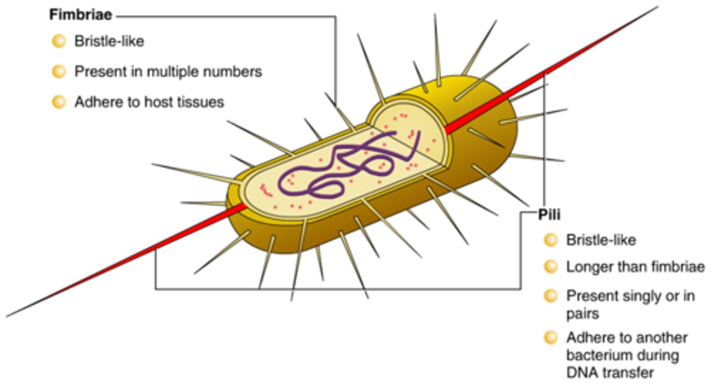
What is the pili?
hairlike structures that help bacteria stick to surfaces
What is the function of pili in prokaryotes?
used to attach prokaryotic cells to other cells or surfaces
What is the flagellum?
a tail-like structure which propels the cell forwards using a corkscrew motion.
What is the function of flagellum in prokaryotes?
movement
What are mesosomes in prokaryotes?
in-foldings of the cell membrane that provide a large surface area for the attachment of enzymes involved in respiration
What does the cytoplasm in a prokaryote contain?
• food store reserves (glycogen granules and oil droplets)
•70S ribosomes
•Circular strand of DNA not associated with histones (proteins)
•Plasmids
What are histones?
a protein that provides structural support for a chromosome
What are plasmids used for in prokaryotes?
Contains genes which can help bacteria survive in adverse conditions (e.g. by producing enzymes which can break down antibiotics) - can also be exchanged between bacteria
How are viruses classified?
Acellular → not made of or able to be divided into cells
Non-living → unable to exist/reproduce without a host cell
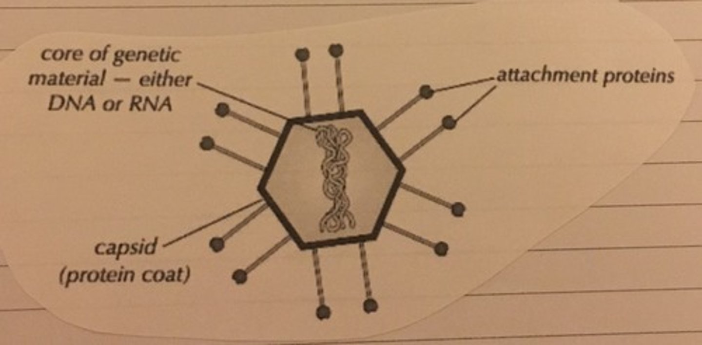
Describe the structure of viruses
All viruses contain nucleic acid, either DNA or RNA, and a protein coat (capsid), which encases the nucleic acid as well as attachment proteins, which let the virus cling onto a host cell
What is cell fractionation used for?
studying cell structure and function
What is cell fractionation?
The process where cells are broken up and the different organelles they contain are separated out.
What are the 2 stages of cell fractionation?
Homogenisation and ultracentrifugation
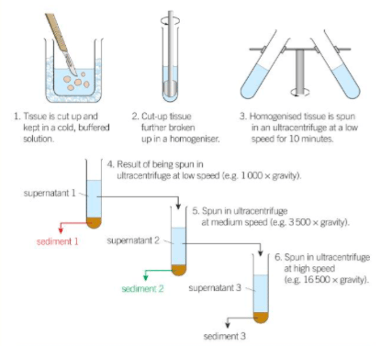
To prepare a tissue sample for cell fractionation, the sample is put in a solution which is:
- Ice cold
- Buffered
- Isotonic
Why is the solution the tissue sample is placed in ice cold?
To reduce enzyme activity which may hydrolyse cell contents
Why is the solution the tissue sample is placed in buffered?
So the pH doesn't fluctuate - pH change could alter the organelle structure or affect enzyme activity
Why is the solution the tissue sample is placed in isotonic?
Ensures same water potential as tissue to prevent organelles bursting or shrinking due to osmotic gain/loss
What occurs in homogenation?
cells are broken open (using a blender) and the different organelles are released
Why is the homogenate filtered?
To remove any complete cells and large pieces of debris
What occurs in ultracentrifugation?
the organelles are separated out to isolate them
Explain the process of ultracentrifugation (3)
1) The filtrate tube is put in a centrifuge and is spun at low speed. The heaviest organelles get flung to the bottom of the tube by the centrifuge.
2) the supernatent (fluid above sediment) is drained off, poured into another tube and spun in the centrifuge at a higher speed. the heaviest organelles form a pellet at the bottom, the supernatant is drained and spun at a higher speed.
3) The process in repeated at higher speeds until all the organelles are separated.
What do the organelles which spin to the bottom of the tube form? (the heaviest organelles)
The pellet
What is the fluid at the top of the test tube called? (the lightest organelles)
the supernatant
List the order of organelles from heaviest to lightest
Nuclei (heaviest)
Chloroplasts
Mitochondria
Lysosomes
Endoplasmic reticulum
Ribosomes (lightest)
Naughty Clever Monkeys Like Eating Red Raspberries
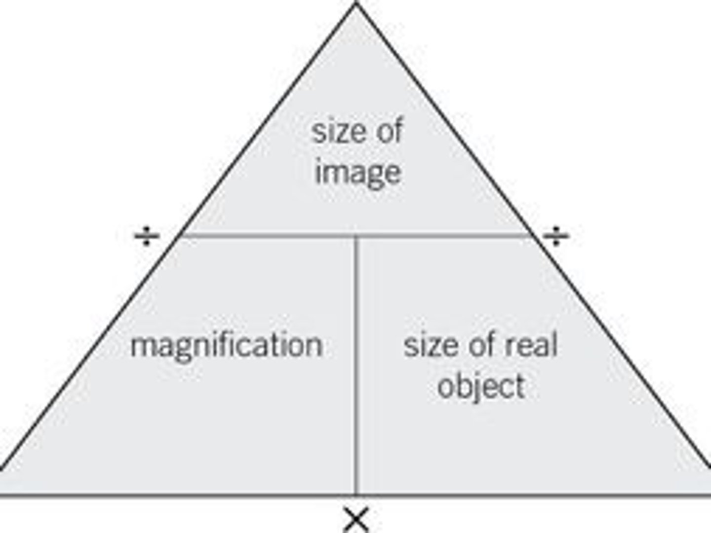
How do we calculate the magnification of a microscope?
Magnification = Size of image/ Size of real object
Define magnification
How much bigger the image is than the specimen
Define resolution
The ability to distinguish between two points
What's the difference between magnification and resolution?
Magnification is how much larger the image is than the specimen while resolution is the ability to distinguish between two separate points.
What is the resolution of an optical microscope?
0.2 um (micrometres)
What is the maximum magnification of an optical microscope?
1500x
What are the advantages of optical microscopes? (3)
- Images in colour.
- Can view living organisms.
- Easy to use and prepare.
What are the disadvantages of electron microscopes? (5)
· Expensive
· Training required to use
· Vacuum required
· Black and white image
· Specimens can't be alive
What are the 2 types of electron microscope?
transmission electron microscope (TEM) and scanning electron microscope (SEM)
How do transmission electron microscopes (TEM) work? (4)
•The electron beam passes through a thin section of the specimen
•Some parts absorb electrons and appear darker than others
•An image is produced on a screen
•A photograph can be taken - photomicrograph
How do scanning electron microscopes (SEM) work?
Scans a beam of electrons across the specimen which knocks electrons off. The electrons gather in a cathode ray to form the image.
What are the principles of a scanning electron microscope? (4)
- 3D image
- Max magnification - 100000x
- Resolution is 0.002 micrometres
- Can be used on thick specimens
What are the principles of a transmission electron microscope? (4)
- 2D image
- Max magnification - 1500000x
- Resolution is 0.0002 micrometres
- Specimens are thinly sliced
Why must electron microscopes be kept in a near vacuum?
electrons are absorbed by air molecules if present, which prevents the electrons from reaching the specimen and creates artefacts
How do prokaryotic cells replicate?
binary fission
Describe the process of binary fission
-The circular DNA and plasmids replicates (circular DNA replicates once, plasmids can be replicated many times)
- Cell gets bigger and DNA moves to opposite ends of cell
- Cytoplasm begins to divide and new cell walls begin to form
- The cytoplasm divides and 2 daughter cells are produced
What does binary fission produce?
two daughter cells
each with a single copy of the circular DNA
and a variable number of copies of plasmids
Describe the lytic cycle
- Virus attaches to host cell (via glycoproteins)
- Virus enters the cell
- Virus then uses host cell mechanisms to replicate, and incorporate their DNA.
- Host cell translates the DNA, which makes replicates of the virus until there are too many in the host cell, causing it to lyse (burst).
What does mitosis produce?
two genetically identical daughter cells that have the same number of chromosomes as the parent cell
What occurs before mitosis?
Interphase - DNA Replication
What are the stages of mitosis? (in order)
prophase, metaphase, anaphase, telophase
What are the stages of interphase?
G1, S, G2
What occurs in the G1 phase of the cell cycle?
Cell growth