Human Bioscience
 Call Kai
 Learn
Learn Practice Test
 Spaced Repetition
 Match
Match Flashcards
 Knowt Play
1/43

Earn XP
Description and Tags
OD 1 Fall Semester Human Bioscience Lecture Flashcards
Name | Mastery | Learn | Test | Matching | Spaced | Call with Kai | Chat |
|---|
No analytics yet
Send a link to your students to track their progress
44 Terms
Basic Principles of Histology?
Body is made of 4 types of tissues: Epithelium, Connective Tissue, Muscle, Nerves… Can also think of Gametes and Embryonic Stem Cells
Epithelium Overview
Covers and Lines tissue, High Cell Density, Little Intercellular Space
Epithelium Functions Overview
Barriers, Absorption, Secretion
Layers of Epithelium
Apical (outer), Basal (inner), Basement Membrane (outside cells - attaches epithelial cells to underlying CT)
Connective Tissue Overview
Low cell density, Excessive Extracellular Matrix (with collagen, elastin, immune cells, etc)
CT Functions Overview
Storage, Structural (holds things tg), Immunological
Types of CT Overview
Connective Tissues (softer), Cartilage, Bone (hardest), Blood (fluid form)
Nerve Tissue Overview
Rapid Cell to Cell communication, Excitable cells that experience changes in membrane potential for either Graded potentials or APs, Synaptic Transmission, Sensory motor integration, Glia - functional support
Muscle Overview
Excitable cells, Contractions, Several types, Effectors, Sensory motor elements
Other effector tissues?
Muscle, Endocrine Glands
Tissue types are categorized by…
Structural and Functional Characteristics
Structural Characteristics
Cell shapes and organizations, Extracellular matrix
Functional characteristics
Storage, protection, absorption, secretion, support, sensory, communication, contraction
Endothelium
specialized subtype of epithelium that lines internal capillaries, blood vessles, and lymphs
Simple Squamous Epithelium
1 single cell layer, flat/packed together, apical/basal side with basement membrane,
Dense Irregular Connective Tissue
Irregular due to unorganized collagen, loaded with collaged, houses capillaries lined with endothelium
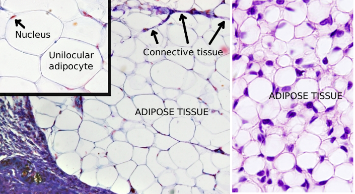
Unilocular Adipose Tissue
Storage connective tissue, adipose filled “bubbles” that has dissolved fats inside. Blood will store and pickup adipose from this connective tissue as needed
Where are all tissues derived from
Embryonic stem cells organized into germinal epithelia
Ectoderm derives…
skin and nerves
Endoderm derives
gut and organs that in gastrointestinal tract, lungs, etc
Mesoderm
Connective tissue, bone, muscle, other organs
Organs are composed of…
many tissue types
Skin layers
Epidermis (epithelium)
Dermis (connective tissue)
Hypodermis (connective tissue)
Ocular tissue can be simply categorized into which three layers?
Fibrous tunic
Vascular tunic
Retina
Fibrous Tunic
Sclera, Cornea
Vascular Tunic
Iris, Ciliary body, Choroid
Retina
Pigmented layer (retinal pigment epithelium - top layer), Neural layer (neural tissue)
Which tissues are found in the eye?
All 4 types: CT, Epithelium, Neural, Muscle
Eye epithelium
Cornea, Ciliary Processes, Lens, RPE
Eye CT
Cornea, Iris, Choroid, Sclera
Eye Neural
Retina, Optic nerve
Eye msucle
ciliary muscles, iris, choroid

Cornea Layers
Corneal Epithelium (5-6 layers and dense) —> Corneal Stroma (CT, less dense with lots of ECM) —> Corneal Endothelium (inner layer with basal, basement, and apical sides)
Rank from most superficial to deep: RPE, Choroid, Sclera, Ganglion Cells, Photoreceptor Cells, Retina
Sclera, Choroid, RPE, Photoreceptor Cells, Ganglion Cells, Retina
Visualization of structure and function: Perspective
Size, Orientation, 3D structures in 2D representation
Red Blood Cell Size
7um
Light Microscopy: Staining
Hematoxylin and Eosin, Special stains, Immunohistochemistry, In situ hybridization
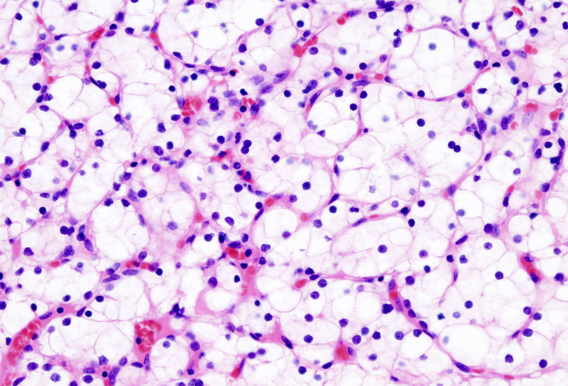
Hematoxylin and Eosin
most common stain - differential
Hemtoxylin - purple color staining nucleus
Eosin - pink color staining ECM/cytoplasm
Special Stain example
Trichrome stain- Keratin on outer skin surface, red dots is nuclei with cytoplasm
Fluorescent stain
antibodies with fluorescent markers:
Dapi (blue) - stains nuclei of cells
Green - stains actin
Red - stains cell/plasma membrane
Immunohistochemistry
use of antibodies —> different muscle fibers staiin different colors depending on speed at which enzymes cause contractions of fibers
in situ hybridization
stains to see function —> red dots usually indicate synthesis of specific proteins
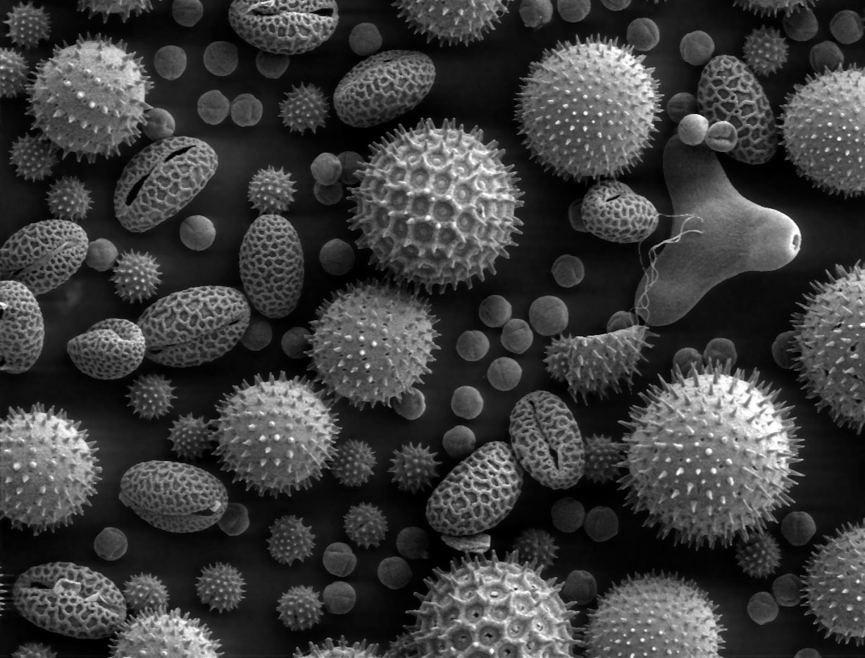
Electron microscopy
higher resolution and magnification visualization
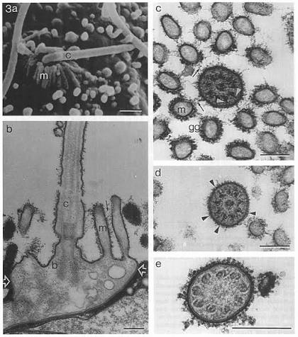
Transmission EM
cut thin section of cells —> scan ultra thin sections